Pharmaceutics 2020, 12, x; doi: FOR PEER REVIEW www.mdpi.com/journal/pharmaceutics A colon-targeted prodrug, 4-phenylbutyric acid-glutamic acid conjugate, ameliorates
2,4-dinitrobenzenesulfonic acid-induced colitis in rats
Soojin Kim1, Seunghyun Lee1, Hanju Lee1, Sanghyun Joo1, Sohee Park1, Doyoung Kwon1, Jin-Wook Yoo1, In-Soo Yoon1, Do Sik Min2, Young-Suk Jung1,*, and Yunjin Jung1,*
1College of Pharmacy, Pusan National University, Busan, 46241 Republic of Korea
2College of Pharmacy, Yonsei University, 85 Songdogwahak-ro, Yeonsu-gu, Incheon, 21983, Republic
of Korea
* Correspondence to Yunjin Jung and Young-Suk Jung
College of Pharmacy, Pusan National University, Busan, 46241 Republic of Korea Tel) 051-510-2527, Fax) 051-513-6754, E-mail) [email protected] (Y. J.)
Tel) 051-510-2816, Fax) 051-513-6754, E-mail) [email protected] (Y-S. J.)
Abstract: An elevated level of endoplasmic reticulum (ER) stress is considered an aggravating factor for inflammatory bowel disease (IBD). To develop an ER stress attenuator that is effective against colitis, 4-phenylbutyric acid (4-PBA), a chemical chaperone that alleviates ER stress, was conjugated with acidic amino acids to yield a 4-PBA-glutamic acid conjugate (PBA-GA) and a 4-PBA-aspartic acid conjugate (PBA-AA). The PBA derivatives were converted to 4-PBA in the cecal contents, where the conversion was greater with PBA-GA. After oral administration of PBA-GA (oral PBA-GA), millimolar levels of PBA were accumulated in the cecum, whereas 4-PBA was not detected in the blood, indicating the targeting of PBA-GA to the large intestine. At concentrations in the cecum achievable by oral PBA-GA, 4-PBA effectively attenuated ER stress in human colon epithelial cells. In 2,4-dinitrobenzenesulfonic acid-induced colitis in rats, oral PBA-GA alleviated the damage and inflammation in the colon. Moreover, oral PBA-GA substantially reduced the elevated levels of ER stress marker proteins in the inflamed colon. Moreover, PBA-GA was as effective as the currently used anti-IBD drug, sulfasalazine. In conclusion, PBA-GA is a colon-targeted prodrug of 4-PBA and is effective against rat colitis probably through the attenuation of ER stress in the inflamed colon.
Keywords: 4-Phenylbutyric acid, Colon-targeted drug delivery, Colitis, Prodrug, ER stress, Chemical chaperone
1. Introduction
Inflammatory bowel disease (IBD), which includes ulcerative colitis and Crohn’s disease, is an incurable and chronic disease of the gastrointestinal (GI) tract, with multifaceted and challenging clinical manifestations. Although the pathogenesis and etiology of IBD remain to be fully elucidated, progress in the past two decades has enhanced our understanding of the immunological and genetic aspects of IBD. Whatever be its fundamental pathological cause, the dysregulated host immune response caused by the gut microflora as well as genetic and environmental factors result in mucosal
inflammation, leading to a vicious cycle of inflammation–mucosal destruction–influx of antigens– immune response–inflammation, thereby perpetuating the unresolved inflammatory state [1,2].
The understanding of the pathogenesis of IBD, although obscure, has been translated to pharmacotherapy using drugs, which include aminosalicylates, glucocorticoids, immunosuppressants, and the burgeoning class of biologics, such as anti-TNF- agents [3,4]. These IBD therapeutics aim at inducing the remission of the destructive inflammation and the maintenance of long-term remission. In addition to the low efficacy (aminosalicylates) and intolerable adverse effects, resistance to these therapies and the induction of tolerance pose a great challenge for the treatment of IBD [5,6]. There is, therefore, an unmet medical need for the development of new IBD drugs with therapeutic and toxicological improvements.
The intestinal epithelium—the physical barrier between the mucosal layer and the gut luminal environment—plays a critical role in mucosal homeostasis through the regulation of the immune response, the composition of microflora, and intestinal pathogens as well as in the formation of the mucus layer, which is mediated by anti-microbial peptides, mucin, cytokines, chemokines, and hormones secreted from four types of intestinal epithelial cells—Paneth cells, goblet cells, enteroendocrine cells, and absorptive enterocytes [7-10]. This has been demonstrated in a number of animal and genome-wide association studies wherein genes closely related to epithelial functions have been identified [11,12]. Together with the physiological importance of the intestinal epithelium for mucosal homeostasis, a growing body of evidence demonstrates that intestinal epithelial dysfunction is associated with the development of various gastrointestinal disorders, including IBD [13-15].
Recent studies have shown that cellular stress signaling, including endoplasmic reticulum (ER) stress signaling and the unfolded protein response (UPR), is linked to the normal function of multiple epithelial cell populations in the gut [16,17]. Accordingly, uncontrolled ER stress impairs the mucosal barrier function, dysregulating the innate or adaptive immune response of the host cells and modulating the intestinal microbiota, which are established pathological conditions for the occurrence of IBD [16-19]. In fact, ER stress is associated with susceptibility to IBD, and signs of ER stress are commonly found in the ileal and/or colonic epithelia of patients with active IBD [17,20]. Therefore, interventions for restoring the ER function in intestinal epithelial cells is considered a therapeutic modality in the treatment of IBD [21].
ER stress involves the increased production of misfolded proteins, leading to the UPR. Under controlled conditions, the activation of the UPR mitigates cellular ER stress by eliminating misfolded proteins by preventing the aggregation of misfolded proteins and promoting their degradation. If cells are subjected to excessive and/or continuousER stress that is beyond the threshold of such stress that these cells can withstand, the cells undergo apoptosis. These cellular responses are mediated by three ER membrane proteins that sense stress; these proteins are protein kinase RNA-like ER kinase, inositol-requiring enzyme 1α, and activating transcription factor (AFT) 6α, each of which is involved in signaling pathways that transduce cellular signals, culminating in homeostatic responses and/or apoptosis [22,23].
restoring ER function are defined as chemical chaperones [25-27]. 4-Phenylbutyric acid (4-PBA), an FDA-approved drug for the treatment of urea cycle disorders, is a representative chemical chaperone. Because of its safety and effectiveness against ER stress [28], the anti-colitic activity of 4-PBA was investigated [29-31]. Consistent with the therapeutic hypothesis, 4-PBA reduces ER stress in the inflamed large intestine and shows considerable anti-colitic effects in animal models. However, its effective doses—500 to 1000 mg/kg—are too high to be translatable for clinical application.
Colon-targeted drug delivery (CTDD) implies the transport of an orally administered drug to the large intestine without loss (absorption and metabolism) in the stomach and small intestine [32,33]. Therefore, in general, the colonic delivery of a drug results in the accumulation of much greater amounts of the drug at the target site (colon) than possible with conventional delivery, indicating that the dose of a drug required for the treatment of colonic diseases, such as IBD, can be reduced by CTDD. Moreover, CTDD tends to have a reduced risk of systemic side effects because of the reduced dose and as the physiological features of the large intestine negatively influence drug absorption. Thus, this delivery technique is usually considered in the development of anti-colitic agents [32].
2. Materials and methods 2.1. Materials
4-PBA, 1,1′-carbonyldiimidazole (CDI), DNBS, glutamic acid dimethyl ester hydrochloride, L-aspartic acid dimethyl ester hydrochloride, and sulfasalazine (SSZ) were purchased from Tokyo Kasei Kogyo Co. (Tokyo, Japan). Phosphate-buffered saline (PBS, pH 7.4) was purchased from Thermo Fisher Scientific (Waltham, MA, USA). The solvents were obtained from Junsei Chemical Co. (Tokyo, Japan). All other chemicals were reagent grade commercially available products. Infrared (IR) spectra were recorded using a Varian FT-IR spectrophotometer (Varian, Palo Alto, CA, USA). 1 H-NMR spectra were recorded using a Varian AS 500 spectrometer (Varian). The chemical shifts in the NMR spectra were reported in ppm, downfield of tetramethylsilane.
2.2. Synthesis of N-(4-phenylbutanoyl) glutamic acid (PBA-GA) and N-(4-phenylbutanoyl)
aspartic acid (PBA-AA)
4-PBA (1.0 mmol) was reacted with CDI (1.3 mmol), dissolved in 10.0 mL of acetonitrile (ACN), for 30 min; this was followed by the addition of L-glutamic acid dimethyl ester hydrochloride (2.0 mmol) at 20 °C for 24 h. The solvent in the reaction mixture was removed by evaporation. The residue was dissolved in ethyl acetate (EA) after the removal of the solvent; it was washed thrice with 0.1 M HCl and 0.1 M NaOH, dried over anhydrous Na2SO4, and subsequently evaporated. The residue, dissolved in 8.0 mL of a 0.5 M NaOH solution, was stirred at 40 °C for 30 min. The resulting solution was acidified using a 1.0 M HCl solution and was extracted with EA. The organic layer was dried over anhydrous Na2SO4 and evaporated to obtain PBA-GA in the oil form. PBA-AA was synthesized using L-aspartic acid dimethyl ester hydrochloride according to the same method. The scheme for the synthesis of the 4-PBA derivatives is shown in Fig. 1A. The formation of PBA-GA and PBA-AA was confirmed via IR and 1H-NMR. PBA-GA; Yield: 67%; IR (nujol mull), νmax (cm−1): 1713 (C=O, -COOH), 1628 (C=O, -CONH); 1H-NMR (DMSO-d6): 1.82–1.72 (m, 2H), 1.99–1.92 (m, 1H), 1.82–1.72 (m, 1H), 2.13 (t, J = 7.3 Hz, 2H), 2.32–2.23 (m, 2H), 2.56 (t, J = 7.7 Hz, 2H), 4.20 (td, J = 9.0, 5.1 Hz, 1H), 7.21–7.15 (m, 3H), 7.27 (t, J = 7.5 Hz, 2H). PBA-AA, Yield: 61%; IR (nujol mull), νmax (cm−1): 1718 (C=O, -COOH), 1625 (C=O, -CONH); 1H-NMR (DMSO-d6): 1.82–1.73 (m, 2H), 2.12 (t, J = 7.3 Hz, 2H), 2.55 (dd, J = 14.5, 7.4 Hz, 2H), 2.69 (dd, J = 16.4, 5.6 Hz, 1H), 2.55 (dd, J = 14.5, 7.4 Hz, 1H), 4.53 (dd, J = 13.4, 7.7 Hz, 1H), 7.21–7.15 (m, 3H), 7.27 (t, J = 7.5 Hz, 2H). The IR and 1H-NMR spectra are shown in Fig. 1B and C.
2.3. High-performance liquid chromatography
The HPLC system consisted of a Gilson model 306 pump, a 151 variable UV detector, and a model 234 autoinjector (Gilson, Middleton, WI, USA). A Symmetry C18 column (HECTOR, RStech, Korea; 250 × 4.6 mm, 5 μm), with an attached guard column (Waters, 20 × 4.6 mm), was used for chromatographic separation. Samples from each experiment were filtered through membrane filters (0.45 μm). HPLC analyses of PBA, PBA-GA, and PBA-AA were conducted at a flow rate of 1.0 mL/min using mobile phases comprising methanol and 0.5% acetic acid solution (6.5:3.5, v/v). 5-Chlorosalicylic acid was used as an internal standard. The absorbance of the eluate was monitored at 214 nm using a UV detector, with a sensitivity of AUFS 0.01. The retention times of 4-PBA, PBA-GA, and PBA-AA were 12.4, 5.9, and 10.5 min, respectively.
2.4. Apparent distribution coefficient and chemical stability
6.8), pre-saturated with 1-octanol, was mixed. The mixture was shaken for 24 h and then allowed to stand for 4 h for phase separation at room temperature. The concentration of each compound in the aqueous phase was determined using a Shimadzu UV-Vis spectrophotometer (Tokyo, Japan) at 214 nm. The apparent distribution coefficients (D6.8) were calculated using the equation:
log 𝐷6.8 = log( 𝐶𝑂𝑐/𝐶𝑊) = log [(𝐶𝑂𝑐/(𝐶𝑂− 𝐶𝑂𝑐)]
CO: initial concentration of compound in 1-octanol
COc: equilibrium concentration of compound in 1-octanol
CW: equilibrium concentration of compound in isotonic phosphate buffer (pH 6.8)
PBA-GA and PBA-AA (1.0 mM) were tested for chemical stability in hydrochloric acid-sodium chloride buffer (pH 1.2) and isotonic phosphate buffer (pH 6.8). Each compound was incubated in these buffers for 24 h, and their concentration was measured by HPLC.
2.5. Cell culture
Human colon epithelial cells, NCM460, were grown in Dulbecco’s modified Eagle’s medium (HyClone, Logan, UT, USA) supplemented with 10% fetal bovine serum (HyClone) and penicillin/streptomycin (HyClone).
2.6. Western blot analysis
To prepare lysates of the distal colon, tissue samples (0.2 g) were disrupted and homogenized in 2.0 mL of ice-cold RIPA buffer (50 mM Tris-HCl [pH 7.4], 1 mM EDTA, 0.7% Na deoxycholate, 1% NP-40, 150 mM NaCl, 0.3 μM aprotinin, 1 μM pepstatin, and 1 mM PMSF) or the ProEXTM CETi protein
extraction solution (Translab, Daejeon, Korea) to determine the levels of ER stress marker proteins. After incubation on ice for 30 min, the homogenates were centrifuged at 10,000 × g at 4 °C for 20 min. The concentrations of proteins in the supernatants were determined using the bicinchoninic acid reagent (Thermo Fisher Scientific, Waltham, MA, USA) according to the manufacturer’s instructions. The cell and tissue lysates were electrophoretically separated on 7.5%, 10.0%, or 15.0% SDS– polyacrylamide gels and transferred onto nitrocellulose membranes (Bio-Rad, Hercules, CA, USA). The membrane was blocked with Tris-buffered saline with 0.1% Tween-20 (TBS-T), containing 5.0% skimmed milk powder, for 30 min at room temperature. Cyclooxygenase (COX)-2, inducible nitric oxide synthase (iNOS), ATF6, and C/EBP homologous protein (CHOP) were detected using anti-COX-2 (sc-376861, Santa Cruz Biotechnology, Santa Cruz, CA, USA), anti-iNOS (NOS-2) (sc-7271, Santa Cruz Biotechnology), anti-ATF6 (NBP1-40256, Novus Biologicals, Centennial, CO, USA), and anti-CHOP (L63F7, Cell Signaling Technology, Danvers, MA, USA) antibodies. The expression levels were normalized to those of α-tubulin or β-actin (Santa Cruz Biotechnology). After incubating the membranes with the appropriate secondary antibodies, they were detected using the EZ-Western Lumi Pico detection kit (DoGenBio, Seoul, Korea). Western blot images were quantified using Image Lab software (version 5.2 build 14, Bio-Rad, Hercules, CA, USA). The results are expressed as the means of quantified blots (n = 3 for cell experiments or 5 for animal experiments).
2.7. ELISA for CINC-3
2.8. Animals
Seven-week-old male Sprague-Dawley (SpD) rats were purchased from Samtako Bio Korea (Kyeong-gi-do, South Korea) and housed in the animal care facility at Pusan National University, Busan, South Korea, under controlled temperature, humidity, and dark/light cycle conditions. The animal protocol used in this study was reviewed and approved by the Pusan National University–Institutional Animal Care and Use Committee (PNU–IACUC) for ethical procedures and scientific care (approval no: PNU-2019-2392).
2.9. Incubation of PBA-GA and PBA-AA with the contents of the small and large intestines of the rats
A male SpD rat (250–260 g) was sacrificed using CO2 euthanasia, and a midline incision was made. The contents of the proximal small intestine (PSI), distal small intestine (DSI) and cecum were collected to prepare a 10% or 20% (w/v) suspension in isotonic phosphate buffer (pH 6.8). The incubation of the drugs with the cecal contents was conducted in a nitrogen gas bag (AtmosBag, Sigma). PBA-GA and PBA-AA (4.0 mL, 10 mM) dissolved in isotonic phosphate buffer (pH 6.8) were individually mixed with 4.0 mL of the 10% (w/v) suspension. The mixture was then incubated at 37 °C. For PBA-GA, an additional experiment was performed with the 20% (w/v) suspension. At appropriate time points, samples were collected and centrifuged at 10,000 × g for 5 min. To 0.1 mL portions of the supernatants, 0.9 mL methanol was added, followed by vortexing and then centrifugation at 20,000 × g at 4 °C for 10 min. The resultant mixture was filtered through a membrane filter (0.45 μm), and the concentration of each drug in the supernatants was measured via HPLC. 2.10. Analysis of drug concentration in blood and cecum
Male SpD rats were fasted for 24 h but had access to water. 4-PBA (30.0 mg/kg) or PBA-GA (53.6 mg/kg) in PBS (1.0 mL) was administered to rats by oral gavage. Blood samples were collected by cardiac puncture at 0.25, 2, and 6 h after the oral gavage. The blood samples were centrifuged at 10,000 × g at 4 °C for 10 min to obtain the plasma (0.3 mL), which was mixed with 10% perchloric acid (75.0 μL) and 5-chlorosalicylic acid (50.0 μM) by vortexing, and centrifuged at 10,000 × g at 4 °C for 5 min. The supernatants (0.25 mL), transferred to centrifuge tubes containing 7.6 μL of a saturated K2CO3 solution, were centrifuged for another 5 min [34]. To the supernatant (0.2 mL), 0.1 M HCl (0.6 mL) was added, which was followed by extraction with EA (0.7 mL). The organic layer (0.5 mL) was evaporated; the residue was dissolved in the mobile phase (0.15 mL) and filtered through a membrane filter (0.45 μm). Then, the filtrate (20.0 μL) was subjected to HPLC analysis.
To determine the drug concentrations in the cecum, the contents of the cecum obtained at 0.25, 2, and 6 h after oral gavage were mixed with isotonic phosphate buffer (pH 6.8) to make 10% suspensions. After centrifuging at 10,000 × g for 10 min at 4 °C, methanol (0.9 mL) was added to 0.1 mL of the supernatant. The mixture was filtered through a membrane filter (0.45 μm) and subjected to HPLC analysis.
2.11. DNBS-induced colitis in rats
2.12. Evaluation of the anti-colitic effect
To evaluate the anti-colitic effect of PBA-GA, two independent animal experiments were performed. In the first experiment, rats were divided into five groups (n = 5 per group) and treated as follows: group 1 (normal group): oral gavage of 1.0 mL PBS; group 2 (colitis group): oral gavage of 1.0 mL PBS; group 3 (4-PBA-treated colitis group): oral gavage of 4-PBA (30.0 mg/kg) in 1.0 mL of PBS; group 4 (PBA-GA-treated colitis group): oral gavage of PBA-GA (17.9 mg/kg, equivalent to 10.0 mg/kg of 4-PBA) in 1.0 mL PBS; and group 5 (PBA-GA-treated colitis group): oral gavage of PBA-GA (53.6 mg/kg, equivalent to 30.0 mg/kg of PBA) in 1.0 mL PBS. In the second experiment, rats were divided into four groups (n = 5 per group) and treated as follows: group 1 (normal group): oral gavage of 1.0 mL PBS; group 2 (colitis group): oral gavage of 1.0 mL PBS; group 3 (PBA-GA-treated colitis group): oral gavage of PBA-GA (53.6 mg/kg) in 1.0 mL PBS; and group 4 (SSZ-treated colitis group): oral gavage of SSZ (30.0 mg/kg) in 1.0 mL PBS. Three days after the induction of inflammation, each drug was administered orally to the rats once per day, and the anti-colitic effects were evaluated after administering the treatments for 7 days. The colonic damage score (CDS) was calculated according to previously reported criteria [36,37]. The modified scoring system was as follows: 0, normal appearance; 1, localized hyperemia but no ulcer; 2, linear ulcers without significant inflammation; 3, 2–4-cm site of inflammation and ulceration; 4, serosal adhesion to other organs, 2–4-cm site of inflammation and ulceration; and 5, stricture, serosal adhesion involving several bowel loops, <4-cm site of inflammation, and ulceration. Four independent observers blinded to the treatment conditions performed the CDS assessment. The myeloperoxidase (MPO) activity in the distal colon (4.0 cm) was measured as described previously [36]. The inflamed colon (0.2 g) was chopped in a vial containing 2.0 mL of a 5.0% hexadecyltrimethylammonium bromide (HTAB, pH 6.0) solution, homogenized, and incubated on ice for 20 min. The homogenate was centrifuged at 10,000 × g at 4 °C for 10 min; 0.1 mL of the supernatant was mixed with 2.9 mL of 0.05 M phosphate buffer (pH 6.0) containing o-dianisidine (16.7 mg) and 30.0% H2O2 (1.7 mL), and the MPO units were measured using a UV spectrophotometer (Shimadzu, Tokyo, Japan) for 5 min at 460 nm. One unit of MPO activity is defined as the amount of enzyme that degrades 1.0 μmol of peroxide per minute at 25 °C.
2.13. Data analysis
The results are expressed as the means ± standard deviation (SD). One-way analysis of variance (ANOVA) followed by Tukey’s HSD test or Mann-Whitney U test (for the CDS) was used to test the differences between the groups. Differences with α or P < 0.05 were considered statistically significant.
3. Results
3.1. Synthesis of 4-PBA conjugated with acidic amino acids
4-PBA was coupled with glutamic acid (GA) and aspartic acid (AA) to prepare the colon-targeted prodrugs, PBA-GA and PBA-AA. The chemical attachment of an amino acid as a carrier to a drug
with a carboxylic functional group is a well-established approach in the design of colon-targeted prodrugs of specific drugs [32]. PBA-GA and PBA-AA were prepared via simple synthetic reactions
as shown in Fig. 1, and the formation of the derivatives was verified by IR and 1H-NMR. In the IR spectra, the carbonyl stretching bands of the amide bonds formed by the conjugation of the amino
acids with 4-PBA were observed at 1628 cm−1 and 1625 cm−1 for PBA-GA and PBA-AA, respectively. In the 1H-NMR spectra, proton signals originating from 4-PBA and the amino acids were detected along with a slight downfield shift of the signals. In addition, the -carbon protons of the amino acids were downfield-shifted from 3.8 to 4.5, and amide protons ascribed to the formation of the amide
bond were newly detected at around 8.1. The IR and 1H-NMR spectra of the conjugates and 4-PBA are shown in supplementary data 1A and B.
.
Fig. 1. Scheme for the synthesis of N-(4-phenylbutanoyl) glutamic acid (PBA-GA) and N
-(4-phenylbutanoyl) aspartic acid (PBA-AA). ACN: acetonitrile, CDI: 1,1′-carbonyldiimidazole
3.2. Colon specificity of PBA-AA and PBA-GA
To examine the colon specificity of PBA-GA and PBA-AA, their DCs were determined. The DCs of
PBA-GA and PBA-AA were -1.56 and -1.86, respectively, and were much lower than that of 4-PBA (2.53). These results suggest that the PBA-amino acid conjugates are more hydrophilic and less efficient than 4-PBA for passive transport through the epithelial layer of the GI tract. To verify the
stability of PBA-GA and PBA-AA in the stomach and small intestine, their chemical stability was
tested in buffers of pH 1.2 and 6.8. The concentrations of the drugs in both buffers did not change over 24 h. Next, PBA-GA and PBA-AA were incubated with the contents of the small intestine and
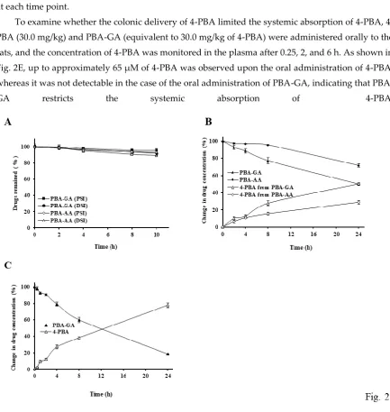
contents, the disappearance rate of PBA-GA was greater than that of PBA-AA; thus, PBA-GA was used in the subsequent in vivo experiments. As shown in Fig. 2C, in the 10% cecal suspension, the
disappearance of PBA-GA and the consequent release of 4-PBA occurred at a faster rate. These results suggest that the amino acid conjugates of 4-PBA are poorly absorbed and are stable in the small
intestine, whereas they are converted to 4-PBA in the large intestine. To ensure that PBA-GA could efficiently deliver 4-PBA to the large intestine, the concentration of 4-PBA in the cecal contents was
measured at 0.25, 2, and 6 h after the oral gavage of PBA-GA (oral PBA-GA, equivalent to 30.0 mg/kg of 4-PBA) and 4-PBA (oral 4-PBA, 30.0 mg/kg) to the rats. As shown in Fig. 2D, both 4-PBA and
PBA-GA were detected in the cecum when PBA-PBA-GA was administered orally, whereas no 4-PBA was detectable in the case of orally administered 4-PBA. The maximum 4-PBA concentration in the cecum
was approximately 2.7 mM at 2 h after the oral administration of PBA-GA. In agreement with the in vitro results for the cecal contents, the concentration of PBA-GA was much lower than that of 4-PBA
at each time point.
To examine whether the colonic delivery of PBA limited the systemic absorption of PBA,
4-PBA (30.0 mg/kg) and 4-PBA-GA (equivalent to 30.0 mg/kg of 4-4-PBA) were administered orally to the rats, and the concentration of 4-PBA was monitored in the plasma after 0.25, 2, and 6 h. As shown in
Fig. 2E, up to approximately 65 μM of 4-PBA was observed upon the oral administration of 4-PBA, whereas it was not detectable in the case of the oral administration of GA, indicating that
Fig. 2. PBA-GA and PBA-AA are colon specific.
(A) PBA-GA and PBA-AA (10.0 mM) were incubated with the contents of the PSI and DSI suspended in PBS (pH 6.8, 10.0%). (B) PBA-GA and PBA-AA were incubated with the cecal contents suspended in PBS (pH 6.8, 5.0%). (C) PBA-GA was incubated with the cecal contents suspended in PBS (pH 6.8, 10.0%). The levels of 4-PBA, PBA-GA, and PBA-AA were analyzed by HPLC. (D) Male SpD rats (250– 260 g) were starved for 24 h and were only provided water. 4-PBA (30.0 mg/kg) or PBA-GA (53.6 mg/kg, equivalent to 30.0 mg/kg of 4-PBA) suspended in PBS (pH 7.4) was administered to rats by oral gavage. The rats were sacrificed 0.25, 2, and 6 h after the oral gavage, and the concentrations of 4-PBA and PBA-GA in the cecum were analyzed via HPLC. (E) Male SpD rats (250–260 g) were starved for 24 h and were only provided water. 4-PBA (30.0 mg/kg) or PBA-GA (53.6 mg/kg, equivalent to 30.0 mg/kg of 4-PBA) suspended in PBS (pH 7.4) was administered to rats by oral gavage. Blood samples were obtained by cardiac puncture. 4-PBA was analyzed in the blood via HPLC. ND: not detectable. The data represent the means ± SD (n = 5).
3.3. PBA-GA mitigates DNBS-induced colitis in rats
To test the possibility that 4-PBA protects the colonic epithelial barrier against ER stress at colonic
concentrations achievable by 4-PBA-GA, human colon epithelial cells, NCM460, were treated with tunicamycin, an inducer of ER stress, in the presence of 1.0, 2.5, 3.0, and 5.0 mM 4-PBA. As shown in
Fig. 3A, 4-PBA dose-dependently reduced the levels of CHOP and ATF6, the ER stress marker proteins that are elevated by tunicamycin, and the degree of reduction at 3.0 mM 4-PBA was
approximately 80–90%. The above data suggest that PBA-GA can deliver 4-PBA at a level sufficient to reduce ER stress in colon epithelial cells. Thus, whether PBA-GA mitigated colonic inflammation
by preventing the induction of ER stress was examined. DNBS was instilled into the distal colon of the rats through the rectal route. Three days after the induction of inflammation, PBA-GA was orally
administered to the colitic rats once a day for 7 days. The same experiment was repeated with 4-PBA to examine whether the anti-colitic effects of PBA-GA were associated with the colonic delivery of
4-PBA. The dose of 4-PBA was 30.0 mg/kg, whereas PBA-GA was administered at two doses equivalent to 10.0 and 30.0 mg/kg of 4-PBA. Seven days after the administration of the drugs, the rats were
sacrificed, and the anti-colitic activity was evaluated. As shown in Fig. 3B and supplementary data 2, DNBS induced severe inflammation and mucosal damage along with tissue edema, stenosis, and
colonic damage at both doses, and there was no significant difference between the doses. In line with this, the MPO activity in the inflamed colonic tissues was decreased from 4.3 to 1.7 unit at 10.0 mg/kg
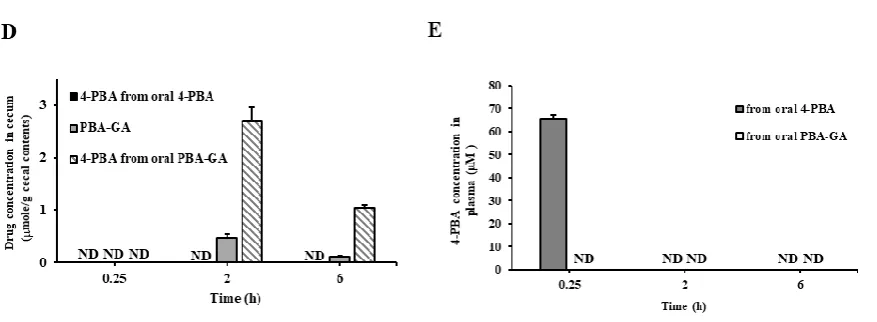
and to 1.4 unit at 30.0 mg/kg of PBA-GA (Fig. 3C). The molecular indices were also measured for the distal colon. As shown in Fig. 3D and E, PBA-GA decreased the production of inflammatory
mediators, such as COX-2, iNOS (Fig. 3D), and CINC-3 (Fig. 3E), which were substantially increased in the inflamed colon tissue. As for the macroscopic indices, no significant difference in the molecular
effects was observed between the doses of PBA-GA. In contrast, no significant macroscopic and molecular effects against colitis were observed with oral 4-PBA. To confirm the relevance of the
prevention of ER stress to the anti-colitic effects of PBA-GA, the levels of CHOP and ATF6 were observed in the inflamed distal colons. As shown in Fig. 3F, the colitis induced by DNBS markedly
elevated the levels of the ER stress marker proteins, and oral PBA-GA reduced the levels of these marker proteins to normal levels, whereas a decrease of approximately 40% was observed for orally
Fig. 3. PBA-GA ameliorates DNBS-induced rat colitis by attenuating ER stress.
(A) Human colon epithelial cells, NCM460, were treated with 4-PBA at various concentrations for 24 h and were then challenged with the ER stress inducer, tumicamycin (TM, 1g/mL), for 24 h. Cells were lysed, and the ER stress marker proteins, ATF6 and CHOP, were detected by western blotting. (B-F) Three days after the induction of colitis, PBA-GA (17.9 (L) and 53.6 mg/kg (H), equivalent to 10 and 30.0 mg/kg of 4-PBA) dissolved in 1.0 mL of PBS (pH 7.4) was administered orally to rats once per day. The rats were sacrificed after the seventh dose. For comparison, 4-PBA (30.0 mg/kg) was administered in the same manner as PBA-GA. (B) The colonic damage score (CDS) was determined for each group, as described in the Materials and methods section. (C) Myeloperoxidase (MPO) activity was measured in the inflamed distal colons (4.0 cm). Inflammatory mediators, such as (D) iNOS and COX-2 and (E) CINC-3, were measured in the inflamed colon. α-Tubulin was used as a loading control to normalize the levels of iNOS and COX-2. (F) The levels of the ER stress marker proteins, ATF6 and CHOP, were monitored in the inflamed colon. β-Actin was used as a loading control to normalize the levels of ATF6 and CHOP. *P < 0.05 vs. DNBS control, NM: not measurable. The data represent the means ± SD (n = 5).
3.4. PBA-GA is equally effective as SSZ in mitigating DNBS-induced rat colitis
activity in the inflamed colonic tissues, and PBA-GA was as effective as SSZ. Moreover, as shown in Fig. 4C and D, the levels of inflammatory mediators, COX-2, iNOS, and CINC-3, were diminished in the inflamed colonic tissues with oral PBA-GA, and there was no significant difference in the anti-inflammatory effects between PBA-GA and SSZ.
Fig. 4. PBA-GA is as effective as SSZ in ameliorating DNBS-induced rat colitis.
(A-D) Three days after the induction of colitis, PBA-GA (53.6 mg/kg, equivalent to 30.0 mg/kg of 4-PBA) dissolved and SSZ (30.0 mg/kg) suspended in 1.0 mL PBS (pH 7.4) were administered orally to rats once per day. The rats were sacrificed after the seventh dose. (A) The colonic damage score (CDS) was determined for each group as described in the Materials and methods section. (B) Myeloperoxidase (MPO) activity was measured in the inflamed distal colons (4.0 cm). Inflammatory mediators, such as (A) iNOS and COX-2 and (B) CINC-3, were assessed in the inflamed colon. α -Tubulin was used as a loading control to normalize the levels of iNOS and COX-2. The data represent the means ± SD (n = 5). *P < 0.05 vs. DNBS control, NM: not measurable
4. Discussion
mg/kg). To circumvent this obstacle, CTDD was employed to yield an orally active colon-targeted prodrug of 4-PBA that can be effective at a practical dose. The conjugation of acidic amino acids with drugs containing a carboxylic group is a well-established strategy in the design of colon-specific prodrugs. Thus, 4-PBA was conjugated with acidic amino acids, GA and AA, to synthesize PBA-GA and PBA-AA; as predicted, the 4-PBA derivatives exhibited colon specificity in vitro and in vivo, and PBA-GA was found to be superior to PBA-AA in the extent of colonic activation, likely leading to the greater availability of 4-PBA in the large intestine, which is more suitable for the purpose of this study, thereby reducing the dose.
Consistent with the general advantages of CTDD [32], PBA-GA is very likely to reduce the risk of systemic side effects due to the absorption of 4-PBA during long-term treatment [38,39]. Oral PBA-GA did not afford 4-PBA to the blood, presumably by effectively preventing the absorption of 4-PBA. In contrast, 4-PBA was detected up to 65.0 M in the blood after 15 min and then became undetectable 2 h after the oral administration of 4-PBA, showing rapid systemic absorption and metabolism, as previously reported [34]. In agreement with this observation, no 4-PBA was detected in the cecum of rats upon the oral administration of 4-PBA (30.0 mg/kg), whereas 4-PBA was accumulated up to about 2.7 mM (0.44 mg/g cecal contents) when PBA-GA (equivalent to 30.0 mg/kg of 4-PBA) was administered orally, further confirming the colon specificity of PBA-GA. A greater concentration of 4-PBA in the large intestine than in the blood, after the administration of equimolar doses, is also a general benefit of CTDD, which makes it possible to reduce the dose of 4-PBA for the treatment of colonic inflammation [32]. In addition, PBA absorbed systemically is rapidly metabolized to 4-phenylacetate, mainly in the liver and kidney, thus necessitating an increase in the dose to maintain active 4-PBA concentrations in the blood [34].
In agreement with these findings, while oral 4-PBA was not effective against rat colitis, oral PBA-GA effectively mitigated the colonic damage and inflammation in rat colitis even at one-third the dose of 4-PBA. Oral PBA-GA at 53.6 mg/kg (equivalent to 30 mg/kg of 4-PBA) was slightly more effective than oral PBA-GA at 17.9 mg/kg (equivalent to 10 mg/kg of 4-PBA), which was not statistically significant. Considering that the SSZ dose required for the treatment of ulcerative colitis is between 1,000 and 4,000 mg/day for human adults [40], the doses of PBA-GA are not impractical. 4-PBA is a chemical chaperone that reduces ER stress, which could be the potential pharmacological mechanism underlying the anti-colitic activity of 4-PBA. The oral administration of PBA-GA at a high dose resulted in the delivery and accumulation of 4-PBA in the cecum up to millimolar concentrations; at these concentrations, 4-PBA significantly reduced the levels of the ER stress marker proteins in human colon epithelial cells, which were elevated upon treatment with tunicamycin. Moreover, oral PBA-GA administration decreased the elevated levels of the ER stress marker proteins in the inflamed colons to their normal levels. These findings may explain why such high doses of 4-PBA (500–1,000 mg/kg) are needed for efficacy against murine colitis [29,31], given that the elimination half-life is very short owing to rapid metabolism, though oral administration of 5,000 mg of 4-PBA could afford about 1.18–1.32 mM in the blood in a human study [41]. Oral PBA-GA administration not only provides higher 4-PBA concentrations at the inflamed site but also avoids such metabolism, thus maintaining active therapeutic concentrations in the inflamed site for longer periods, even at much lower doses.
manifestation of the anti-colitic activity ascribed to the prevention of ER stress requires a greater reduction of the ER stress imposed on the inflamed colonic tissue. The oral administration of PBA-GA indeed reduced the levels of the ER stress markers to almost normal levels and was substantially effective against rat colitis. However, the manner in which oral 4-PBA reduces the levels of the ER stress marker proteins is not clear as the blood concentration obtained after oral 4-PBA administration was not sufficient to decrease the levels of the proteins, at least in human colon epithelial cells. Although, for now, no suitable explanation is available on this question, it is possible that the 4-PBA-mediated reduction of ER stress might be cell type-specific. Other than colon epithelial cells, other types of cells in the mucosal layers, such as immune cells, may be more sensitive to 4-PBA, which needs to be tested using immune cells isolated from the colonic mucosal layer.
In the DNBS-induced rat colitis model, PBA-GA was observed to be as effective against colitis as SSZ. Unlike the suppression of inflammatory signals, such as NF-B [42], by SSZ, PBA-GA is considered to exert its anti-colitic effects by protecting and fortifying the epithelial barrier. In addition, long-term NF-B inhibition may impair the integrity of the epithelium [43,44]. Thus, the combination of PBA-GA with a conventional drug may provide greater therapeutic benefit than that obtained with the use of such drugs alone. PBA-GA may reduce the risk of SSZ toxicity, and the distinct anti-colitic mechanisms may enable the combination therapy to elicit synergistic effects against colitis. For this reason, it is worth developing a colon-targeted mutual prodrug comprising 4-PBA and 5-ASA, which would be more beneficial and patient-friendly than the physical combination of SSZ and PBA-GA.
In conclusion, PBA-GA, a colon-targeted prodrug of the chemical chaperone, 4-PBA, effectively mitigated colonic damage and inflammation in a DNBS-induced rat colitis model without significant systemic absorption of the drug and was as effective as SSZ. Moreover, the anti-colitic activity of PBA-GA was observed at a lower dose than that achievable with 4-PBA. These results suggest that the colonic delivery of 4-PBA is a feasible strategy to reduce the effective dose and potential side effects of 4-PBA, thus allowing better compliance by patients.
Supplementary Materials: The following are available online
Figure S1: Instrumental analysis of PBA-GA and PBA-AA Figure S2: Photos of the serosal and luminal sides of the colons Figure S3: Photos of the serosal and luminal sides of the colons
Author Contributions
Soojin Kim: Performing most experiments, data curation, analysis, and interpretation, writing original draft
Seunghyun Lee, Hanju Lee, Sanghyun Joo, Sohee Park, and Doyoung Kwon: HPLC analysis and assistance in animal experiments
Jin-Wook Yoo, In-Soo Yoon, and Do Sik Min: HPLC analysis, data interpretation, and writing-review Yunjin Jung and Young-Suk Jung: Conceptualization, supervision, funding acquisition (Y.J.), data interpretation and writing-review and editing
Acknowledgments
This research was supported by Basic Science Research Program through the National Research Foundation of Korea (NRF) funded by the Ministry of Education (2018R1D1A3B07045694)
Conflicts of Interest:
5. References
1. Xavier, R.J.; Podolsky, D.K. Unravelling the pathogenesis of inflammatory bowel disease. Nature 2007, 448, 427-434, doi:10.1038/nature06005.
2. Abraham, C.; Cho, J.H. Inflammatory bowel disease. N Engl J Med 2009, 361, 2066-2078, doi:10.1056/NEJMra0804647.
3. Bryant, R.V.; Brain, O.; Travis, S.P. Conventional drug therapy for inflammatory bowel disease. Scand J Gastroenterol 2015, 50, 90-112, doi:10.3109/00365521.2014.968864.
4. Ko, J.K.; Auyeung, K.K. Inflammatory bowel disease: etiology, pathogenesis and current therapy. Curr Pharm Des 2014, 20, 1082-1096, doi:10.2174/13816128113199990416.
5. Randall, C.W.; Vizuete, J.A.; Martinez, N.; Alvarez, J.J.; Garapati, K.V.; Malakouti, M.; Taboada, C.M. From historical perspectives to modern therapy: a review of current and future biological treatments for Crohn's disease. Therap Adv Gastroenterol 2015, 8, 143-159, doi:10.1177/1756283X15576462.
6. Pithadia, A.B.; Jain, S. Treatment of inflammatory bowel disease (IBD). Pharmacol Rep 2011, 63, 629-642, doi:10.1016/s1734-1140(11)70575-8.
7. Peterson, L.W.; Artis, D. Intestinal epithelial cells: regulators of barrier function and immune homeostasis. Nat Rev Immunol 2014, 14, 141-153, doi:10.1038/nri3608.
8. Kim, Y.S.; Ho, S.B. Intestinal goblet cells and mucins in health and disease: recent insights and progress. Curr Gastroenterol Rep 2010, 12, 319-330, doi:10.1007/s11894-010-0131-2. 9. Gallo, R.L.; Hooper, L.V. Epithelial antimicrobial defence of the skin and intestine. Nat Rev
Immunol 2012, 12, 503-516, doi:10.1038/nri3228.
10. Henderson, P.; van Limbergen, J.E.; Schwarze, J.; Wilson, D.C. Function of the intestinal epithelium and its dysregulation in inflammatory bowel disease. Inflamm Bowel Dis 2011, 17, 382-395, doi:10.1002/ibd.21379.
11. Vaishnava, S.; Behrendt, C.L.; Ismail, A.S.; Eckmann, L.; Hooper, L.V. Paneth cells directly sense gut commensals and maintain homeostasis at the intestinal host-microbial interface. Proc Natl Acad Sci U S A 2008, 105, 20858-20863, doi:10.1073/pnas.0808723105.
12. Kobayashi, K.S.; Chamaillard, M.; Ogura, Y.; Henegariu, O.; Inohara, N.; Nunez, G.; Flavell, R.A. Nod2-dependent regulation of innate and adaptive immunity in the intestinal tract. Science 2005, 307, 731-734, doi:10.1126/science.1104911.
13. Salim, S.Y.; Soderholm, J.D. Importance of disrupted intestinal barrier in inflammatory bowel diseases. Inflamm Bowel Dis 2011, 17, 362-381, doi:10.1002/ibd.21403.
14. Zuo, T.; Ng, S.C. The Gut Microbiota in the Pathogenesis and Therapeutics of Inflammatory Bowel Disease. Front Microbiol 2018, 9, doi:ARTN 224710.3389/fmicb.2018.02247.
15. Atreya, R.; Neurath, M.F. IBD pathogenesis in 2014: Molecular pathways controlling barrier function in IBD. Nat Rev Gastroenterol Hepatol 2015, 12, 67-68, doi:10.1038/nrgastro.2014.201. 16. Coleman, O.I.; Haller, D. ER Stress and the UPR in Shaping Intestinal Tissue Homeostasis
and Immunity. Front Immunol 2019, 10, 2825, doi:10.3389/fimmu.2019.02825.
17. Cao, S.S. Endoplasmic reticulum stress and unfolded protein response in inflammatory bowel disease. Inflamm Bowel Dis 2015, 21, 636-644, doi:10.1097/MIB.0000000000000238. 18. Ma, X.; Dai, Z.; Sun, K.; Zhang, Y.; Chen, J.; Yang, Y.; Tso, P.; Wu, G.; Wu, Z. Intestinal
Pathogenesis: An Update Review. Front Immunol 2017, 8, 1271, doi:10.3389/fimmu.2017.01271.
19. Eri, R.D.; Adams, R.J.; Tran, T.V.; Tong, H.; Das, I.; Roche, D.K.; Oancea, I.; Png, C.W.; Jeffery, P.L.; Radford-Smith, G.L., et al. An intestinal epithelial defect conferring ER stress results in inflammation involving both innate and adaptive immunity. Mucosal Immunol 2011, 4, 354-364, doi:10.1038/mi.2010.74.
20. Cao, S.S. Epithelial ER Stress in Crohn's Disease and Ulcerative Colitis. Inflamm Bowel Dis 2016, 22, 984-993, doi:10.1097/MIB.0000000000000660.
21. Hetz, C.; Chevet, E.; Harding, H.P. Targeting the unfolded protein response in disease. Nat Rev Drug Discov 2013, 12, 703-719, doi:10.1038/nrd3976.
22. Liu, C.Y.; Kaufman, R.J. The unfolded protein response. J Cell Sci 2003, 116, 1861-1862, doi:10.1242/jcs.00408.
23. Kadowaki, H.; Nishitoh, H. Signaling Pathways from the Endoplasmic Reticulum and Their Roles in Disease. Genes-Basel 2013, 4, 306-333, doi:10.3390/genes4030306.
24. Ryno, L.M.; Wiseman, R.L.; Kelly, J.W. Targeting unfolded protein response signaling pathways to ameliorate protein misfolding diseases. Curr Opin Chem Biol 2013, 17, 346-352, doi:10.1016/j.cbpa.2013.04.009.
25. Burrows, J.A.; Willis, L.K.; Perlmutter, D.H. Chemical chaperones mediate increased secretion of mutant alpha 1-antitrypsin (alpha 1-AT) Z: A potential pharmacological strategy for prevention of liver injury and emphysema in alpha 1-AT deficiency. Proc Natl Acad Sci U S A 2000, 97, 1796-1801, doi:10.1073/pnas.97.4.1796.
26. Powers, E.T.; Morimoto, R.I.; Dillin, A.; Kelly, J.W.; Balch, W.E. Biological and Chemical Approaches to Diseases of Proteostasis Deficiency. Annu Rev Biochem 2009, 78, 959-991, doi:10.1146/annurev.biochem.052308.114844.
27. Ozcan, U.; Yilmaz, E.; Ozcan, L.; Furuhashi, M.; Vaillancourt, E.; Smith, R.O.; Gorgun, C.Z.; Hotamisligil, G.S. Chemical chaperones reduce ER stress and restore glucose homeostasis in a mouse model of type 2 diabetes. Science 2006, 313, 1137-1140,
doi:10.1126/science.1128294.
28. Kolb, P.S.; Ayaub, E.A.; Zhou, W.; Yum, V.; Dickhout, J.G.; Ask, K. The therapeutic effects of 4-phenylbutyric acid in maintaining proteostasis. Int J Biochem Cell Biol 2015, 61, 45-52, doi:10.1016/j.biocel.2015.01.015.
29. Ono, K.; Nimura, S.; Hideshima, Y.; Nabeshima, K.; Nakashima, M. Orally administered sodium 4-phenylbutyrate suppresses the development of dextran sulfate sodium-induced colitis in mice. Exp Ther Med 2017, 14, 5485-5490, doi:10.3892/etm.2017.5251.
30. Ono, K.; Nimura, S.; Nishinakagawa, T.; Hideshima, Y.; Enjyoji, M.; Nabeshima, K.; Nakashima, M. Sodium 4-phenylbutyrate suppresses the development of dextran sulfate sodium-induced colitis in mice. Exp Ther Med 2014, 7, 573-578, doi:10.3892/etm.2013.1456. 31. Cao, S.S.; Zimmermann, E.M.; Chuang, B.M.; Song, B.; Nwokoye, A.; Wilkinson, J.E.; Eaton,
K.A.; Kaufman, R.J. The unfolded protein response and chemical chaperones reduce protein misfolding and colitis in mice. Gastroenterology 2013, 144, 989-1000 e1006, doi:10.1053/j.gastro.2013.01.023.
33. Sharma, S.; Sinha, V.R. Current pharmaceutical strategies for efficient site specific delivery in inflamed distal intestinal mucosa. J Control Release 2018, 272, 97-106,
doi:10.1016/j.jconrel.2018.01.003.
34. Wang, C.L.; Hsueh, P.R.; Sun, M.J.; Leu, Y.L.; Chang, F.S.; Yang, S.W.; Lian, J.F.; Wang, H.P. PBA-omega-Lys as Sustained Phenylbutyrate-Releasing Prodrug. J Food Drug Anal 2010, 18, 371-379.
35. Morris, G.P.; Beck, P.L.; Herridge, M.S.; Depew, W.T.; Szewczuk, M.R.; Wallace, J.L. Hapten-induced model of chronic inflammation and ulceration in the rat colon. Gastroenterology 1989, 96, 795-803.
36. Hong, S.; Yum, S.; Yoo, H.J.; Kang, S.; Yoon, J.H.; Min, D.; Kim, Y.M.; Jung, Y. Colon-targeted cell-permeable NFkappaB inhibitory peptide is orally active against experimental colitis. Molecular pharmaceutics 2012, 9, 1310-1319, doi:10.1021/mp200591q.
37. Yano, H.; Hirayama, F.; Kamada, M.; Arima, H.; Uekama, K. Colon-specific delivery of prednisolone-appended alpha-cyclodextrin conjugate: alleviation of systemic side effect after oral administration. J Control Release 2002, 79, 103-112,
doi:10.1016/s0168-3659(01)00532-6.
38. Camacho, L.H.; Olson, J.; Tong, W.P.; Young, C.W.; Spriggs, D.R.; Malkin, M.G. Phase I dose escalation clinical trial of phenylbutyrate sodium administered twice daily to patients with advanced solid tumors. Invest New Drugs 2007, 25, 131-138, doi:10.1007/s10637-006-9017-4.
39. Batshaw, M.L.; MacArthur, R.B.; Tuchman, M. Alternative pathway therapy for urea cycle disorders: twenty years later. J Pediatr 2001, 138, S46-54; discussion S54-45,
doi:10.1067/mpd.2001.111836.
40. Azad Khan, A.K.; Howes, D.T.; Piris, J.; Truelove, S.C. Optimum dose of sulphasalazine for maintenance treatment in ulcerative colitis. Gut 1980, 21, 232-240, doi:10.1136/gut.21.3.232. 41. Berry, S.A.; Vockley, J.; Vinks, A.A.; Dong, M.; Diaz, G.A.; McCandless, S.E.; Smith, W.E.;
Harding, C.O.; Zori, R.; Ficicioglu, C., et al. Pharmacokinetics of glycerol phenylbutyrate in pediatric patients 2months to 2years of age with urea cycle disorders. Mol Genet Metab 2018, 125, 251-257, doi:10.1016/j.ymgme.2018.09.001.
42. Kim, W.; Nam, J.; Lee, S.; Jeong, S.; Jung, Y. 5-Aminosalicylic Acid Azo-Linked to Procainamide Acts as an Anticolitic Mutual Prodrug via Additive Inhibition of Nuclear Factor kappaB. Molecular pharmaceutics 2016, 13, 2126-2135,
doi:10.1021/acs.molpharmaceut.6b00294.
43. Nenci, A.; Becker, C.; Wullaert, A.; Gareus, R.; van Loo, G.; Danese, S.; Huth, M.; Nikolaev, A.; Neufert, C.; Madison, B., et al. Epithelial NEMO links innate immunity to chronic intestinal inflammation. Nature 2007, 446, 557-561, doi:10.1038/nature05698.