IJSRR, 8(2) April. – June., 2019 Page 4007
Research article Available online www.ijsrr.org
ISSN: 2279–0543
International Journal of Scientific Research and Reviews
Designing Different Types of Cantilever Prosthesis: Multiple Case
Study
Sharma Arun Kumar
11
General Dentist, Absolute Dental Clinic, Laxmi Nagar, Delhi 110092
ABSTRACT
A critical edentulous case for any dentist is placing a Cantilever prosthesis. Preparing 2 crowns for a single tooth replacement is a common practice, when it can be done with only one crown preparation with similar results. Advancement in implant prosthesis have proven to be a better solution in these kinds of cases. In cases where implant prosthesis cannot be performed or are contraindicated are the prime contenders for cantilever prosthesis. This article provides 3 different types of these cases – (1) Maxillary Canine supported Lateral Incisor (2) Maxillary 2nd Premolar supported 1st Premolar (3) Mandibular 1st Molar and 2nd Premolars supported 2nd Molar.
KEYWORDS:
Cantilever, Fixed Prosthesis, Crown, Edentulous, Ante’s Law.*Corresponding Author
Dr. Arun Kumar Sharma
BDS, Reg. No. A-12431 General Dentist,
Absolute Dental Clinic, Laxmi Nagar, Delhi -110092. Email- [email protected]
IJSRR, 8(2) April. – June., 2019 Page 4008
INTRODUCTION
Single missing teeth with normal adjacent tooth are one of most common cases that are seen by a general dentist. For these types of cases the most common treatment planning is a conventional fixed partial denture, taking support from both sides. A common situation that a dentist finds himself in is the crown preparation of two completely normal tooth for the replacement of single teeth. Other treatment options for consideration include Implants, Removable Partial dentures, Resin bonded bridge and Cantilever bridge. All of these options have their own indications as well as limitations.The treatment plan for the patient is based on factors such as the clinical site, bone, age, costetc.
To combat the above-mentioned indications, Cantilever proves to be a more appropriate option. Cantilevers are less invasive, less expensive and still achieves similar results as compared to Implants and Conventional Fixed bridges. A Cantilever bridge in dental reference is a bridge that is supported only from one side and left unsupported from other side. There is an important correlation between the biology and mechanics that comes into play when the dentist chooses the option for a Cantilever bridge.The most common places where a Cantilever is placed is missing lateral supported by canine.
This article will be presenting different types of Cantilever bridges under different clinical circumstances.
CASE 1- Maxillary Canine supported Lateral Incisor
Patient 1
Sex- Male
Age – 40 years
Chief complaint – Replacement of missing tooth
History – Non-contributory to treatment planning
Material Used – Porcelain Fused to Metal
Treatment – 2 Unit Cantilever Bridge.
Recall - every 6 months follow up for first 5 years.
IJSRR, 8(2) April. – June., 2019 Page 4009
the missing space to maintain an adequate esthetics and function. We can manipulate the size and shape in posteriors where esthetics is not much of a concern.
Figure 1.Missing Lateral incisor replaced by Cantilever bridge(Front view), Lower picture presents the tooth
preparation on Canine and upper picture depicts the restored canine with lateral Incisor.
Figure 2.Missing Lateral incisor replaced by Cantilever bridge. (Lateral view)
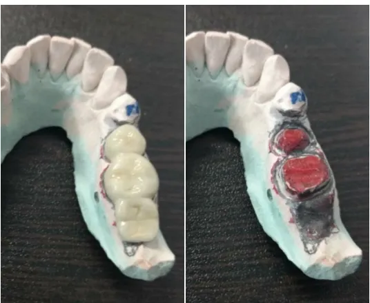
CASE 2- Mandibular 2
ndPremolar and 1
stMolar supported Mandibular 2
ndMolar
Patient 2
Sex- Female
Age – 45 years
Chief complaint – Replacement of missing tooth
History –Non-contributory to treatment planning
IJSRR, 8(2) April. – June., 2019 Page 4010
Treatment – 3 Unit Cantilever Bridge.
Recall - every 6 months follow up for first 5 years.
In this case 2nd mandibular molar has been replaced with premolar and molar. According to Ante’s Law, Peri cemental area of Mandibular 2nd molar is 282 sq.mm (Table 1), the peri cemental area of 2nd premolar is 135(Table 1) and for 1st molar it is 352(Table 1). So,consequently the total for the abutments is larger than missing teeth. Also, you can notice in Figure 3, the size of 2nd molar has been kept small both mesio-distally as well as bucco-lingually thus decreasing the amount of force it will exert on the abutment. As this is the last teeth in the arch, the size can be easily manipulated without any significant change in occlusion and mastication. The third molar in the clinical picture (Figure 4) was not considered in the occlusion as it was decayed and indicated for extraction.
Figure 3. Replacing 2nd molar with 2nd premolar and 1st molar abutment
IJSRR, 8(2) April. – June., 2019 Page 4011
CASE 3 –Maxillary 2
ndPremolar supported 1
stPremolar
Patient 3 (2015)
Sex- Male
Age – 48 years
Chief complaint – Replacement of missing tooth
History – Non-contributory to treatment planning
Material Used – Porcelain Fused to Metal
Treatment – 2 Unit Cantilever Bridge.
Recall - every 6 months follow up for first 5 years.
In this case, we can see that it does not follow Ante’s law. The Peri cemental area for 1st premolar is
149 sq.mm.(Table 1) and that of 2nd Premolar is 140 sq.mm (Table 1), whereas Ante’s Law dictates that the peri cemental area of abutment (2nd Premolar) should be equal or more than that of the missing tooth (1st
premolar). However,the decision to place a Cantilever bridge was taken because of the lack of opposing forces
against Maxillary 2nd premolar. So, the only force 2nd Premolar has to adjust to is the force coming from the
Maxillary 1stPremolar. Prospectively,If the patient wants to replace the mandibular missing teeth,the option
for the fabrication of a Removable Partial Denture(RPD) can be presented considering it is a free end saddle.
An RPD would not cause much pressure or force on the opposing teeth as free end RPDs are tissue supported
and they distribute the forcesover a larger surface area. Subsequently, If the patient is interested in an implant,
the Maxillary 1st molar is supra-erupted, so the implant in any case would not make contact with the Maxillary
2nd Premolar.
IJSRR, 8(2) April. – June., 2019 Page 4012
DISCUSION
Cantilever prosthesis may contribute to the initiation and progression of periodontal diseases, since
risk with a cantilever design is certain 2, 3.The treatment planning with cantilever bridges have been reported successful in the past4, 5. The most important factor to be considered and maintained is occlusion.It was mentioned in the literature that minimal functional contact6, reduction of the occlusa l table7,8 and emphasis was placed on different design considerations are the prime factors for the success.
Even though, Implants would be the best solution for these kind of single teeth replacements, they have their own limitations too. The most important aspect in an implant placement is Osteointegration. Considering the fact that optimum bone quality plays a vital role in osteointegration, the elderly or people who have been edentulous for a long period of time often present with low bone quality issues. Implant placement is a surgical procedure, hence factors such as medical and oral conditions, immunity, habits (Smoking, alcohol, tobacco) etc. play an important role in the success of an implant. Also, the cost of the procedure and material is comparatively higher than the rest of its counterparts, making it financially difficult for the patient.
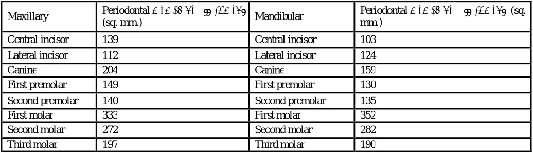
Table 1: The root surface area of periodontal membrane attachments of average normal teeth 1
Maxillary Periodontal membrane attachment
(sq. mm.) Mandibular
Periodontal membrane attachment (sq. mm.)
Central incisor 139 Central incisor 103
Lateral incisor 112 Lateral incisor 124
Canine 204 Canine 159
First premolar 149 First premolar 130
Second premolar 140 Second premolar 135
First molar 333 First molar 352
Second molar 272 Second molar 282
Third molar 197 Third molar 190
CONCLUSION
IJSRR, 8(2) April. – June., 2019 Page 4013
BIBLIOGRAPHY
1. Henderson D, Blevins WR, Wesley RC, Seward T. The cantilever type of posterior fixed partial dentures: a laboratory study. The Journal of Prosthetic Dentistry. 1970;24(1):47– 67. [PubMed] [Google Scholar]
2. Stockton LW. Cantilever fixed partial denture-a literature review. J Can Dent Assoc. 1997; 63(2): 118–21.
3. Dykema RW, Goodacre CJ, Phillips RW.Johnson’s Modern practice in Fixed Prosthodontics. 4th Ed. Philadelphia, PA:WB Saunders Co; 1986: 188, 380.
4. Leempoel PJB, Kayser AF, Van Rossum GMJM, De Haan AFJ. The survival rate of bridges. A study of 1674 bridges in 40 Dutch general practices. J Oral Rehabil. 1995; 22: 327–30. 5. Lindquist E, Karlsson S. Success rate and failures for fixed partial dentures after 20 years’
service: Part I. Int J Prosthodont. 1998; 11(2): 133–38.
6. Hammerle CH, Ungerer MC, Fantoni PC, Bragger U, Burgin W, Lang NP. Long-term analysis of biological and technical aspects of fixed partial dentures with cantilevers. Int J Prosthodont. 2000; 13:409–15.
7. Decock V, De Nayer K, De Boever JA, Dent M. 18-year longitudinal study of cantilevered fixed restorations. Int J Prosthodont. 1996; 9: 331–40.