WEAK ACID EFFECTS ON GENE
ACTIVATION IN YEAST
IAN JONATHAN SEYMOUR
THESIS SUBMITTED FOR THE DEGREE OF DOCTOR OF
PHILOSOPHY
UNIVERSITY COLLEGE LONDON (UNIVERSITY OF LONDON)
ProQuest Number: U643559
All rights reserved
INFORMATION TO ALL USERS
The quality of this reproduction is dependent upon the quality of the copy submitted.
In the unlikely event that the author did not send a complete manuscript and there are missing pages, these will be noted. Also, if material had to be removed,
a note will indicate the deletion.
uest.
ProQuest U643559
Published by ProQuest LLC(2016). Copyright of the Dissertation is held by the Author.
All rights reserved.
This work is protected against unauthorized copying under Title 17, United States Code. Microform Edition © ProQuest LLC.
ProQuest LLC
789 East Eisenhower Parkway P.O. Box 1346
ABSTRACT
Weak lipophilic acids have been commonly used as antifungal agents to
preserve low pH foods. In low pH cultures weak acids rapidly enter the cell,
dissociating to cause intracellular acidification. The plasma membrane H^-ATPase
maintains pH homeostasis by the active extrusion of protons, but this is energetically
expensive causing reduced growth rates and biomass yield. All cells show rapid
responses when exposed to unfavourable environmental conditions which appear to
increase their ability to grow under moderately stressful conditions, or survive
conditions of more extreme stress. Yeasts, especially Zygosaccharomyces bailii, and to
a lesser extent, Saccharomyces cerevisiae, can adapt to growth in weak acids at low pH.
In S. cerevisiae, weak acids elicit strong induction of two plasma membrane proteins,
Hsp30 and Pdrl2 which play a major role in this adaptive response.
Discrete gene promoter elements responsive to stress have been identified in S.
cerevisiae. Investigations carried out for this thesis were aimed at characterising the
effects of weak acids on the expression of stress gene promoters and stress promoter
elements.
Research described in chapter 3 revealed that weak acids can both inhibit the
heat shock activation of stress gene promoters and stress promoter elements in low pH
cultures or act as chemical inducers of these same sequences in the absence o f heat
shock, possibly by a mechanism involving intracellular pH.
Studies on the HSP30 promoter and stress-responsive transcription factor
mutants (chapter 4) demonstrated that none of the previously characterised stress
signalling pathways are involved in the stress-induced activation of this gene. This is
Chapter 5 outlined a strategy to isolate homologues of S. cerevisiae HSP30 in
other yeast species. Although the approach used was unsuccessful Candida albicans
and Hansenula polymorpha were shown to contain DNA sequences that hybridized to
ACKNOWLEDGEMENTS
I would like to thank my supervisor. Dr Peter Piper, whose intelligence,
guidance and scientific insight structured my research. Thanks also to all members of
Lab G 18, past and present, especially Richard Braley and Barry Panaretou for their
helpful advice and friendship. I would also like to thank my mother, father and Maria
for their continued support throughout the years. Finally I would like to thank the
A absorbance
AR analytical reagent
ATP adenosine-5'-triphosphate
bp base pairs
cAMP cyclic adenosine monophosphate
Ci(^Ci) curies (microcuries)
DEPC diethylpyrocarbonate
dH20 deionised plus 1 x distilled water
EDTA ethylenediamine tetra acetic acid
Fig Figure
5-FOA 5-fluoro-orotic acid
HSE heat shock element
HSF heat shock transcription factor
Hsp heat shock protein
kb kilobase
kDa kilodaltons
lacZ P-galactosidase
MES 2-(N-morpholino) ethane sulphonic acid
OD optical density
ONPG 0-nitrophenyl-P-D-galactoside
PDRE pleiotropic drug resistance element
PDSE post-diauxic shift element
pHi intracellular pH
RNase ribonuclease
SIR E stress response element
TABLE OF CONTENTS
TITLE PAGE ... 1
ABSTRACT ...2
ACKNOWLÈDGÎËMENTS ...4
ABBREVIATIONS ...5
TABLE OF CONTENTS... 6
CHAPTER 1... 11
1. INTRODUCTION... 11
1.1 The stress response o f yeast...11
1.2 Stress proteins...14
1.2.1 H s p l0 4 ... 15
1.2.2 Hsp90 17 1.2.3 HspTO...18
1.2.4 Hsp60...19
1.2.5 Small Hsps: H spl2 and Hsp26...20
1.2.6 Ubiquitin... 20
1.2.7 Other proteins involved in the yeast stress response... .21
1.3 Stress-induced changes in yeast gene expression...23
1.3.1 Heat shock elements (HSEs)... 24
1.3.2 Stress response elements (STREs)...26
1.3.2.1 The role of the high osmolarity glycerol (HOG) NIAJP kinase pathway in STRE regulation... 27
1.3.2.2 The role of the RAS-protein kinase A (PKA) pathway in STRE regulation... 28
1.3.3 Pleiotropic drug resistance elements (PDREs) and AP-1 responsive elements (AREs)... 29
1.3.4 The unfolded protein response element (UPRE)... 31
1.4 Weak acid preservatives ...33
1.4.1 Mode of action of weak acid preservatives... .34
1.4.1.1 Plasma membrane H+-ATPase... 35
1.4.2 The weak acid stress response... 37
1.4.2.1 The role of Hsp30...37
1.4.2.2 The role of Pdrl2... 40
1.4.2.3 Other effects of weak acid stress on gene expression... 43
1.4.3 Resistance of yeast to weak acid preservatives... .43
1.5 Aims o f this study...46
CHAPTER 2... 47
2. MATERIALS AND METHODS... 47
2.1 Materials...47
2.2 Yeast strains...48
2.3 E. coli strains^...49
2.4 Plasmids...49
2.5 Yeast Methods...^0
2.5.1 Growth media and culture conditions... 50
2.5.2 Monitoring of cell growth...50
2.5.3 Yeast transformation... 50
2.5.4 Assaying for stress tolerance... 51
2.5.5 Selection of ura3 mutants...51
2.6 Biochemical assays ...51
2.6.1 P-galactosidase assay... 51
2.7 Recombinant DNA techniques...52
2.7.1 Restriction enzyme digests...52
2.7.3 Purification and precipitation of DNA... 52
2.7.4 Agarose gel electrophoresis of DNA...52
2.7.5 Isolation and purification of DNA fragments from agarose gels...53
2.7.6 Dephosphorylation of 5* end of DNA and oligonucleotides...53
2.7.7 Phosphorylation of 5* end of DNA and oligonucleotides... 53
2.7.8 Ligation of fragments of foreign DNA to plasmid vectors... 53
2.7.9 Site-directed mutagenesis... 53
2.7.10 E. coligrowth media and culture conditions...53
2.7.10.1 Preparation of competent E. coli_...53
2.7.10.2 E. coli transformation...54
2.7.10.3 High efficiency transformation by electroporation... 54
2.7.10.4 E. coli miniprep... 54
2.7.10.5 E. coli maxiprep... 54
2.8 Procedures fo r nucleic acid analysis...55
2.8.1 Isolation of yeast genomic D N A ... 55
2.8.2 Isolation of yeast RNA... 55
2.8.3 Quantitation of DNA and RNA... 55
2.8.4 Southern analysis...55
2.8.5 Northern analysis...56
2.8.6 In vitro labelling of single stranded DNA probes...56
2.8.7 Hybridization analysis of DNA probes to membrane bound nucleic acid...56
2.8.8 Autoradiography... 57
2.8.9 Removal of probe from hybridized membrane... 57
2.8.10 DNA sequence analysis... 57
CHAPTERS...58
3. INVESTIGATIONS OF WEAK ACID EFFECTS ON STRESS GENE PROMOTER AND STRESS PROMOTER ELEMENT-L/ICZ ACTIVATION...58
3.1 Introduction...58
3.2 Results... 3.2.1 Stress gene promoter and stress promoter element-lacZ vectors... 3.2.2 Investigations of weak acid stress on gene promoter and stress promoter element-/acZ induction... 3.2.2.1 The effect of heat shock, medium pH and sorbate on HSE-/acZ expression... 3.2.2.2 The effect of heat shock, medium pH and sorbate on STRE-/acZ expression... 3.2.2.3 The effect of heat shock, medium pH and sorbate on HSP12-/<3cZ expression... 3.2.2.4 The effect of heat shock, medium pH and sorbate on HSP26-/ûicZ expression... 3.2.2.5 Construction of an HSP30 promoter-/acZ fusion... 3.2.2.6 Stress activation of HSP30-/acZ expression... 3.3 Discussion... 3.3.1 STRE sequences are induced by weak acid treatment in low pH cultures... 3.3.2 Weak acid preservatives inhibit heat induction of stress gene promoter and stress promoter element-/acZ fusions in low pH cultures... 3.3.3 An HSP30-/flcZ fusion was not stress-inducible... 3.4 Conclusions 59 59 62 62 64 66 66 69 69 71 71 72 73 74 CHAPTER 4...75
4. INVESTIGATION OF THE ROLES OF PUTATIVE STRESS-RESPONSIVE PROMOTER ELEMENTS ON STRESS ACTIVATION OF THE HSP30 GENE...75
4.1 Introdu ction...75
4.2 Results...80
4.2.1 Site-directed mutagenesis of the STREl m otif... 80
4.2.2 Site-directed mutagenesis of the PDSE motifs... 83
4.2.3 Attempted transplacement of mutant alleles of the HSP30gene into the hsp30::URA3locus of strain KT3... 86
4.2.5 Construction of YIplac204-derived vectors containing mutant
alleles of ihtHSPSOgene... 90 4.2.6 Integration of mutant alleles of the HSP30gene in the TRPllocus... 4.2.7 Influence of weak acid preservatives, ethanol, nitrogen starvation and heat
stress on mutant promoter HSP30genes in KT3-YIplac204-30 strains... 4.2.8 Comparison of the stress-induced expression of HSP30and HSP12_...
4.2.9 Effects of the hsfl-m 3and msn2 ntsn4double mutations on the
stress-induced expression of HSP30and HSP12...
4.2.10 Effects o f the y a p l and p d r l pdr3double mutations on the
sorbate-induced expression of HSP30...
4.3 Discussion...
4.3.1 The putative STRE, PDSEs and Msn2 , 4 are not essential for the
induction o f HSP30gene expression in stressed cells... 4.3.2 H sfl , Yapl , Pdrl and Pdr3 are not essential for the induction
of HSP30gene expression in stressed cells...
4.4 Conclusions 92 94 97 99 101 103 103 105 106
CHAPTER 5 107
5. A SEARCH FOR HSP30 GENE HOMOLOGUES IN OTHER YEASTS 107
5.1 Introduction... 107
5.2 Results...
5.2.1 Analysis of low stringency hybridization of a S. cerevisiaeHSP30
gene probe to genomic DNA of different yeast species... 5.2.2 Cloning of the C. albicansand H. polymorphagenomic DNA
fragments hybridizing to the S. cerevisiae HSP30gene... 5.2.3 Restriction mapping and southern hybridization to the insert
fragment of pBluescript-CAl... 5.2.4 Sequencing strategy of the 1.1 kb C. albicansgenomic DNA fragment... 5.2.5 Nucleotide sequence analysis of the 1.1 kb C. albicans
genomic DNA fragment... 5.2.6 Comparison of the C. albicans1.1 kb fragment to the S. cerevisiae
HSP30gene... 5.3 Discussion 108 108 110 110 112 114 118 122 CHAPTER 6...124
6. DISCUSSION...124
6.1 Introduction...124
6.2 Intracellular p H is a possible trigger fo r the weak acid-induced
activation o f stress gene promoters and stress promoter elements...125
6.3 The S. cerevisiae HSP30 gene is regulated by a novel stress
response pathway...127
6.4 Future studies leading to dissection o f the novel weak acid response pathway 130 6.4.1 Identification of genes involved in the weak acid stress response pathway... 130 6.4.2 Identification of the cis and trans acting components of the weak acid
stress regulatory pathway acting on the S. cerevisiae HSP30promoter... 131
TABLES AND FIGURES
CHAPTER 1...11
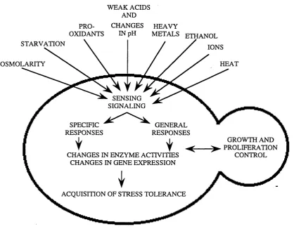
Fig. 1.1 Different stress conditions are sensed by the yeast cell and trigger both specific and general molecular responses...15
Table 1.1 Heat shock proteins o f yeast...16
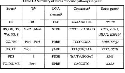
Table 1.2 Summary o f stress response pathways in yeast...32
Fig. 1.2 Plasma membrane H^-ATPase activity in purified plasma membrane from hspSO mutant and wild-type cells...39
Fig. 1.3 Schematic model o f how Pdrl2 action may help acidified yeast cultures counteract the effect o f weak organic acids...4\
CHAPTER 2...47
Table 2.1 Yeast strains...48
Table 2.2 E. coli strains_...49
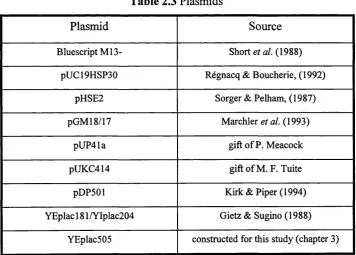
Table 2.3 Plasmids...49
CHAPTER 3 58 Fig. 3.1 (a) pHSE2 59 Fig. 3.1 (b) pGM18/17 60 Fig. 3.1 (c) pUC41a .. . 61
Fig. 3.1 (d) PÜCK4Ï4...61
Fig. 3.2 Influences o f medium pH and sorbate on the expression o f a HSE-lacZfusion at (a) 25 °C or (b) heat shocked to 39 T7...63
Fig. 3.3 Influences o f medium pH and sorbate on the expression o f a STRE-lacZfusion at (a) 25 °C or (b) heat shocked to 39 T7...65
Fig. 3.4 Influences o f medium pH and sorbate on the expression o f a HSP12-lacZ fusion at (a) 25 °C or (b) heat shocked to 39 67 Fig. 3.5 Influences o f medium pH and sorbate on the expression o f a HSP26-lacZ fusion at (a) 25 °C or (b) heat shocked to 39 °C...68
Table 3.1 Primers used for the PCR synthesis o f HSP30 promoter...69
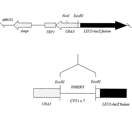
Fig. 3.6 YEplac505 (HSP30promoter-lacZ) vector construction...70
CHAPTER 4...75
Fig. 4.1 Northern analysis o f total lU^A from yeast cells subjected to different stresses,...76
Table 4.1 Putative regulatory elements in the HSPSO and PDR12 promoters... 77
Fig. 4.2 Nucleotide sequence o f the proximal HSPSO 5 ^-flanking region...78
Fig. 4.3 Restriction map o f the pUC19HSP30 plasmid,...79
Fig. 4.4 Strategy fo r generating specific base changes using the Clontech site-directed mutagenesis kit,...80
Table 4.2 Primers fo r the site-directed mutagenesis o f the STREl motif...81
Fig. 4.5 DNA sequence analysis o f wild-type and mutant pUC19HSP30...83
Table 4.3 Primers fo r the site-directed mutagenesis o f the PDSE motifs,...83
Fig. 4.6 Strategy fo r generating specific base changes using the Stratagene® site-directed mutagenesis kit,...84
Table 4.4 The complete mutagenisedset o f pUC19HSP30 vectors incorporating mutant alleles o f the HSPSO gene...85
Fig. 4.7 Map o f chromosome integration o f mutant alleles o f the HSPSO promoter,...87
Fig. 4.8 Southern blot o f PstI digested genomic DNA...88
Table 4.5 The YIplac204-30 series o f vectors incorporating different promoter mutations within the HSPSO gene,...90
Fig, 4.10 Map o f integration o f YIplac204-30 derivatives in
the TRPl locus... 93 Fig. 4.11 Southern blot o f PstI digested genomic DNA...
Fig. 4.12 Stress-induced expression o f HSP30 sequences in RNA from
wild-type and mutant HSP30 strains...
Fig. 4.13 Comparison o f stress-induced expression o f HSP30 and HSP12...
Fig. 4.14 Effect o f the hsfl-m3 and msn2 msn4 double mutations on the
stress-induced expression o f HSP30 and HSP12...
Fig. 4.15 Effect o f the ya p l and p drl pdr3 double mutations on the sorbate-induced expression o f HSp30...
93
95
100
102
CHAPTERS...107
Fig. 5.1 Southern blot o f restriction enryme digested genomic DNA from
Z. bailii, C. albicans, H. polymorpha and S. pombe...109 Fig. 5.2 (a) Electrophoresis o f restriction digests ofpBluescript-CAl (b)
Restriction map ofpBluescript-CAl 1.6 kb fragment insert (c) Southern
blot o f (a) probed with HSP30...I l l Fig. 5.3 Outline o f the strategy fo r sequencing the C. albicans genomic
DNA insert o f pBluescript-CA2...113 Table 5.1 Primers usedfor the DNA sequence analysis o f pBluescript-CA2...113 Fig. 5.4 Nucleotide and peptide sequence o f the 1.1 kb C. albicans
genomic DNA fragment...115 Fig. 5.5 Diagram o f the putative ORFs present in the 1.1 kb C. albicans
genomic DNA fragment...116 Fig. 5.6 Hydropathy plot o f the predicted C. albicans ORF product ...117 Table 5.2 S. cerevisiae genes sharing weak similarities to the C. albicans
1.1 kb genomic DNA fragment^...118 Fig. 5.7 (a) Electrophoresis o f restriction digests o f pBluescript-CA2 (b)
Restriction map ofpBluescript-CA21.6 kb fragment insert (c) Southern
blot o f (a) probed with HSP30...120 Fig. 5.8 Martinez/Needleman-Wunsch DNA aiignment,...121
CHAPTER 6...124
Fig. 6.1 Possible model o f a weak acid stress signalling pathway in
CHAPTER 1
1. INTRODUCTION
1.1 The stress response of yeast
The survival of living cells is dependent on their ability to sense changes in the
environment and to respond appropriately to the new situation. Cells can be challenged
by variations in temperature, pH, the availability of nutrients, osmolarity or by the
presence of pro-oxidants or cytotoxic compounds (Ruis & Schuller, 1995). Exposure to
many adverse environmental conditions evokes rapid molecular responses which assist
survival. These responses are commonly designated stress responses. They can either
increase the ability o f cells for growth under moderately stressful conditions or enhance
survival under conditions o f even more severe stress. Stress responses lead to the
acquisition of increased stress tolerance by enhancing the repair o f molecular damage
and establishing mechanisms that limit stress-induced damage.
A large number of cellular responses to different chemical or physical stress
conditions have been described in S. cerevisiae (reviewed by Hohmann & Mager,
1997). These responses include the immediate production of compounds which protect
cellular components and the activation of signalling pathways which increase the
expression of a wide variety o f protective genes (Lindquist, 1986; Mager & Moradas-
Ferreira, 1993; Mager & de Kruijff, 1995). A fundamental aspect of the yeast stress
response is the phenomenon of acquired resistance whereby cells pre-exposed to a
comparatively mild non-damaging stress can withstand a more severe form of the same
stress. For example, a mild heat shock induces the highly conserved heat shock
response which increases the capacity for the cell to withstand potentially lethal
temperatures (Coote et al., 1991; Piper, 1993). The same has been described for pre
treatment with sorbate (Bills et al., 1982; Holyoak et al., 1996), 0.7 M NaCl (Varela et
al., 1992) and hydrogen peroxide (Collinson & Dawes, 1992; Jamieson, 1992).
Consequently, mild stress conditions probably trigger the appropriate cellular responses
to prepare cells for more severe exposure to the same stress. Sometimes, pre-exposure
to a mild stress condition will induce resistance not only to this stress, but also tolerance
against the severe stress caused by other agents. For example, a mild treatment with
either sorbate (Coote et al., 1991), NaCl (Varela et al., 1992) or ethanol (Piper et al.,
partly reflect the inherent overlap between certain stress responses, whereby exposure
to a particular stress may lead to tolerance against another type of stress.
In addition to specific stress responses, S. cerevisiae also has what has been
termed the "general stress response", primarily triggered by low protein kinase A levels
(Thevelein, 1994; Siderius & Mager, 1997). Some enzymes already present in non
stressed cells, such as protein kinases and enzymes involved in trehalose metabolism,
are activated by the stress of nutrient limitation and provide an immediate stress
response (Ruis & Schuller, 1995). The general stress response switches cells to a more
quiescent state in which they are much more resistant to practically all stresses.The
stress response of yeast is summarised in Fig. 1.1. The effect of each stress response at
the level of gene expression is complex but generally the expression of most genes is
lowered or switched off under stress conditions while the induction of a small number
of specific stress genes is enhanced (section 1.2). Miller et al. (1982) found that more
than 80 of 500 S. cerevisiae proteins examined were induced by a temperature upshift
from 23°C to 37°C, while the synthesis of a further 300 proteins was reduced. This
suggests that while the majority of genes possess stress-sensitive transcriptional
repression, stress genes contain positive control elements which enhance their
activation upon stress exposure (section 1.3). This thesis attempts to investigate in
greater detail the effects of a weak acid preservative stress on stress gene activation in
Fig. 1.1 Different stress conditions are sensed by the yeast cell and trigger both specific
and general molecular responses. These stress responses result in changes at the level of
enzyme activities and gene expression and lead to the acquisition of stress tolerance
(modified fi-om Siderius & Mager, 1997).
WEAK ACIDS AND
PRO- CHANGES HEAVY
OXIDANTS IN pH METALS ETHANOL
STARVATION
IONS
OSMOLARITY HEAT
SENSING SIGNALING
SPECIFIC RESPONSES
GENERAL RESPONSES
GROWTH AND ^ P R O L I F E R A T I O N
CONTROL CHANGES IN ENZYME ACTIVITIES
CHANGES IN GENE EXPRESSION
1.2 Stress proteins
When cells from a wide variety of species are exposed to an upshift in ambient
temperature, they respond by synthesising a small number o f highly conserved proteins,
the heat-shock proteins (Hsps). Hsps have been implicated in all major growth related
processes such as cell division, DNA synthesis, transcription, translation, protein
folding and transport (molecular chaperones) and membrane function (Craig et al.,
1993; Mager & Moradas Ferreira, 1993). This response is universal and has been called
the heat shock response (reviewed by Lindquist, 1986; Nagao et al., 1990; Piper, 1997).
An almost identical response is induced by ethanol exposure (Piper et ah, 1994), while
the responses to osmostress (reviewed by Serrano et al., 1997) or pro-oxidants
(Jamieson, 1992; Jamieson et al., 1994) are quite distinct from each other and from this
heat shock response, although these mechanisms cause a transient increase in the
synthesis of at least some of the Hsps. As shovm in this thesis, the response to weak
acid exposure is yet another stress response (section 1.4.2).
The heat shock response was first reported as a dramatic increase in
transcriptional activity induced by a brief heat treatment of Drosophilia larvae (seen as
a new set of puffs on the salivary gland polytene chromosomes; Ritossa, 1962).
Tissières et al. (1974) proved the existence of heat shock proteins and soon afterwards
the heat shock genes of Drosophiliawere among the first eukaryotic genes to be cloned
(Livak et al., 1978; Craig et al., 1979; Voellmy et al., 1981). Similar findings with
chicken fibroblasts (Kelley & Schlesinger, 1978), E. coli (Lemaux et al., 1978), yeast
(McAlister & Finkelstein, 1980), plants (Barnett et al., 1980) and in many other
organisms suggested that the heat shock response represents an evolutionarily
conserved genetic system which might be beneficial for the living cell. Although
Lindquist (1981) revealed that in Drosophilia a sudden heat shock both specifically
represses the translation of pre-existing mRNAs and induces the synthesis of mRNAs
encoding Hsps, a translational control mechanism is not
j
as prevalent in yeast.The range of Hsps synthesised in yeast upon a stress challenge is similar to that
produced in other cells. The major Hsps can be classified into five families according to
their average apparent molecular mass, hsp 100 (hsp 104 in yeast; section 1.2.1), hsp90
(hsp82 in yeast; section 1.2.2), hsp70 (DnaK in E. coli', section 1.2.3), hsp60 (the
chaperonin or groEL-family; section 1.2.4) and small-size Hsps (hsp 12, hsp26 and
hsp30 in yeast; sections 1.2.5 and 1.4.2.1). Several proteins homologous to Hsps are
synthesised constitutively, such as hsc82 (Parsell & Lindquist, 1993), reflecting the
addition, the rate of synthesis of several other proteins such as ubiquitin (section 1.2.6),
some glycolytic enzymes and catalase (section 1.2.7) are strongly induced upon stress
exposure and should therefore be considered as heat shock proteins (see Table 1.1 for
summary). In S. cerevisiae, heat shock also induces the secretory glycoprotein Hsp 150
which has no known function (Russo et al., 1992).
1.2.1 Hspl04
Yeast Hsp 104 is a member of the heat shock protein family with a molecular
mass greater than 100 kDa (Parsell & Lindquist, 1993; Piper, 1997). The HSP 104 gene
was first isolated by Sanchez & Lindquist (1990) and sequenced by Parsell et al.
(1991). Hsp 104 is thought to play an important role in tolerance to extreme stress
conditions including severe heat stress and ethanol (Sanchez & Lindquist, 1990;
Sanchez et al., 1992; Lindquist & Kim, 1996). Hsp 104 is not expressed during normal
growth on fermentable carbon sources, but constitutively synthesised in respiring cells
and strongly induced during heat shock, transition to stationary phase and early in
sporulation (Sanchez et al., 1992). Cells carrying mutations in the HSP 104 gene grow
at the same rate as wild-type cells between 25-37°C, demonstrating that Hsp 104 is
dispensable under normal growth conditions. However, within a few minutes exposure
to 50°C (after a sublethal pre-treatment at 37°C), mutant cells begin to die at 100 to
1000 times the rate of the wild-type cells, indicating that Hsp 104 is essential for
induced thermotolerance (Sanchez & Lindquist, 1990). Mutant cells are not sensitive to
copper and cadmium and are only slightly more sensitive to arsenite, suggesting that
damage by these agents is different to that caused by heat (Sanchez et al., 1992).
As in other eukaryotes, Hsp 104 is thought to have a nuclear localisation
(Subjeck et a l, 1983). Some clues to the possible functions of Hsp 104 come from the
analysis o f its primary structure. The heat inducible members o f the Hsp 100 family
jfrom yeast, plants, animals and bacteria share approximately 60% homology across
their entire lengths and even higher similarity in regions of two ATP-binding site
consensus elements, suggesting that they share a conserved heat-related function
(Parsell & Lindquist, 1993). Site-directed mutagenesis revealed that these binding sites
are essential for the stress protective function of Hsp 104 (Parsell et al., 1991). Yeast
Hsp 104 is closely related to the heat-inducible E. coli ClpB protein (Squires et al.,
Table 1.1 Heat shock proteins of yeast (from Mager & Moradas Ferreira, 1993)
Designation Cellular localisation Function
Hspl50 (Secretory) Unknown r
Hsp104 Nucle(ol)us Stress tolerance
Hsp83 Cystol/nucleus Chaperone
HspTO
Ssal Cystol? Chaperone
Ssa2 Cystol Chaperone
Ssa3 Cystol Chaperone
Ssa4 Cystol Chaperone
Ssbl Unknown Chaperone
Ssb2 Unknown Chaperone
Sscl Mitochondria Chaperone
Ssdl (Kar2) Endoplasmic reticulum Chaperone
Hsp60 Mitochondria Chaperone
Hsp30 Plasma membrane ATPase regulator?
Hsp26 Cystol/nucleus Unknown
Hspl2 Cystol Unknown
Ubiquitin Cystol Protein degradation
Enzymes
Enolase Cystol Glycolysis
Glyceraldehyde-3 -P- Cystol Glycolysis
dehydrogenase
Phosphoglycerate kinase Cystol Glycolysis
Parsell et al. (1994) revealed that vegetative cells lacking Hsp 104 show enhanced
accumulation of heat-induced aggregates of vital cell structures, implying that Hsp 104
may act as a catalyst of protein disaggregation or reactivation. The HspTO Ssal isoform
assumes an important role in tolerance to extreme temperatures in the absence of
Hsp 104, while in cells with low levels of Ssal, Hsp 104 assumes an important role in
growth at normal temperatures, suggesting that HspTO Ssal and Hsp 104 have
complementary roles (Parsell et ah, 1994). Lindquist & Kim (1996) showed that
HSP 104 could not be deleted in h sfl-m3 cells (which have a nonsense mutation in the
HSFl gene, section 1.3.1) because expression of heat shock factor (and the viability of
the strain) requires nonsense suppression mediated by the yeast prion [psi+], which in
turn depends upon Hsp 104. The self-modifying psi+ factor becomes lost in yeast strains
with overexpression or inactivation of HSP 104, suggesting a chaperone role for Hsp 104
(Chemoff et al., 1995).
1.2.2 Hsp90
Like the HspTOs, members of the Hsp90 class of Hsps are highly conserved in
bacteria, yeasts and mammals and show among eukaryotes at least 50% sequence
identity (Craig & Lindquist, 1988). Hsp90s are abundant chaperone proteins, localised
primarily in the cytoplasm; a small fraction translocates to the nucleus upon heat shock
(Schlessinger, 1990). Hsp90s differ from both Hsp60s (section 1.2.4) and HspTOs
(section 1.2.3) in that they regulate the function of specific, substantially folded
proteins. In vertebrates Hsp90s interact with a variety of cellular proteins, including
steroid hormone receptors, several kinases and the cytoskeleton proteins actin and
tubulin (Lindquist, 1986; Mager & Moradas Ferreira, 1993; Hartl, 1996).
Glucocorticoid receptor proteins are kept in the cytoplasm in an inactive conformation
through interaction with Hsp90 which serves as a cytoplasmic anchoring protein (Hunt,
1989). In S. pombe Hsp90 participates in cell cycle control by regulating Weel protein
tyrosine kinase activity (Parsell et al., 1991). However, in S. cerevisiae. a demonstrable
function for Hsp90 has yet to be identified.
S. cerevisiae contains two genes encoding Hsp90: HSP82 and HSC82
(Borkovich, 1989) whose products are 9T% identical (Parsell & Lindquist, 1993).
HSC82 (heat shock cognate) is constitutively expressed and is only weakly induced
upon stress exposure while HSP82has a low basal level of expression and is strongly
activated during heat shock (Borkovich, 1989), transition to stationary phase (Kurtz &
levels by heat shock appears to reflect a need for higher levels o f Hsp90 during high
temperature growth. If either of these genes is inactivated, the cell is unable to grow at
high temperature while inactivation of both genes renders the cell inviable. The three-
dimensional structure of the 27 kDa N-terminal domain of the yeast protein containing
three o f the four highly-conserved regions of Hsp90 structure and the ATP-binding
domain has recently been solved (Prodromou et ah, 1997).
1.2.3 Hsp70
Hsp70 proteins are the most highly conserved Hsps among all species. A 50%
identity exists between Hsp70 proteins found in higher eukaryotes and the E. coli
Hsp70 DnaK, while eukaryotic proteins are between 50 and 97% identical (Lindquist &
Craig, 1988). The genes encoding Hsp70 in S. cerevisiae constitute a multigene family
consisting of eight members (Table 1.1) which are subdivided into four subfamilies,
SSA^ SSB, SSC and SSD (Stress Seventy). SSA (SSAl-4) and SSB {SSBJ-2) constitute
the cytoplasmic Hsp70s and are thought to be functionally distinct (Boorstein & Craig,
1990 a and b; Parsell & Lindquist, 1993; Mager & Moradas-Ferreira, 1993; Hartl,
1996). SSA gene products are important for protein translocation across the ER and
mitochondrial membranes while SSB products are associated with polysome-associated
nascent peptides (Hartl, 1996). Three of the four SSA genes are induced by heat shock
and the SSA subfamily is indispensable for growth. The SSB genes are repressed by heat
shock and mutations in SSBgene products result in a cold-sensitive phenotype (Parsell
& Lindquist, 1993; Mager & Moradas-Ferreira, 1993; Hartl, 1996). The SSC and SSD
subfamilies (reviewed by Craig et al., 1993) encode two organelle localised proteins:
Sscl (SSCI) in the matrix of the mitochondrion (Craig et al, 1987; 1989) and
Ssdl/Kar2 (KAR2; section 1.3.4) in the lumen of the ER (Normington et al., 1989).
It is likely that Hsp70 proteins induced upon stress exposure perform functions
similar to those under normal growth conditions, namely molecular chaperones. Hsp70
induced by heat shock is thought to mainly function in sequestering partially heat-
damaged protein until this protein can either be degraded or reactivated. In unstressed
cells a major role of the different cytoplasmic forms of the Hsp70 protein is to associate
and control the folding states of newly synthesised polypeptide chains for translocation
across the ER or mitochondrial membranes (Piper, 1997). Hsp70 proteins of both
eukaryotes and prokaryotes possess both ATPase and peptide binding activities which
are crucial for their chaperone activity (Ziegelhoffer et al., 1995). Sequence similarity
N-terminal regions where ATP binding sites are located. ATPase activity is stimulated by
binding to either unfolded proteins, synthetic peptides or a DnaJ homologue, Ydjl
(Ziegelhoffer et al., 1995). The peptide-binding domain is thought to be located near the
C-terminus (Hartl, 1996). HspTO may also interact with the heat shock factor (section
1.3.1), thereby modulating its transcription activating potency (reviewed by Mager &
Moradas Ferreira, 1993).
1.2.4 Hsp60
The highly conserved Hsp60 protein family (chaperonins, Cpn60s) facilitate
post-translational assembly of polypeptides at normal temperatures, a cellular function
similar to that of HspTO (Mager & Moradas Ferreira, 1993). Hsp60s play a crucial role
in binding to unfolded proteins, catalysing ATP-dependent folding o f these proteins and
assisting their assembly into higher-order protein structures (reviewed by Craig et al.,
1993; Parsell & Lindquist, 1993; Piper, 199T). Yeast Hsp60 (encoded by the essential
nuclear MIF4 gene) was first identified as a mitochondrial protein (Cheng et ah, 1989),
showing 54% homology to the heat-inducible groEL of E. coli, a protein involved in
bacteriophage head assembly (McMullin & Hallberg, 1988). Hsp60 is assembled into a
typical chaperonin structure o f two stacked heptameric rings (reviewed by Ellis & van
der Vies, 1991). Mutations in the MIF4 gene were first identified as conditional-lethal
mutations which prevented the correct folding and assembly o f FI-ATPase, cytochrome
b2 and the Rieske FeS protein of complex III (Cheng et ah, 1989). Although Hsp60
clearly functions in the folding and assembly of mitochondrial proteins (Ostermann et
al., 1989; Koll et al., 1992), its role in the translocation of proteins passing from the
matrix into the inner membrane space remains to be resolved. At high temperatures,
Hsp60 associates with a wide variety o f proteins in yeast mitochondria. This association
presumably prevents protein aggregation and promotes refolding when cells are
returned to lower temperatures (Parsell & Lindquist, 1993). Hsp60 chaperonins have
also been implicated in the cytoplasmic protein-folding machinery but, unlike
mitochondrial Hsp60 of S. cerevisiae, they are not heat-inducible. In yeast, this
cytoplasmic complex is comprised o f the essential Tcpl protein (tailless complex
polypeptide) which plays an important role in the biogenesis of tubulin and actin (Hartl,
1.2.5 Small Hsps: H spl2 and Hsp26
Yeast cells contain two major small Hsps: Hsp 12 (Praekelt & Meacock, 1990)
and Hsp26 (Bentley et al., 1992) for which demonstrable functions have not been
shown (Petko & Lindquist, 1986; Susek & Lindquist, 1989). Although both of these
proteins are synthesised under many stress conditions (Praekelt & Meacock, 1990;
Bentley et al., 1992; Parsell & Lindquist, 1993; Mager & Moradas Ferreira, 1993;
Varela et ah, 1995), loss of both Hsp 12 and Hsp26 in S. cerevisiae produces no
apparent phenotype. However, an Hsp 12 homologue of Schizosaccharomyces pombe
suppressed a mutational defect in the cdc4gene (late septation), suggesting that Hsp 12
could play a role in the formation of the F-actin contractile ring at cytokinesis (Jang et
al., 1996). The small Hsps represent a very diverse group of Hsps, which nevertheless
display conserved structural features (Lindquist & Craig, 1988) and share the ability to
form high molecular weight polymeric aggregates called heat shock granules (Tuite et
al., 1990; Bentley et al., 1992). Hsp26 shows a significant sequence similarity to a-
crystallin proteins, particularly with respect to a highly conserved hydrophobic domain
located at the C-terminus (Tuite et al., 1990). Although Jakob et al., (1993) have
recently demonstrated that mammalian small Hsps and a-crystallin molecules exhibit
chaperone activity, a function has yet to be assigned to hsp26. Unlike Hsp26, Hsp 12
does not show homology to a-crystallin (Praekelt & Meacock, 1990), but shares 47%
identity to the N-terminal region of W h ll, a 7.8 kDa polypeptide encoded by a gene
differentially expressed in the budding phase and hyphal-forming cells of Candida
albicans (Srikantha & Soil, 1993). A universal property o f the small Hsps may also be
their developmental regulation, such that HSP 12and HSP26are strongly stress-induced
while expression is dramatically increased following transition o f cells to stationary
phase and upon induction of sporulation. The heat shock, weak acid and low pH-
induced expression of HSP 12 and HSP26 are investigated in Chapter 3. Hsp30
(Régnacq & Boucherie, 1992; Panaretou & Piper, 1992) is another small Hsp, which is
I found in the plasma membrane.
1.2.6 Ubiquitin
Conjugation of ubiquitin (Ub) to short-lived or damaged proteins mediates their
selective degradation (Finley et al., 1987). Ubiquitin, a highly conserved 76 amino acid
protein, acts by becoming covalently attached to the free amino acid groups of target
ligases (reviewed by Finley & Chau, 1991). Proteins targeted by ubiquitination are then
degraded by the proteosome (proteinase YscE) (reviewed by Heinemeyer et al., 1991).
In yeast, Ub is encoded by four genes (UBll-4). UBIl-3 code for hybrid proteins in
which Ub is fused to unrelated amino acid sequences. Polyubiquitin, a protein encoded
by the UBI4 gene in yeast (containing five tandem Ub repeats), is thought to be a heat
shock protein since it displays a strongly enhanced rate of synthesis under stress
conditions (Finley et al., 1987). Also, production of the Ubc4/5 Ub conjugating
enzymes is heat shock-inducible (Seufert & Jentsch, 1990). This probably indicates a
much greater requirement for turnover o f unfolded and non-functional proteins in cells
recovering from heat shock and other forms of stress. In several cases, it has been
shown that the proteolytic signal takes the form of a multi-Ub chain in which successive
Ub molecules are linked tandemly at various lysine residues, a process strongly
dependent on the presence of Ubc4/5 (Amason & Ellison, 1994).
Strains carrying deletions in UBI4, or UBC4plus UBC5 display a considerably
reduced resistance to starvation, an increased sensitivity to high temperatures, amino
acid analogues and alkylating agents, and a block on the sporulation of a/a ubi4/ubi4
diploid cells (Finley et al., 1987; Tanaka et al., 1988; Treger et al., 1988; Fraser et al.,
1991). This reveals the importance of ubiquitination in the cellular response to stress.
Also, the proteosome is important in the stress response since deletion of a gene
encoding a subunit of this protease causes sensitivity to stress conditions and
accumulation of ubiquitin-protein conjugates (Heinemeyer et al., 1991)
1.2.7 Other proteins involved in the yeast stress response
Apart from the classical Hsps, proteins o f unstressed cells of yeast and other
organisms play a part in the stress response. Some o f these exhibit significantly
increased levels of expression following a stress treatment (Mager & Moradas-Ferreira,
1993). Heat shock increases the activities of at least two enzymes important for
protection against oxidative damage. These are cytoplasmic catalase T (encoded by
CTTl) and the mitochondrial manganese superoxide dismutase, MnSOD (encoded by
S0D2) (Wieser et al., 1991; Costa et al., 1993). Heat shock-induced transcriptional
activation of CTTl and SOD2 is controlled by the stress response element, STRE
(section 1.3.2). Strains carrying a deletion in CTTl show reduced thermotolerance in
both proliferating and stationary cells except when PKA levels are high (Wieser et a l,
1991). Loss o i S0D2 also renders the cell more sensitive to the lethal effects of heat
The effects o f these antioxidant defence enzymes on thermotolerance are thought to
reflect the more severe oxidative damage to cellular proteins, nucleic acids and lipids
caused by reactive oxygen species at higher temperatures, especially in respiratory
cultures (Moradas-Ferreira et al., 1996).
Several enzymes of the glycolytic pathway are induced upon heat treatment of
yeast cells. One o f the genes encoding glyceraldehyde-3-phosphate dehydrogenase
(Lindquist & Craig, 1988), enolase (lida & Yahara, 1985) and phosphoglycerate kinase
(Piper et a l, 1986) are induced fblloAving a heat shock. Heat stress imposes large
demands for energy (ATP) generation by the cell (Findley et a l, 1983). Increased
glycolytic flux may assist stressed cells to restore intracellular ATP levels, although
these enzymes are present in such large amounts in unstressed cells, they are not
limiting for glycolytic flux. A further consequence o f heat shock and other stress
challenges on yeast cells is the transient dissipation of the electrochemical pH gradient
across the plasma membrane, leading to a decrease in intracellular pH, activation of
plasma membrane H"*’-ATPase (section 1.4.1.1) and induction o f a 30 kDa plasma
membrane heat shock protein (section 1.4.2.1).
It is well known that glycogen and trehalose accumulate in yeast under nutrient
starvation and entry to stationary phase, while high levels of trehalose are found in heat
shocked cells (reviewed by François et al., 1997). In yeast, trehalose is a stress-
protectant rather than a reserve carbohydrate since it is one of the most effective
substances known for in vitro preservation o f membrane structures and enzyme
activities during desiccation, freezing or heating (Hottiger et al., 1994). Heat shock
causes the rapid accumulation of a large cytoplasmic pool of trehalose (up to 100-fold)
(Hottiger et al., 1987). There is a good correlation between trehalose levels and
thermotolerance in stationary phase and nonfermentative yeast cultures, although no
such correlation exists in fermentative yeasts (De Virgilio et al., 1994; van Dijck et al.,
1995). The mechanism of heat-induction of trehalose is readily reversible since the
trehalose accumulated with heat shock is rapidly mobilised ^vith a subsequent
temperature downshift (Neves & François, 1992). The genes involved in glycogen and
trehalose metabolism exhibit stress regulation, being induced by nutrient starvation,
temperature, osmotic and oxidative stresses (Winderickx et al., 1996; Parrou et al.,
1997). Almost all genes encoding the enzymes involved in the metabolism of these two
Recently, Parrou et a l, (1997) demonstrated that the stress activation of the genes
encoding glycogen synthase (GSY2) and trehalose-6-phosphate synthase (TPSl) is
dependent on STREs, although the levels of transcription varies considerably.
Mobilisation of trehalose with temperature-downshift is defective in strains with low
HspTO levels (Hottiger et al., 1992) and mutants defective in TPSl do not show normal
levels o f Hsp synthesis with heat shock (Hazell et al., 1995), suggesting that the
induction of Hsps and trehalose in the heat shock response may be linked.
Finally, recent evidence suggests that Ca^^-activated enzymes anrf signal
transduction pathways are strongly stimulated by heat stress (reviewed by Piper, 1997).
Weakness of cell walls at high temperatures might be detected by systems responding
to plasma membrane stretch, such as ion channels (Kamada et al., 1995), which in turn
lead to Ca^+ influx. Cystolic Ca^+ may then activate Ca^+-regulated enzymes such as
phospholipase C (PI-PLC), protein kinase C (PKC) and Ca^^/calmodulin-dependent
protein kinase. Strains that lack PI-PLC progressively lose viability and eventually lyse
after a shift to 37°C (Payne & Fitzgerald-Hayes, 1993; Flick & Thomer, 1993; Yoko-o
et al., 1993). Mutants defective in the PKC pathway (Kamada et al., 1995) and mutants
lacking Ca^+Zcalmodulin-dependent protein kinase (lida et al., 1995) all show impaired
acquisition of thermotolerance with heat shock. There is no evidence that induction of
Hsps is impaired in these mutants, suggesting that these pathways contribute to a Hsp-
independent mechanism of thermotolerance. Also, the stress-activated cell membrane
H+-ATPase possesses potential phosphorylation sites for the Ca^+Zcalmodulin-
dependent protein kinase (section 1.4.1.1).
1.3 Stress-induced changes in yeast gene expression
The promoter regions of yeast stress genes contain various positive
transcriptional control elements that are activated by stress conditions: heat shock
elements (HSEs; section 1.3.1), stress response elements (STREs; section 1.3.2),
pleiotropic drug resistance elements and AP-1 responsive elements (PDREs and AREs
respectively; section 1.3.3) and unfolded protein response elements (UPREs; section
1.3.4). Some stress proteins encoded by HSE-regulated genes are necessary for growth
of yeast at high temperatures (37-39°C), products of STRE-activated genes seem to be
involved in survival under severe stress, PDRE-controlled genes confer tolerance to a
range of cytotoxic compounds, ARE-induced genes mainly function during stresses
which generate H%0% free radicals and in response to heavy metal ions, while
accumulation of unfolded secretory and transmembrane proteins in the endoplasmic
reticulum (ER).
1.3.1 Heat shock elements (HSEs)
In prokaryotes, the heat shock response is mediated by a specific heat-induced
32 kDa sigma factor, which is a product of the rpoH gene (Grossman et al., 1985).
This factor binds to the RNA polymerase holoenzyme (RNAP) and directs it to heat
inducible promoters located upstream o f heat shock genes. The promoters of these heat
shock genes are not recognised by the RNAP carrying the subunit, which
undertakes most transcription in the cell at normal growth temperatures. At least 13 heat
shock promoters are known to be transcribed by RNAPc^^. These differ from regular
promoters in their -35 region (consensus sequence, TCTCNCCCTTGAA), their -10
region (consensus sequence, CCCCATNTA), and the length of the spacer (13 to 17
nucleotides) that separate these two regions (Cowing et al., 1985). In general, these
promoters are recognised by RNAPa^^ and not by RNAPa^^in vitro (Zhou et al.,
1988). The mechanisms of heat shock-induced transcription in prokaryotes has been
reviewed by Mager & De Kruijff, 1995.
In contrast, transcriptional activation of (most but not all) eukaryotic heat-shock
genes by elevated temperature and other forms of physiological stress is mediated by
the binding of a transcriptional transactivator, heat shock factor (Hsfl ), to a short
highly conserved DNA sequence, the heat shock element (HSE), (reviewed by Sorger,
1991; Mager & Moradas Ferreira, 1993). The HSFl gene encoding the 833 amino acid
protein, Hsfl was first isolated from S. cerevisiae, and shown to be essential for
viability at all temperatures (Wiederrecht et al., 1988; Sorger & Pelham, 1988). In
higher eukaryotes activation of the Hsfl requires induction of DNA-binding activity
(Sorger, 1991). In yeast, however, Hsfl exists as a trimer which is constitutively
bound to HSEs of target genes irrespective of their transcriptional state (Sorger &
Pelham, 1987). Hsfs are composed of a DNA-binding domain at their N-terminus, an
adjacent cluster of hydrophobic amino acids (leucine zippers) and a distally located
heptad repeat near the C-terminus (Mager & De Kruijff, 1995). Hsfs from different
species show only limited sequence homology, the similarity being mainly confined to
the DNA-binding and trimérisation domains. In S. cerevisiae, Hsfl becomes highly
phosphorylated following heat shock and although this correlates with the
determines activity (Flick et aL, 1994). Also, under nonstress conditions Hsfl remains
in an inactive conformation, possibly through the interaction of Hsp70 with a conserved
heptapeptide element, RXLLKNR, located near the activator region (Jakobsen &
Pelham, 1991). It has been proposed that upon stress exposure Hsp70 is either released
from the complex or causes a conformational change in the Hsfl binding domain, thus
enabling the factor to change into an active transcription complex (Mager & Moradas
Ferreira, 1993). At present, the mechanism by which the Hsfl stimulates transcription
of heat shock genes remains poorly understood.
HSEs, the DNA binding sites for Hsfl that are essential for the heat shock
activation o f many heat shock genes, are contiguous repeats of at least three copies of
the 5 bp sequence, nGAAn arranged in alternating orientations (n denotes less strongly
conserved nucleotides that may be involved in DNA-protein interactions). Each repeat
comprises at least one half-tum o f the DNA double helix (Sorger, 1991). The distance
between HSEs can differ considerably as well as their location from the transcriptional
start site. Also, the degree of homology of the bases in each 5 bp unit to the standard
nGAAn motif can influence the affinity with which Hsfl binds to HSEs (Mager & De
Kruijff, 1995). Bonner et aL, (1994) investigated the interactions between DNA-bound
trimers of the yeast Hsfl. They found that Hsfl can bind DNA with the sequence
nGAAnnTTCn or with the sequence nTTCnnGAAn, with little preference for either
sequence over the other. Therefore, a single Hsfl multimer can establish contact with
an HSE containing a minimum o f two 5 bp units. However, (nGAAnnTTCn)] was
found to be considerably less active as a HSE than (nTTCnnGAAn)]. This difference
was attributed to the fact that (nGAAnnTTCn)] is capable o f binding only one Hsfl
trimer while (nTTCnnGAAn)] is capable of binding two trimers. HSE/Hsfl has also
been implicated in activation of the yeast metallothionein gene (CUPl) in response to
glucose starvation (Tamai et al., 1994) and oxidative stress (Lin & Thiele, 1996). Also,
Boorstein & Craig, (1990 a) demonstrated that mutations in two overlapping heat shock
elements 156 bp upstream of the SSA3 gene reduced the diauxic-shifl-induced
expression of the promoter by 71% compared to the wild-type sequence. Although the
HSE alone exhibited no diauxic shift activation, the element was shown to act
positively with a post-diauxic shift upstream activating sequence similar to the general
1.3.2 Stress response elements (STREs)
It has become apparent that the Hsfl -HSE cis-trans combination is not the
only pathway mediating stress-induced transcription. Smith & Yaffe (1991) proved that
the S. cerevisiae Hsfl is not essential for the induction of resistance to severe heat
stress. A nonsense mutation in the HSFgene {hsfl-m3) causing temperature sensitivity
was found to block the induction of the major heat shock proteins at 37°C, but had no
effect on the acquisition of thermotolerance at 50°C. This suggests that Hsfl is needed
for growth during moderate stress but is not required for the induction of tolerance
against severe stress. In S. cerevisiae, Hsfl -independent control elements have been
identified in the promoter regions of a DNA damage-responsive gene DDR2
(Kobayashi & McEntee, 1990; 1993), a gene encoding the cytoplasmic catalase T
C7T7, (Wieser et aL, 1991; Marchler et aL, 1993), a gene encoding a small heat shock
protein of unknovm fimction HSP12 (Varela et aL, 1995) and genes involved in
trehalose synthesis (Winderickx et aL, 1996). This alternative promoter element is
activated by multiple stress conditions including heat shock, low external pH, weak acid
preservatives, ethanol, osmotic and oxidative stress and nitrogen starvation (Belazzi et
aL, 1991; Marchler et aL, 1993; Schuller et aL, 1994) and was therefore called the
general stress response element (STRE). Its core consensus is AGGGG or CCCCT
(Kobayashi & McEntee, 1990; Wieser et aL, 1991). From analysis of promoter
sequences for the AGGGG element, many putative STRE-controlled genes activated by
multiple stresses have now been identified (Varela et aL, 1995; Mager & De Kruijff,
1995; Siderius & Mager, 1997) but the STRE sequences in their promoter regions have
mostly yet to be proven fimctional. A few years ago, Boorstein & Craig, (1990 a)
revealed that the SSA3 gene for Hsp70 contains a variant o f the STRE element which is
activated during diauxic growth and under stationary phase conditions. This post-
diauxic shift element (PDSE) (consensus T(A/T)AGGGAT) contains the AGGGA
sequence compared to the AGGGG core consensus displayed in STREs. However, in
contrast to the STRE, the PDSE is not activated by heat stress.
Kobayashi & McEntee (1993) revealed that the CCCCT element bound a single
140 kDa polypeptide distinct from Hsfl in yeast crude extracts, but these results were
not reproducible. Recently, two zinc finger proteins, Msn2 and Msn4 (Estruch &
Carlson, 1993) were shown to bind specifically to STREs (Martmez-Pastor et aL, 1996;
Schmitt & McEntee, 1996). Zinc fingers are a 30 amino acid sequence motif (arranged:
0-X-Cys-X2_5-Cys-X3-0-X$-0-X2-His-X2_5-His, where X=any amino acid, 0 = a
folded around a central zinc ion with tetrahedral arrangement of cysteine and histidine
residues (reviewed by Klug & Schwabe, 1995). Tandem repetition of structurally
similar small finger domains with different DNA recognition sites is widely used in
biological systems for modular recognition of specific DNA sequences. Disruption of
both the MSN2 and MSN4 genes results in an increased sensitivity to carbon source
starvation, heat shock and severe osmotic and oxidative stresses (Martmez-Pastor et al.,
1996). Also, Northern analysis indicated that both MSN2and MSN4 aiQrequired for the
stress activation o f the CTTl, DDR2, HSP12 and TPS2 genes whose induction is
mediated via STREs (Martmez-Pastor et aL, 1996; Schmitt & McEntee, 1996). In
contrast, MSN2 and MSN4 are not required for the activation of the SSA3 gene under
identical stress conditions (Martmez-Pastor et aL, 1996). This suggests that the PDSE
requires an as yet, unidentified DNA-binding protein and does not behave as a
fimctional STRE. Also, Msn2 and Msn4 are specific to the AGGGG motif of the
STRE. At present, the factor(s) binding to PDSEs remain to be elucidated. In addition,
it is possible that more factors can bind to the STRE and may compete with Msn2 and
Msn4 under certain conditions. Recent computer analyses by Bohm et aL (1997)
detected a possible 53 yeast C2H2 zinc finger proteins, most of those being of unknown
fimction. The mechanisms by which cells sense stress conditions and transmit signals to
stress genes remain poorly understood. It has been proposed that the STRE may
function as the element whereby all these signals are integrated with the response
leading to general stress resistance (Ruis and Schuller, 1995). Two key pathways have
been identified which influence the expression of STRE-controlled genes: the high
osmolarity glycerol (HOG) mitogen-activated protein (MAP) kinase pathway (section
1.3.2.1) and the RAS-protein kinase A (PKA) pathway (section 1.3.2.2).
1.3.2.1 The role of the high osmolarity glycerol (HOG) MAP kinase pathway in
STRE regulation
When yeast cells are confronted with increases in external osmolarity, they
induce the synthesis of glycerol to increase their internal osmolarity (Varela et aL,
1992). Osmotic stress inactivates the Slnl -Ypdl -Sskl two-component membrane-
bound osmosensor (Maeda et aL, 1994; Posas et aL, 1996) which in turn leads to
activation of the high osmolarity glycerol (HOG) MAP kinase cascade composed of the
Ssk2 and Ssk22 MAP kinase kinase kinases (MAPKXKs), the Pbs2 MAPKK and
the Hogl MAPK (Brewster et aL, 1993). Osmotic stress also activates a second
Stel 1 MAPKKK (Posas & Saito, 1997). Although Stel 1 is an integral component of
the mating pheromone-responsive MAPK cascade (reviewed in Herskowitz, 1995),
there was no detectable cross talk between these two pathways (Posas & Saito, 1997).
Ruis & Schuller, (1995) have suggested that two different osmosensors with different
concentration dependence and response kinetics may be required.
Recent investigations have revealed that defects in PBS2 and HOGl almost
completely abolish the transcriptional activation o f CTTl, DDR2, HSP12 or a STRE-
lacZ reporter gene by osmotic stress, shovsdng that STREs are specific targets of the
HOG pathway (Schuller et aL, 1994; Varela et aL, 1995). This group further
demonstrated that induction o f STREs by other stress factors (section 1.3.2) appears to
be HOG pathway independent. //0G7-dependent accumulation of CTTl transcripts
occurred independently of protein synthesis and could be detected rapidly after an
increase of tyrosine phosphorylation of Hogl triggered by high osmolarity. This is
consistent with transcriptional activation being triggered directly by Hogl. At present,
there is no evidence to suggest that Hogl interacts directly with the Msn2 or Msn4
proteins. Furthermore, STRE-mediated transcription in msn2 msn4 cells reaches
induction levels compaiable to that in wild type cells in response to high osmolarity
stress. However, elimination of HOG pathway activity in a msn2 msn4 mutant
background completely eliminates high osmolarity induction of STRE-dependent
transcription, suggesting that additional factors may bind the STRE (Martinez-Pastor et
a/., 1996).
1.3.2.2 The role of the RAS-protein kinase A (PKA) pathway in STRE regulation
S. cerevisiae cells are able to modulate their metabolic activity, their growth rate
and the cell cycle in response to the nutritional conditions (reviewed by De Winde et
aL, 1997). cAMP has been implicated as an important secondary messenger in
transduction of the nutrient signal to various intracellular sites (Matsumoto et aL, 1985;
Bollag & McCormick, 1991). The nutrient sensing mechanisms of yeasts have not been
fully elucidated. It has been demonstrated that the S. cerevisiae Rasl and Ras2 proteins
are activated by glucose, these then activate adenylate cyclase and trigger an increase in
cellular cAMP levels. cAMP then activates the PKA (encoded by TPKl, TPK2 and
TPK3) by binding to its regulatory subunit (encoded by BCYl). The PKA cascade then
phosphorylates a number o f target proteins which then trigger a variety of responses at
the transcriptional and metabolic level, stimulating cell growth when nutrient status is
favourable (Thevelein, 1994). PKA can also be activated in a cAMP-independent