R E S E A R C H
Open Access
Clinicopathological and prognostic
significance of PD-L1 expression in
colorectal cancer: a systematic review and
meta-analysis
Zefeng Shen
1, Lihu Gu
2, Danyi Mao
3, Manman Chen
4and Rongjia Jin
1*Abstract
Objective:To analyze the prognostic value of programmed death factor ligand 1 (PD-L1) in colorectal cancer.
Methods:Electronic databases, such as PubMed, Web of Science, Embase, and Cochrane library, were searched to
identify studies evaluating the PD-L1 expression and overall survival (OS) in these patients. Afterwards, the relevant data were extracted to perform the meta-analysis.
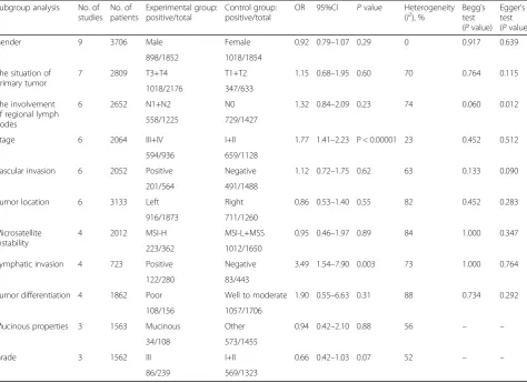
Results:A total of 3481 patients were included in 10 studies. The combined hazard ratio (HR) was 1.22 (95%CI = 1. 01–1.48,P= 0.04), indicating that high expression of PD-L1 was significantly correlated with poor prognosis of colorectal cancer. Apropos of clinicopathological features, the merged odds ratio (OR) exhibited that highly expressed PD-L1 was firmly related to lymphatic invasion (OR = 3.49, 95%CI = 1.54–7.90,P= 0.003) and advanced stage (OR = 1.77, 95%CI = 1. 41–2.23,P< 0.00001), but not correlative with patients’gender, microsatellite instability, or tumor location.
Conclusion:The expression of PD-L1 can be utilized as an independent factor in judging the prognosis of colorectal cancer, and patients with advanced cancer or lymphatic invasion are more likely to express PD-L1. This conclusion may lay a theoretical foundation for the application of PD-1/PD-L1 immunoassay point inhibitors but still needs verifying by sizeable well-designed cohort studies.
Keywords:Colorectal cancer, PD-L1 expression, Prognosis, Clinicopathological features, Meta-analysis
Introduction
Among the most common cancers worldwide, colorectal cancer ranks third, accounting for 10% of all tumor cases [1]. In 2012, the disease engendered 1,400,000 new cases and nearly 700,000 deaths [2]. According to relevant re-search, 4.96% of the population born in the USA are suf-fering from colorectal cancer [3]. Even in Asia, where the incidence rate is reported to be the lowest [4], the threat posed by colorectal cancer cannot be underestimated. Taking China as an example, the incidence and mortality of colorectal cancer there have kept rising. China’s cancer statistics manifest that the incidence and mortality of colo-rectal cancer ranked fifth among all malignant tumors in
China, bringing about 380,000 new cases and 190,000 deaths annually. When they are seeking medical examin-ation, most patients have already been in the advanced stage [5,6]. Despite the continuous development of treat-ment technology, the 5-year survival rate of patients with metastatic disease is still less than 10% [7], which is prob-ably due to the inability to diagnose early and the lack of specific markers to determine tumor development or pa-tients’ prognosis. Therefore, to enhance the prognosis of patients with colorectal cancer, it is indispensable to ex-plore effective diagnostic and therapeutic methods.
Programmed cell death protein 1 (PD-1), a sort of inhibitory checkpoint molecule, was discovered and named by Japanese scholar Ishida in 1992 [8]. It belongs to the CD28 family and is expressed on the surface of activated T cells to regulate proliferation and activation [9]. PD-L1 (also known as B7-H1) is the dominant * Correspondence:jrj@zcmu.edu.cn
1The Second Clinical Medical College, Zhejiang Chinese Medical University,
Hangzhou, Zhejiang, China
Full list of author information is available at the end of the article
ligand for PD-1 and expressed in activated T cells, B cells, dendritic cells, macrophages, endothelial cells, and a significant number of tumor cells [10]. In the healthy immune system, the activation of the PD-1/PD-L1 path-way can inhibit the immune function of T lymphocytes and promote the inhibitory function of regulatory T cells, which can reduce the immune response of the body to normal peripheral tissues. Consequently, it can inhibit autoimmune responses, prevent the development of autoimmune diseases, and maintain autoimmune tol-erance in healthy individuals [11]. When cancer occurs, the tumor cells will reduce their immunogenicity by ex-pressing PD-L1. Hence, they will not be recognized by the immune system and will evade immune attack [12]. In a variety of tumors, the PD-L1 expression is usually associated with poor prognosis [13,14].
Current theories on the expression of PD-L1 in colo-rectal cancer and tumor prognosis are limited and con-troversial. Some studies have manifested the palpable connection between PD-L1 expression and overall sur-vival rate of colorectal cancer patients [15–18], but the others utter the contradictory statement [19, 20]. So, we used meta-analysis to analyze the prognostic value of programmed death factor ligand 1 (PD-L1), which will also lay a theoretical foundation for the application of PD-1/PD-L1 immunoassay point inhibitors in colo-rectal cancer.
Materials and methods Bibliographic search
Two authors independently searched PubMed, Web of Science, Embase, and Cochrane Library for published lit-erature on PD-L1 and colorectal cancer. Publication time of the included articles ranges from the time when the database was established until August 2018. All pub-lications are in English. Search strategies are (“colorectal neoplasms” OR “colorectal cancer” OR “colorectal car-cinoma” OR “colorectal cancers” OR “colonic neo-plasms” OR “rectal neoplasms”) AND (“PD-1” OR “PD-L1” OR “programmed death 1” OR “programmed death ligand 1” OR “programmed cell death ligand 1” OR “programmed death 1 ligand 1” OR “B7-H1” OR “CD274”).
Inclusion criteria
The following are the inclusion criteria:
1. The clinical and pathological data of all cases are complete, and all were diagnosed as colorectal cancer by pathological examination;
2. Detecting the PD-L1 expression in colorectal cancer tissues by immunohistochemical staining;
3. The literature provides the relationship between PD-L1 expression and overall survival (OS) in patients with colorectal cancer;
4. The literature provides the relationship between PD-L1 expression and clinicopathological features, such as primary tumor size, clinical stage, and differentiation;
5. The literature provides sufficient information to estimate the hazard ratio (HR).
Exclusion criteria
The following are the exclusion criteria:
1. The included literature is not an original study; 2. The data contained in the research is wrong, or the
quality of the incorporated literature is low; 3. The included literature is based on animal or cell
experiments;
4. Cannot use the data provided in the literature to calculate the hazard ratio (HR) associated with PD-L1;
5. The included literature did not analyze the expression of PD-L1 in tumor cells.
Data extraction and quality evaluation
Two authors independently screened and extracted data found on inclusion and exclusion criteria and discussed together or adjudicated by third parties in case of disagreement. For the lack of information, we contacted the original author as much as possible. Ex-tracted contents included author, publication year, country, positive threshold, follow-up period, baseline and clinicopathological information of patients, hazard ratio (HR), and 95% confidence interval (95%CI) re-lated to PD-L1 expression.
Methodological quality assessment of the included data was carried out using the Newcastle-Ottawa Scale (NOS). Scores of the NOS are split into three aspects: object selection, inter-group comparability, and outcome measurement. The highest rating is 9 points, and the study with more than 6 points is considered as a high-quality one [21].
Statistical analysis
fixed-effect model or the random-effect model according to heterogeneity. All the analyses above were presented by Revman 5.3 software.
All the studies are retrospective cohort studies, whose heterogeneity is often inevitable. Therefore, based on the analyses above, we carried out publication bias test and sensitivity analysis with the Stata 12.0 software to ex-plore the sources of heterogeneity. And according to Begg’s or Egger’s test,P> 0.05 manifested that there was no publication bias in the study.
Results
Data collection and characteristics
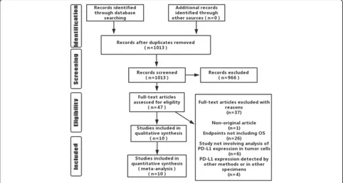
A total of 1013 related articles were initially retrieved. After the layer-by-layer screening, 10 items were ultim-ately included, totaling 3481 cases (Fig. 1). The basic characteristics of the included studies were presented in Table 1. The NOS was used to estimate the quality of the included studies, and all were proved to be high-quality ones (Table2).
Relationship between PD-L1 expression and prognosis of colorectal cancer
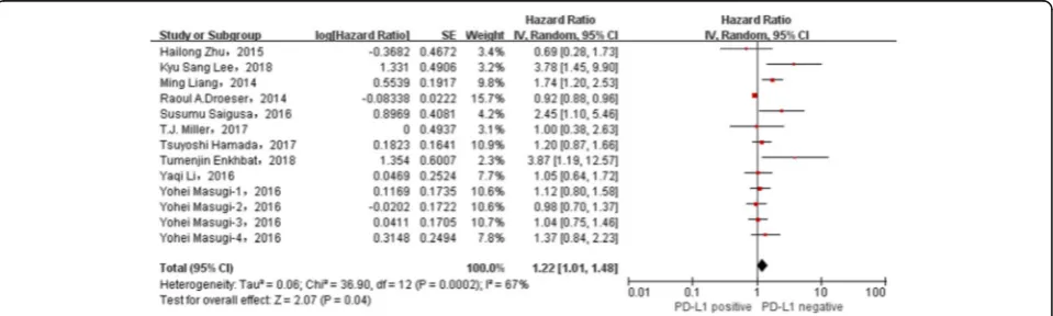
The relationship between the overexpression of PD-L1 and the poor prognosis of colorectal cancer patients was evaluated, and the consequence displayed a significant correlation (HR = 1.22, 95%CI = 1.01–1.48, P= 0.04, ran-dom effect) (Fig.2).
Relationship between PD-L1 expression and clinicopathological features of colorectal cancer
Apropos of clinicopathological features, the merged odds ratio (OR) exhibited that highly expressed PD-L1 was firmly related to lymphatic invasion (OR = 3.49, 95%CI = 1.54–7.90, P= 0.003) and advanced stage (OR = 1.77, 95%CI = 1.41–2.23, P< 0.00001), but not correlative with patients’ gender, microsatellite in-stability, or tumor location (Table3) (Additional file2: Figure S1).
Subgroup analysis of heterogeneity sources
As for subgroup analysis of heterogeneity sources, the het-erogeneity of each subgroup decreased in varying degrees (Additional file3: Figure S2). Among them, the non-Asian group had the minimum heterogeneity (I2 = 12% < 50%). In addition, the results of the Asian group (HR = 1.73, 95%CI = 1.10–2.73, P= 0.02, I2 = 60%), the non-Asian group (HR = 0.93, 95%CI = 0.89–0.97,P= 0.001,I2= 12%), and the tumor stages I–IV group (HR = 1.32, 95%CI = 1.06–1.63, P= 0.01, I2 = 52%) were still statistically significant, but other subgroup analyses failed to ar-rive at such a statistically significant conclusion (Table 4).
Publication bias analysis and sensitivity analysis
The funnel plot is a conventional method to evaluate whether there is a“publication bias”in the meta-analysis, but as a qualitative judgment, its subjectivity makes differ-ent observers come to differdiffer-ent conclusions [22]. Given
this, Begg’s test [23] and Egger’s test [24] were created to evaluate “publication bias” quantificationally. In this meta-analysis, according to Begg’s test (P= 0.428 > 0.05), there was no publication bias in the included literature in-volving PD-L1 and OS (Fig. 3). The detection results of publication bias in subgroup analyses are shown in Table2 and Additional file 4: Figure S3. Sensitivity analysis pointed out that the conclusions were generally stable (Fig.4and Additional file5: Figure S4).
Discussion
PD-1 and PD-L1 are inhibitory costimulatory molecules. When tumor cells express PD-L1 to combine with the PD-1 provided by tumor-infiltrating lymphocytes, the immune effect of T cells in the tumorous microenviron-ment is inhibited, which mediates the occurrence of tumor immune escape and promotes the progress of cancer [25]. At present, a lot of research has been done on this pair of inhibitory costimulatory molecules, but the regulatory mechanism of the signaling pathway in colorectal carcinoma has not been clarified, and theories in many fields are controversial. Although some system-atic reviews focused on the prognostic value of PD-L1 in
all types of solid tumors also mentioned the relationship between PD-L1 and prognosis of colorectal cancer in passing [26–28], yet their results in this regard had limita-tions because of the lack of in-depth research. Wu et al. suggested that PD-L1 overexpression was positively corre-lated with 5-year OS deterioration in colorectal cancer, but they only included two papers and used OR value to evaluate the results, meaning the existence of great bias [28]. Different from the above conclusion, Pyo et al. ap-plied HR involving PD-L1 to assess the relevance between PD-L1 expression and prognosis of colorectal cancer and then concluded that there was no connection between them. However, they merely contained four retrospective studies, which lacked persuasiveness [27]. Xiang et al. also argued that PD-L1 could not be used as a prognostic indi-cator of colorectal cancer, but they misapplied risk ratio (RR), a specific measure for evaluating prospective studies, to the analysis of retrospective studies, so the findings should be treated cautiously [26]. Therefore, to resolve the controversy and deficiency mentioned above, this meta-analysis comprehensively collected relevant litera-ture based on the inclusion criteria and adopted the haz-ard ratio (HR) associated with PD-L1 to estimate the Table 1Basic characteristics of included studies
Author, year
Country No. Stage Follow-up Preoperative
chemoradiotherapy
Japan 116 II–III 52 months (mean) NA YES 57 52/116 3.873 Score > 3
(intensity + area)
Masugi, 2016 [35]
America 450 I–IV > 5 years NA YSE NA 121/450 1.124 Score = 1
(intensity)
117/450 0.980 Score = 2
(intensity)
139/450 1.042 Score = 3
(intensity)
26/450 1.370 Score = 4
(intensity)
China 120 NA 39 months (mean) NA YES NA 28/120 0.692 Score > 4
(intensity + area)
Switzerland 1420 NA > 5 years NA YES NA 495/1420 0.92 Subjective
evaluation
China 276 NA 61 months (mean) NA YES 189 138/276 1.048 Score > 4
(intensity + area)
Miller, 2017 [38]
Australia 104 III 82.5 months (mean) NA YES 89 60/104 1.00 Subjective
evaluation
prognostic value of PD-L1 in colorectal cancer. In addition to that, we also explored the relationship be-tween the expression of PD-L1 and the clinicopatho-logical characteristics of colorectal cancer to make the outcome more convincing.
This meta-analysis demonstrated that PD-L1 expres-sion could be utilized as an independent factor in judg-ing the prognosis of colorectal cancer (HR = 1.22, 95%CI = 1.01–1.48, P= 0.04, random effect). Neverthe-less, there was inevitable heterogeneity among the retro-spective studies included in this meta-analysis (P= 0.0002,I2= 67%). In order to make the conclusion more persuasive and scientific, we adopted the subgroup ana-lysis to explore the heterogeneity sources. And as the findings suggested, the heterogeneity of each subgroup decreased in varying degrees, indicating that these factors have certain degrees of influence. Among them, the non-Asian group had the minimum heterogeneity (I2= 12% < 50%), which implied the essential role the re-gional or ethnic differences played on engendering heterogeneity.
As the subgroup analysis of PD-L1 and clinicopatho-logical features indicated, PD-L1 overexpression in colo-rectal cancer cells was associated with lymphatic invasion. Previous experimental studies have shown that they are indeed relevant. Epithelial-to-mesenchymal transition (EMT) leads to lymphatic invasion [29], and the expression of PD-L1 in tumor cells facilitates im-munosuppression, both of which contribute to tumor progression and metastasis. MiR-200/Zinc finger E-box-binding homeobox 1 (ZEB1) was initially known as EMT regulatory axis, and the bidirectional negative feed-back regulation mechanism between ZEB1 and miR-200 makes the corresponding cells actualize EMT [30]. But recently, the mechanism that miR-200/ZEB1 axis can also regulate PD-L1 to facilitate immunosuppression has been proved by experiments [31]. Therefore, the rela-tionship between lymphatic invasion and PD-L1 overex-pression can be considered to be mutually “parallel.” Also, consistent with another inference of this meta-ana-lysis that the inhibition of the PD-1/PD-L1 signaling pathway in advanced colorectal cancer could achieve Table 2Newcastle-Ottawa Scale for quality assessment
Author, year Selection Comparability Outcome Total
score Exposed
cohort
Non-exposed cohort
Ascertainment of exposure
Outcome of interest
Control for factor
Assessment of outcome
Follow-up long enough
Adequacy of follow-up
Enkhbat, 2018 [15] * * * ** * * 7
Masugi, 2016 [35] * * * ** * * 7
Saigusa, 2016 [16] * * * ** * * 7
Zhu, 2015 [36] * * * ** * * 7
Liang, 2014 [17] * * * * ** * * 8
Droeser, 2014 [37] * * * ** * * 7
Hamada, 2017 [20] * * * ** * * 7
Lee, 2018 [18] * * * ** * * 7
Li, 2016 [19] * * * ** * * 7
Miller, 2017 [38] * * * ** * * 7
*The article scored one point in the project **The article scored two points in the project
remarkable results, existing clinical trials have exhibited the high security and activity of the treatment with PD-1/PD-L1 immunocheckpoint inhibitors [32].
Limitations
To mention first, all the included articles were retro-spective studies, whose bias could not be eliminated, so the consequences were generally stable. Also, it should be noted that all the articles included are in English,
meaning the lack of research especially those negative studies in non-English speaking countries, which leads to the absence of representativeness and the production of bias.
Secondly, it has been reported that the expression of PD-L1 in tumor cells is a critical factor in making the monoclonal antibody against PD-1/PD-L1 effective. However, in every article, the threshold of PD-L1 posi-tive is different and brings about a tremendous impact Table 3The relationship between PD-L1 expression and clinicopathological characteristics in subgroup analysis
Subgroup analysis No. of
studies No. of patients
Experimental group: positive/total
Control group: positive/total
OR 95%CI Pvalue Heterogeneity
(I2), % Beggtest ’s
(Pvalue) Egger’s test (Pvalue)
Gender 9 3706 Male Female 0.92 0.79–1.07 0.29 0 0.917 0.639
898/1852 1018/1854
The situation of primary tumor
7 2809 T3+T4 T1+T2 1.15 0.68–1.95 0.60 70 0.764 0.115
1018/2176 347/633
The involvement of regional lymph nodes
6 2652 N1+N2 N0 1.32 0.84–2.09 0.23 74 0.060 0.012
558/1225 729/1427
Stage 6 2064 III+IV I+II 1.77 1.41–2.23 P < 0.00001 23 0.452 0.512
594/936 659/1128
Vascular invasion 6 2052 Positive Negative 1.12 0.72–1.75 0.62 63 0.133 0.090
201/564 491/1488
Tumor location 6 3133 Left Right 0.86 0.53–1.40 0.55 82 0.452 0.283
916/1873 711/1260
Microsatellite instability
4 2012 MSI-H MSI-L+MSS 0.95 0.46–1.97 0.89 84 1.000 0.347
223/362 1012/1650
Lymphatic invasion 4 723 Positive Negative 3.49 1.54–7.90 0.003 73 1.000 0.764
122/280 83/443
Tumor differentiation 4 1862 Poor Well to moderate 1.90 0.55–6.63 0.31 88 0.734 0.292
108/156 1057/1706
Mucinous properties 3 1563 Mucinous Other 0.94 0.42–2.10 0.88 56 – –
34/108 573/1455
Grade 3 1562 III I+II 0.66 0.42–1.03 0.07 52 – –
86/239 569/1323
Table 4Subgroup analysis of heterogeneity sources
No. of studies HR 95%CI Pvalue Heterogeneity (I2), %
Country Asian 6 1.73 1.10–2.73 0.02 60
Non-Asian 4 0.93 0.89–0.97 0.001 12
Stages I–IV 5 1.32 1.06–1.63 0.01 52
Follow-up period ≥5 years 4 1.12 0.94–1.34 0.20 65
< 5 years 6 1.62 0.93–2.82 0.09 61
Postoperative adjuvant chemotherapy 4 1.61 0.88–2.94 0.12 54
Sample size ≥200 5 1.09 0.92–1.28 0.32 54
on the experimental results. According to published articles, patients with PD-L1 positive can obtain a bet-ter outcome in the treatment with immunocheckpoint inhibitors as the threshold increases [33]. So, uni-formly applying the most suitable PD-L1 positive threshold to the following research should be a top priority. Besides that, using of different immunohisto-chemical antibodies in various studies leads to specific errors.
Furthermore, the latest experiments indicate that cancer patients [34] with intestinal flora disorders have a worse prognosis in the treatment with anti-PD-1/ PD-L1 antibodies, which can be concluded that intes-tinal flora balance plays a vital role in the efficacy of
PD-1/PD-L1 immunocheckpoint inhibitors. Therefore, the following study in this field should take intestinal flora into consideration.
Conclusions
This study analyzed all the available interrelated in-formation in the published literature and exhibited that the expression of PD-L1 was significantly corre-lated with the overall survival rate of colorectal cancer. The more the PD-L1 was expressed, the worse prognosis the colorectal cancer patients would undergo. Concerning clinicopathological features, the expression of PD-L1 was bound up with lymphatic
invasion and tumor stage, but not gender,
Fig. 3Begg’s funnel plot for publication bias test including PD-L1 expression and prognosis in colorectal cancer
microsatellite instability, or tumor differentiation. In other words, the expression of PD-L1 could be uti-lized as an independent factor in judging the prog-nosis of colorectal cancer, and patients with advanced cancer or lymphatic invasion were more likely to express PD-L1. This conclusion may lay a theoretical foundation for the application of PD-1/ PD-L1 immunoassay point inhibitors but still need to be verified by sizeable well-designed cohort studies.
Additional files
Additional file 1:Table S1.PRISMA 2009 Checklist. (DOC 53 kb)
Additional file 2:Figure S1.Forest plots assessing the relationship between PD-L1 and clinicopathological characteristics: (a) gender; (b) grade; (c) lymphatic invasion; (d) microsatellite instability; (e) mucinous properties; (f) stage; (g) the involvement of regional lymph nodes; (h) tumor differentiation; (i) tumor location; (j) the situation of primary tumor; (k) vascular invasion. (PNG 220 kb)
Additional file 3:Figure S2.Subgroup analysis of heterogeneity sources: (a) Asian; (b) non-Asian (c) stages I–IV; (d) follow-up more than 5 years; (e) follow-up less than 5 years (f) postoperative adjuvant chemotherapy; (g) sample size≥200; (h) sample size < 200. (PNG 287 kb)
Additional file 4:Figure S3.Detection of publication bias in subgroup analysis: (a) gender; (b )lymphatic invasion; (c) microsatellite instability; (d) stage; (e) the involvement of regional lymph nodes; (f) the situation of primary tumor; (g) tumor location; (h) tumor differentiation; (i) vascular invasion. (PNG 99 kb)
Additional file 5:Figure S4.Sensitivity analysis of subgroup analysis: (a) gender; (b) lymphatic invasion; (c) microsatellite instability; (d) stage; (e) the involvement of regional lymph nodes; (f) the situation of primary tumor; (g) tumor location; (h) tumor differentiation; (i) vascular invasion. (PNG 147 kb)
Abbreviations
CI:Confidence interval; EMT: Epithelial-to-mesenchymal transition; HR: Hazard ratio; NOS: Newcastle-Ottawa Scale; OR: Odds ratio; OS: Overall survival; PD-1: Programmed cell death protein 1; PD-L1: Programmed death factor ligand 1; PRISMA: Preferred Reporting Items for Systematic Review and Meta-Analysis; ZEB1: Zinc finger E-box-binding homeobox 1
Acknowledgements
Not applicable
Funding
The study received no fund support.
Availability of data and materials
The datasets supporting the conclusions of this article are included within the article and its additional files.
Authors’contributions
ZFS designed the research process. LHG and MMC searched the database for corresponding articles. DYM and ZFS extracted the useful information from the articles above. ZFS and RJJ used the statistical software for analysis. ZFS and LHG drafted the meta-analysis. The final draft came into being after the careful examination by all the authors. All authors read and approved the final manuscript.
Ethics approval and consent to participate
Not applicable
Consent for publication
Not applicable
Competing interests
The authors declare that they have no competing interests.
Publisher’s Note
Springer Nature remains neutral with regard to jurisdictional claims in published maps and institutional affiliations.
Author details
1The Second Clinical Medical College, Zhejiang Chinese Medical University,
Hangzhou, Zhejiang, China.2Department of General Surgery, Ningbo No. 2
Hospital, Ningbo, Zhejiang, China.3Basic Medical College, Zhejiang Chinese
Medical University, Hangzhou, Zhejiang, China.4Affiliated Hospital of Medical School Ningbo University and Ningbo City Third Hospital, Ningbo, Zhejiang, China.
Received: 16 September 2018 Accepted: 11 December 2018
References
1. Calon A, Espinet E, Palomoponce S, et al. Dependency of colorectal cancer on a TGF-beta-driven programme in stromal cells for metastasis initiation. JAK-STAT. 2013;22(2):571–84.
2. Stewart B, Wild C. World cancer report 2014. Lyon: International Agency for Research on Cancer; 2014. p. 19-20.
3. Howlader N, Noone AM, Krapcho M, et al. SEER Cancer Statistics Review, 1975–2009 (Vintage 2009 Populations). Bethesda: National Cancer Institute; 2012.
4. Merika E, Saif M W, Katz A, et al. Review. Colon cancer vaccines: an update. Vivo 2016; 24(5):607.
5. Chen W, Zheng R, Baade PD, et al. Cancer statistics in China, 2015. CA Cancer J Clin. 2016;66(2):115–32.
6. Yang J, Du XL, Li ST, et al. Characteristics of differently located colorectal cancers support proximal and distal classification: a population-based study of 57,847 patients. PLoS One. 2016;11(12):e0167540.
7. Weitz J, Koch M, Debus J, et al. Colorectal cancer. Lancet. 2005; 365(9454):153–65.
8. Ishida Y, Agata Y, Shibahara K, et al. Induced expression of PD-1, a novel member of the immunoglobulin gene superfamily, upon programmed cell death. EMBO J. 1992;11(11):3887–95.
9. Zou W, Wolchok JD, Chen L. PD-L1 (B7-H1) and PD-1 pathway blockade for cancer therapy: mechanisms, response biomarkers, and combinations. Sci Transl Med. 2016;8(328):328rv4.
10. Hansen JD, Pasquier LD, Lefranc MP, et al. The B7 family of
immunoregulatory receptors: a comparative and evolutionary perspective. Mol Immunol. 2009;46(3):457–72.
11. Li B, Vanroey M, Wang C, et al. Anti-programmed death-1 synergizes with granulocyte macrophage colony-stimulating factor--secreting tumor cell immunotherapy providing therapeutic benefit to mice with established tumors. Clinical Cancer Research An Official Journal of the American Association for Cancer Research. 2009;15(5):1623.
12. Mittal D, Gubin MM, Schreiber RD, et al. New insights into cancer immunoediting and its three component phases—elimination, equilibrium and escape. Curr Opin Immunol. 2014;27(1):16–25.
13. Velcheti V, Schalper KA, Carvajal DE, et al. Programmed death ligand-1 expression in non-small cell lung cancer. Lab Investig. 2014;94(1):107. 14. Shi F, Shi M, Zeng Z, et al. PD-1 and PD-L1 upregulation promotes CD8(+)
T-cell apoptosis and postoperative recurrence in hepatocellular carcinoma patients. Int J Cancer. 2011;128(4):887–96.
15. Enkhbat T, Nishi M, Takasu, et al. Programmed cell death ligand 1 expression is an independent prognostic factor in colorectal cancer. Anticancer Res. 2018;38(6):3367–73.
16. Saigusa S, Toiyama Y, Tanaka K, et al. Implication of programmed cell death ligand 1 expression in tumor recurrence and prognosis in rectal cancer with neoadjuvant chemoradiotherapy. Int J Clin Oncol. 2016;21(5):946–52. 17. Liang M, Li J, Wang D, et al. T-cell infiltration and expressions of T
lymphocyte co-inhibitory B7-H1 and B7-H4 molecules among colorectal cancer patients in Northeast China’s Heilongjiang province. Tumor Biol. 2014;35(1):55–60.
19. Li Y, Lei L, Dai W, et al. Prognostic impact of programed cell death-1 (PD-1) and PD-ligand 1 (PD-L1) expression in cancer cells and tumor infiltrating lymphocytes in colorectal cancer. Mol Cancer. 2016;15(1):55.
20. Hamada T, Cao Y, Qian ZR, et al. Aspirin use and colorectal cancer survival according to tumor CD274 (programmed cell death 1 ligand 1) expression status. J Clin Oncol. 2017;35(16):1836–44.
21. Stang A. Critical evaluation of the Newcastle-Ottawa Scale for the assessment of the quality of nonrandomized studies in meta-analyses. Eur J Epidemiol. 2010;25(9):603–5.
22. Greenland S. Invited commentary: a critical look at some popular meta-analytic methods. Am J Epidemiol. 1994;140:290–6.
23. Begg CB, Mazumdar M. Operating characteristics of a rank correlation test for publication bias. Biometrics. 1994;50(4):1088–101.
24. Egger M, Smith GD, Schneider M, et al. Bias in meta-analysis detected by a simple, graphical test. BMJ. 1997;315:629–34.
25. Sun C, Mezzadra R, Schumacher TN. Regulation and function of the PD-L1 checkpoint. Immunity. 2018;48(3):434.
26. Xiang X, Yu PC, Long D, et al. Prognostic value of PD-L1 expression in patients with primary solid tumors. Oncotarget. 2018;9(4):5058–72. 27. Pyo JS, Kang G, Kim JY. Prognostic role of PD-L1 in malignant solid tumors:
a meta-analysis. Int J Biol Markers. 2017;32(1):e68-e74.
28. Wu P, Wu D, Li L, et al. PD-L1 and survival in solid tumors: a meta-analysis. PLoS One. 2015;10(6):e0131403.
29. Ahn YH, Gibbons DL, Chakravarti D, et al. ZEB1 drives prometastatic actin cytoskeletal remodeling by downregulating miR-34a expression. J Clin Investig. 2012;122(9):3170–83.
30. Burk U, Schubert J, Wellner U, et al. A reciprocal repression between ZEB1 and members of the miR-200 family promotes EMT and invasion in cancer cells. EMBO Rep. 2008;9(6):582–9.
31. Chen L, Gibbons DL, Goswami S, et al. Metastasis is regulated via microRNA-200/ZEB1 axis control of tumour cell PD-L1 expression and intratumoral immunosuppression. Nat. Commun. 2014;5:5241.
32. O’Neil BH, Wallmark JM, Lorente D, et al. Safety and antitumor activity of the anti-PD-1 antibody pembrolizumab in patients with advanced colorectal carcinoma. PLoS One. 2017;12(12):e0189848.
33. Jia L, Zhang Q, Zhang R. PD-1/PD-L1 pathway blockade works as an effective and practical therapy for cancer immunotherapy. Cancer Biol Med. 2018;15(2):116-23.
34. Gopalakrishnan V, Spencer CN, Nezi L, et al. Gut microbiome modulates response to anti-PD-1 immunotherapy in melanoma patients. Science. 2018; 359(6371):97.
35. Masugi Y, Nishihara R, Yang J, et al. Tumour CD274 (PD-L1) expression and T cells in colorectal cancer. Gut. 2016;66(8):1463.
36. Zhu H, Qin H, Huang Z, et al. Clinical significance of programmed death ligand-1 (PD-L1) in colorectal serrated adenocarcinoma. Int J Clin Exp Pathol. 2015;8(8):9351–9.
37. Droeser RA, Hirt C, Viehl CT, et al. Clinical impact of programmed cell death ligand 1 expression in colorectal cancer. Eur J Cancer. 2013;49(9):2233–42. 38. Miller TJ, Mccoy MJ, Hemmings C, et al. The prognostic value of cancer