C A S E R E P O R T
Open Access
Pancreatic metastases from renal cell carcinoma:
a case report and literature review of the clinical
and radiological characteristics
Yoshinori Hoshino
1*, Hiroharu Shinozaki
1, Yuki Kimura
1, Yohei Masugi
2, Homare Ito
1, Toshiaki Terauchi
1,
Masaru Kimata
1, Junji Furukawa
1, Kenji Kobayashi
1and Yoshiro Ogata
1Abstract
Metastatic pancreatic cancer is rare, accounting for approximately 2% of all pancreatic malignancies, and most cases arise from renal cell carcinoma. We report the case of a 63-year-old woman, who presented with a pancreatic tumor detected during her annual health examination. She had undergone left nephrectomy 13 years previously for renal cell carcinoma. Computed tomography (CT) revealed two tumors in the head and body of the pancreas, a
hypervascular tumor and a hypovascular tumor with an enhanced rim, respectively. She underwent pylorus-preserving pancreaticoduodenectomy, and metastatic pancreatic tumors arising from the kidney with clustered clear cell carcinoma immunohistochemically positive for CD10 were diagnosed. This report presents the different enhancement features of different lesions on CT scans. Because the enhancement features of lesions have been reported to vary according to the size of the metastatic tumor, a knowledge of the history of renal cell carcinoma is crucial for diagnosis.
Keywords:Pancreatic metastasis, Surgery, Renal cell carcinoma, Imaging, Radiological characteristics
Background
Isolated metastasis to the pancreas is rare, ranging in inci-dence from 2% to 5% in clinical studies [1-6]. Renal cell carcinoma (RCC), melanoma, lung cancer, colorectal can-cer and breast cancan-cer are known to metastasize to the pancreas [7-11]. Most patients with pancreatic metastases are asymptomatic, whereas some exhibit jaundice or ab-dominal pain [12]. RCC has an annual incidence of more than 30,000 a year in the United States, and localized disease is treated via nephrectomy. Of patients with pancreatic metastases, 12% present with synchronous extrapancreatic metastasis, and they have a poor prog-nosis [13,14]. However, surgical treatment for isolated metachronous pancreatic metastases from RCC has been reported in recent years to improve prognosis [6,13-17]. In this study, we report a case of pancreatic metastases from RCC with different radiographic pat-terns for each lesion and review the radiographic
patterns of pancreatic metastases using computed tom-ography (CT) and fluorodeoxyglucose (FDG)-positron emission tomography (PET).
Case presentation
A 63-year-old woman had undergone left nephrectomy for RCC at our hospital 13 years previously. After 5 con-secutive years of follow-up, she underwent an annual medical examination. Abdominal ultrasonography (US) revealed an abnormal mass in the body of the pancreas. CT revealed two lesions: a low-density mass (15 mm in diameter) in the pancreatic body that displayed rim en-hancement and a homogeneously enhanced mass (8 mm in diameter) in the head (Figure 1). Magnetic resonance imaging (MRI) did not show enhancement in either lesion. FDG-PET did not show any abnormal metabolic activity in the pancreas. To allow a pathological diagnosis, endo-scopic ultrasonography (EUS)-guided fine-needle aspir-ation biopsy was performed, but only necrotic tissue was obtained from the specimen. Although the radiographic features of the lesions were different, pancreatic metasta-ses from RCC were strongly suspected because of the * Correspondence:ymailbiz@gmail.com
1
Department of Surgery, Saiseikai Utsunomiya Hospital, 911-1 Takebayashi, Utsunomiya 321-0974, Japan
Full list of author information is available at the end of the article
patient’s history of RCC. We noted the following from the la-boratory findings: DUPAN-2, <25 U/ml (normal, <25 U/ml); Span-1, 2.3 U/ml (normal, <30 U/ml); carcinoembryonic antigen, 1.7 ng/ml (normal, <5.0 ng/ml); carbohydrate antigen 19-9, 2.6 U/ml (normal, <37 U/ml) and gastrin, 480 pg/ml (normal, <200 pg/ml). The patient underwent pylorus-preserving pancreaticoduodenectomy using the Imanaga method [18]. For the R0 resection, an extended pancreaticoduodenectomy was required rather than a classical resection. Intraoperative US revealed a low echoic mass with a bright halo and peripherally enriched blood flow in the body and a low echoic mass with homoge-neously enriched blood flow in the head. Gross patho-logical examination revealed a 15 mm × 13 mm tumor occupying the body of the pancreas and another 8-mm tumor in the uncinate process of the pancreas. The head lesion was soft, whereas the body lesion was firm in consistency. The cut surface of the head lesion was yellow, whereas that of the lesion in the pancreatic body was grayish-white. Metastatic tumor cells homogeneously oc-cupied the tumor in the head of the pancreas, and the firm lesion in the body of the pancreas showed a necrotic change in the center, which was surrounded by viable tumor cells and a fibrous capsule, identified as a low-density area on a CT scan (Figure 2). Immunohistochemically,
the tumors were positive for CD10 and negative for chromogranin A and synaptophysin (Figure 2). Microscopic examination revealed large epithelial cells with clear cyto-plasm and eosinophilic nuclei arranged in alveolar structures with abundant vascularity (Figure 3a). In addition, histo-logical examination revealed another 1-mm occult micro-metastatic lesion in the head of the pancreas (Figure 3b). The harvested lymph nodes and surgical margins were free of malignancy. Taken together, the pathological findings in-dicated that the lesions were metastases from RCC, and the thick enhanced rim of the body lesion was believed to be composed of viable RCC cells with high vascularity. The postoperative course was uneventful, and to date the patient has survived for 6 months without any evidence of recur-rence or metastasis.
Discussion
RCC accounts for approximately 2% of all adult malig-nances. Among kidney-limited diseases, RCC has a high overall survival rate (up to 95%) [19]. However, 20% to 30% of patients have metastases at presentation, and the 5-year survival rate is less than 10% once metastases spread [20]. In studies of resected specimens, RCC was the most common primary tumor leading to isolated pancreatic metastases [6,15]. RCC recurrence is classi-fied as early or late recurrence. Late recurrence after nephrectomy is not common: recurrence is seen in 10% of patients after more than 10 years after surgery [21]. In most studies, the development of pancreatic metastasis was observed after a disease-free period after nephrec-tomy exceeding 10 years (maximum, 32.7 years) [22]. The relation between the metastatic location of the pan-creas and the primary RCC is controversial [23] and can be either hematogeneous or via lymphatics [24]. Hema-togeneous spread may occur along the draining collat-eral vein of a hypervascular renal tumor, and the spread through lymphatics may occur by retrograde lymph flow secondary to tumor infiltration of the retroperitoneal lymph nodes [25].
The symptoms of pancreatic metastasis are often non-specific. Sellner et al. [13] reported in a review of 236 cases that 35% of patients were asymptomatic, whereas other patients had abdominal pain (20%), gastrointes-tinal tract bleeding (20%), obstructive jaundice (9%), weight loss (9%), pancreatitis (3%) or diabetes mellitus (3%). The diameter of the metastatic lesion was reported to have some association with patient symptoms. Bassi et al. [26] reported that the median tumor diameter in asymptomatic patients was 25 mm, compared to 45 mm in symptomatic patients. Conversely, Reddy et al. [27] reported that 93% (42/45) of patients had symptoms such as abdominal pain, jaundice or weight loss.
In general, the preoperative diagnosis of pancreatic metastases begins with a suspicion based on the patient’s
history [28]. Imaging modalities such as CT, MRI, FDG-PET and EUS support the diagnosis. Muranaka et al. [29] reviewed the CT findings of pancreatic metastases from 28 metastatic carcinomas and classified these into 3 types according to their configuration: (1) a single local-ized metastasis (50% to 73%) [29-31]; (2) a diffuse enlarge-ment with homogeneous attenuation of the pancreas (15% to 44%) [29-31] and (3) multifocal metastases (5% to 10%) [29-31]. Metastases from RCC are usually hypervascular and consequently display homogeneous contrast medium enhancement in the arterial phase of CT. Hyperenhance-ment of pancreatic metastases from RCC plays an import-ant role in both the detection of tumor locations and the distinction of metastases from primary adenocarcinoma of the pancreas [32]. When hypervascular pancreatic tumors are identified on enhanced CT scans, differentiation from primary pancreatic endocrine tumors, intrapancreatic accessory spleens and vascular lesions is difficult. Palmowskiet al. [4] observed two types of enhancement on CT scans of pancreatic metastases, namely lesions with either a homogeneous enhancement or a highlighted rim and nonenhancing internal components, depending on the size. In lesions greater than 15 mm in size, rim en-hancement with hypodense central areas of necrosis has
been observed on CT scans [32]. This hypodense aspect is associated with colonic metastases, hyperdense attenu-ation with RCC, or breast cancer [33,34]. We reviewed the radiological features of pancreatic metastases from RCC shown by dynamic CT scans (Table 1). Of 66 patients, 45 (68%) had homogeneous hypervascular enhancement fea-tures, whereas 21 (32%) had central hypovascularity with rim enhancement. Of these, 31 patients had a metastatic configuration, 5 patients had multifocal metastases to the pancreas and 26 patients had solitary tumors. No patients with pancreatic metastasis from RCC developed diffuse enlargement of the pancreas. In the present review, the size of pancreatic metastases from RCC has no particular relation with the presence of central hypodense areas be-cause even tumors > 5 cm in size displayed hypervascular attenuation. The present case was classified as one with multifocal metastases in terms of the metastatic location. To our knowledge, this is the first case of concomitant multifocal metastases featuring two different enhance-ment characteristics, hypodense areas and homogenous hypervascularity.
FDG-PET has not been established for the diagnosis of metastatic RCC. Ramdaveet al. [45] reported that FDG-PET was useful for identifying distant metastases from
RCC in all six of the patients in the study. Majhailet al. [46] calculated the sensitivity and specificity of FDG-PET for identifying distant metastases from RCC. They revealed that the sensitivity of the procedure was linked to the size of the metastases (83% for lesions larger than 15 mm; 93% for those larger than 20 mm). In the present patient with 15- and 8-mm lesions, FDG did not indicate any abnormal metabolic activity in either lesion.
Surgical resection of the pancreas is associated with substantial morbidity after surgery, and the survival benefit of surgery for metastatic lesions of the pancreas remains questionable since randomized control trials have not been conducted. However, surgical resection of metastatic deposits of RCC remains the most effective treatment because chemotherapy, immunotherapy and radiotherapy have generally proved to be ineffective for metastatic RCC [14,22,25,47]. We reviewed studies published from 1998 to 2013 that focused on surgical
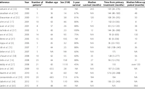
resection of the pancreas for metastases from RCC. The survival rates and features are listed in Table 2. The me-dian 5-year overall survival rate after metastasectomy was reported to be 75% to 88%, compared to 0% to 50% without metastasectomy [13,14,17]. In a review, Masetti et al. [48] analyzed data for 159 patients who underwent metastasectomy of the pancreas for RCC: the median survival and 5-year survival rate were 5.8 years and 63.5%, respectively. Tanis et al. [49] reviewed 170 arti-cles, and data for a total of 411 patients who underwent resection of pancreatic metastases were analyzed. They reported a pancreatic recurrence rate of 4.0% after a me-dian of 42 months and an extrapancreatic recurrence rate of 17.1%. The 5-year survival rate was 72.6%. The surgical mortality rate after pancreaticoduodenectomy for pancreatic metastases was reported to be 2.6% [23]. Sellner et al. [13] compared the 5-year overall survival rate of patients with a solitary metastasis with those with multiple metastases (solitary metastasis, 64%; multiple metastases, 78%). Combining the findings outlined in several reports [50-52], radical surgery for multifocal metastases from RCC in the pancreas appears to be as justified as that for a solitary metastasis. Radical surgery with the R0 resection is the only curative option for pa-tients with pancreatic metastases. Therefore, surgical re-section for pancreatic metastases should be considered under individualized conditions including the manage-ment of comorbidities.
Conclusions
Multifocality of pancreatic metastasis has been re-ported to be in the range 20% to 45% [13,14]. In one report, a preoperative multifocality detection of 17.4% increased up to 34.8% on pathological examin-ation of resected specimens [14]. Here, we report a rare case of pancreatic metastases with micrometas-tasis, which was not detected preoperatively, in the resected pancreas specimen on pathological examin-ation. Moreover, pancreatic metastases from RCC can show both hypervascular attenuation and are central hypodense on CT scans although they are generally hypervascular tumors. Careful examination with multiple modalities for the diagnosis of the meta-static configuration and follow-up are recommended after surgery.
Consent
Written informed consent was obtained from the patient for publication of this case report and any accompanying images. A copy of the written consent is available for review by the Editor-in-Chief of this journal.
Table 2 Literature review of surgical treatment for pancreatic metastases from RCC
Reference Year Number of
patientsa Median age Sex (F:M) survival5-year survival (months)Median treatment (months)Time from primary Median follow-upperiod (months)
Butturiniet al.[53] 1998 5 63 2:3 NA 24.5 120 (24–276) 19
Kassabianet al.[54] 2000 5 56 1:4 67% NA 144 (48–180) 48
Ghavamianet al.[55] 2000 11 68 3:8 81% 120 108 (18–295) 50
Sohnet al.[17] 2001 10 63 4:6 80% 7 102 (0–336) 8
Faureet al.[36] 2001 8 57 2:6 88% NA 83 (12–120) 38
Yachidaet al.[37] 2002 5 60 2:3 100% 12 144 (36–288) 18
Lawet al.[56] 2003 14 64 9:5 75% NA 78 (0–300) 130
Wenteet al.[47] 2005 15 63 10:5 NA NA 85 (0–258) 10
Crippaet al.[6] 2006 5 65 3:2 80% NA 36 (22–192) 41
Eidtet al.[15] 2007 7 64 2:5 88% NA 160 (108–240) 36
Varkeret al.[57] 2007 5 NA NA 60% NA 175 NA
Schaueret al.[58] 2008 10 62 5:5 60% 33 128 (5–277) 56
Zerbiet al.[14] 2008 23 64 15:8 88% 27 96 (12–276) 31
Reddyet al.[27] 2008 21 60 11:10 45% 58 110 over 120
Taniset al.[49] 2009 10 63.5 2:8 NA NA 107 (5–228) NA
Masettiet al.[48] 2010 6 62 6:0 NA NA 57.6 (20–288) 3
Konstantinidiset al.[59] 2010 20 68.5 7:13 61% 104 104 NA
Yazbeket al.[44] 2012 11 73 1:10 90% 84 136 (12–240) NA
Gardiniet al.[60] 2012 8 68 4:4 NA NA NA 38.6
NA: not available.
a
Studies that included less than four patients were excluded.
Table 1 Radiologic features of pancreatic metastases from renal cell carcinoma shown by computed tomography
Reference Year Number of patients
Enhancement features Imaging
configuration
Homogeneous hypervascularity
Central hypodense areas with rim enhancement
Total Solitary Multiple
Marunakaet al.[29] 1989 1 0 1 1 0
Boudgheneet al.[33] 1994 3 2 5 NA NA
Scatarigeet al.[35] 2001 1 1 2 2 0
Faureet al.[36] 2001 5 3 8 NA NA
Yachidaet al.[37] 2002 1 0 1 1 0
Zacharouliset al.[38] 2003 3 0 3 2 1
Davidet al.[25] 2006 0 1 1 1 0
Palmowskiet al.[4] 2008 12 10 22 NA NA
Mechoet al.[39] 2009 4 2 6 6 0
Youet al.[40] 2011 2 0 2 0 2
Katsourakiset al.[41] 2012 1 0 1 1 0
Atiqet al.[42] 2012 2 2 4 3 1
Comunogluet al.[43] 2012 1 0 1 0 1
Yazbeket al.[44] 2012 9 0 9 9 0
Totals 45 21 66 26 5
Abbreviations
CT:Computed tomography; EUS: Endoscopic ultrasonography; FDG: Fluorodeoxyglucose; HE: Hematoxylin and eosin; MRI: Magnetic resonance imaging; PET: Positron emission tomography; RCC: Renal cell carcinoma; US: Ultrasonography.
Competing interests
The authors declare that they have no competing interests.
Authors’contributions
YH performed the majority of this study and drafted the manuscript. YK, HI, YM, and TT surveyed the literature. JF and MK critically revised the manuscript. HS, KK, and YO participated in the design and interpretation of this study under supervision. All authors read and approved the final manuscript.
Acknowledgements
We are grateful to Nobuhiro Nishizawa for his dedicated work in the patient management.
Author details 1
Department of Surgery, Saiseikai Utsunomiya Hospital, 911-1 Takebayashi, Utsunomiya 321-0974, Japan.2Department of Pathology, Keio University, School of Medicine, 35 Shinanomachi, Tokyo 165-8582, Japan.
Received: 29 May 2013 Accepted: 25 October 2013 Published: 9 November 2013
References
1. Bonapasta SA, Gregori M, Lanza R, Sangiorgi E, Menghi A, Scarpini M, Modesti M:Metastasis to the pancreas from breast cancer: difficulties in diagnosis and controversies in treatment.Breast Care (Basel)2010,5:170–173. 2. Blazer DG 3rd, Ramirez PT, Wang H, Fleming JB:Distal pancreatectomy for
isolated metastasis of endometrial carcinoma to the pancreas.JOP2008, 9:56–60.
3. Hernandez S, Martin-Fernandez J, Lasa I, Busteros I, Garcia-Moreno F: Pancreaticoduodenectomy for metastasis of uterine leiomyosarcoma to the pancreas.Clin Transl Oncol2010,12:643–645.
4. Palmowski M, Hacke N, Satzl S, Klauss M, Wente MN, Neukamm M, Kleeff J, Hallscheidt P:Metastasis to the pancreas: characterization by morphology and contrast enhancement features on CT and MRI. Pancreatology2008,8:199–203.
5. Sperti C, Pasquali C, Berselli M, Frison L, Vicario G, Pedrazzoli S:Metastasis to the pancreas from colorectal cancer: is there a place for pancreatic resection?Dis Colon Rectum2009,52:1154–1159.
6. Crippa S, Angelini C, Mussi C, Bonardi C, Romano F, Sartori P, Uggeri F, Bovo G:Surgical treatment of metastatic tumors to the pancreas: a single center experience and review of the literature.World J Surg 2006,30:1536–1542.
7. Roland CF, van Heerden JA:Nonpancreatic primary tumors with metastasis to the pancreas.Surg Gynecol Obstet1989,168:345–347. 8. Lopez-Cantarero Ballesteros M, Fuentes Porcel O, Perez Cabrera B, Perez
Benitez F, Bustos De Abajo M, Jean B:Melanoma metastasis to the pancreas.Rev Esp Enferm Dig1992,82:61–62.
9. Brodish RJ, McFadden DW:The pancreas as the solitary site of metastasis from melanoma.Pancreas1993,8:276–278.
10. Pereira-Lima JC, Coral GP, Bayer LR, da Silva CP:Metastasis from colon carcinoma in the dorsal pancreas of a patient with pancreas divisum: report of a case.Hepatogastroenterology2000,47:554–555.
11. Bachmann J, Michalski CW, Bergmann F, Buchler MW, Kleeff J, Friess H: Metastasis of rectal adenocarcinoma to the pancreas. Two case reports and a review of the literature.JOP2007,8:214–222.
12. Sweeney AD, Wu MF, Hilsenbeck SG, Brunicardi FC, Fisher WE:Value of pancreatic resection for cancer metastatic to the pancreas.J Surg Res 2009,156:189–198.
13. Sellner F, Tykalsky N, De Santis M, Pont J, Klimpfinger M:Solitary and multiple isolated metastases of clear cell renal carcinoma to the pancreas: an indication for pancreatic surgery.Ann Surg Oncol2006, 13:75–85.
14. Zerbi A, Ortolano E, Balzano G, Borri A, Beneduce AA, Di Carlo V:Pancreatic metastasis from renal cell carcinoma: which patients benefit from surgical resection?Ann Surg Oncol2008,15:1161–1168.
15. Eidt S, Jergas M, Schmidt R, Siedek M:Metastasis to the pancreas–an indication for pancreatic resection?Langenbecks Arch Surg2007,392:539–542. 16. Karimi KM, McFadden DW:Pancreatic resection for metastatic renal cell
carcinoma to the pancreas.Am Surg2007,73:1158–1160.
17. Sohn TA, Yeo CJ, Cameron JL, Nakeeb A, Lillemoe KD:Renal cell carcinoma metastatic to the pancreas: results of surgical management.J Gastrointest Surg2001,5:346–351.
18. Imanaga H:A new method of pancreaticoduodenectomy designed to preserve liver and pancreatic function.Surgery1960,47:577–586. 19. Pantuck AJ, Zisman A, Belldegrun AS:The changing natural history of
renal cell carcinoma.J Urol2001,166:1611–1623.
20. Motzer RJ, Bander NH, Nanus DM:Renal-cell carcinoma.N Engl J Med1996, 335:865–875.
21. Sahin M, Foulis AA, Poon FW, Imrie CW:Late focal pancreatic metastasis of renal cell carcinoma.Dig Surg1998,15:72–74.
22. Thompson LD, Heffess CS:Renal cell carcinoma to the pancreas in surgical pathology material.Cancer2000,89:1076–1088. 23. Hung JH, Wang SE, Shyr YM, Su CH, Chen TH, Wu CW:Resection for
secondary malignancy of the pancreas.Pancreas2012,41:121–129. 24. Machado NO, Chopra P:Pancreatic metastasis from renal carcinoma
managed by Whipple resection. A case report and literature review of metastatic pattern, surgical management and outcome.JOP2009, 10:413–418.
25. David AW, Samuel R, Eapen A, Vyas F, Joseph P, Sitaram V:Pancreatic metastasis from renal cell carcinoma 16 years after nephrectomy: a case report and review of the literature.Trop Gastroenterol2006,27:175–176. 26. Bassi C, Butturini G, Falconi M, Sargenti M, Mantovani W, Pederzoli P:High
recurrence rate after atypical resection for pancreatic metastases from renal cell carcinoma.Br J Surg2003,90:555–559.
27. Reddy S, Edil BH, Cameron JL, Pawlik TM, Herman JM, Gilson MM, Campbell KA, Schulick RD, Ahuja N, Wolfgang CL:Pancreatic resection of isolated metastases from nonpancreatic primary cancers.Ann Surg Oncol2008, 15:3199–3206.
28. Hirota T, Tomida T, Iwasa M, Takahashi K, Kaneda M, Tamaki H:Solitary pancreatic metastasis occurring eight years after nephrectomy for renal cell carcinoma. A case report and surgical review.Int J Pancreatol1996, 19:145–153.
29. Muranaka T, Teshima K, Honda H, Nanjo T, Hanada K, Oshiumi Y:Computed tomography and histologic appearance of pancreatic metastases from distant sources.Acta Radiol1989,30:615–619.
30. Ferrozzi F, Bova D, Campodonico F, Chiara FD, Passari A, Bassi P:Pancreatic metastases: CT assessment.Eur Radiol1997,7:241–245.
31. Maeno T, Satoh H, Ishikawa H, Yamashita YT, Naito T, Fujiwara M, Kamma H, Ohtsuka M, Hasegawa S:Patterns of pancreatic metastasis from lung cancer.Anticancer Res1998,18:2881–2884.
32. Klein KA, Stephens DH, Welch TJ:CT characteristics of metastatic disease of the pancreas.Radiographics1998,18:369–378.
33. Boudghene FP, Deslandes PM, LeBlanche AF, Bigot JM:US and CT imaging features of intrapancreatic metastases.J Comput Assist Tomogr1994, 18:905–910.
34. Ng CS, Loyer EM, Iyer RB, David CL, DuBrow RA, Charnsangavej C:Metastases to the pancreas from renal cell carcinoma: findings on three-phase contrast-enhanced helical CT.AJR Am J Roentgenol1999,172:1555–1559. 35. Scatarige JC, Horton KM, Sheth S, Fishman EK:Pancreatic parenchymal
metastases: observations on helical CT.AJR Am J Roentgenol2001, 176:695–699.
36. Faure JP, Tuech JJ, Richer JP, Pessaux P, Arnaud JP, Carretier M:Pancreatic metastasis of renal cell carcinoma: presentation, treatment and survival. J Urol2001,165:20–22.
37. Yachida S, Fukushima N, Kanai Y, Nimura S, Shimada K, Yamamoto J, Sakamoto M:Pancreatic metastasis from renal cell carcinoma extending into the main pancreatic duct: a case report.Jpn J Clin Oncol2002,32:315–317.
38. Zacharoulis D, Asopa V, Karvounis E, Williamson RC:Resection of renal metastases to the pancreas: a surgical challenge.HPB (Oxford)2003,5:137–141. 39. Mecho S, Quiroga S, Cuellar H, Sebastia C:Pancreatic metastasis of renal
cell carcinoma: multidetector CT findings.Abdom Imaging2009,34:385–389. 40. You DD, Choi DW, Choi SH, Heo JS, Kim WS, Ho CY, Lee HG:Surgical resection
41. Katsourakis A, Noussios G, Hadjis I, Alatsakis M, Chatzitheoklitos E:Late solitary pancreatic metastasis from renal cell carcinoma: a case report. Case Rep Med2012,2012:464808.
42. Atiq M, Bhutani MS, Ross WA, Raju GS, Gong Y, Tamm EP, Javle M, Wang X, Lee JH:Role of endoscopic ultrasonography in evaluation of metastatic lesions to the pancreas: a tertiary cancer center experience.Pancreas2012,42:516–523. 43. Comunoglu C, Altaca G, Demiralay E, Moray G:Multiple metastatic renal
cell carcinoma isolated to pancreas.Malays J Pathol2012,34:63–66. 44. Yazbek T, Gayet B:The place of enucleation and enucleo-resection in the
treat-ment of pancreatic metastasis of renal cell carcinoma.JOP2012,13:433–438. 45. Ramdave S, Thomas GW, Berlangieri SU, Bolton DM, Davis I, Danguy HT,
Macgregor D, Scott AM:Clinical role of F-18 fluorodeoxyglucose positron emission tomography for detection and management of renal cell carcinoma.J Urol2001,166:825–830.
46. Majhail NS, Urbain JL, Albani JM, Kanvinde MH, Rice TW, Novick AC, Mekhail TM, Olencki TE, Elson P, Bukowski RM:F-18 fluorodeoxyglucose positron emission tomography in the evaluation of distant metastases from renal cell carcinoma.J Clin Oncol2003,21:3995–4000.
47. Wente MN, Kleeff J, Esposito I, Hartel M, Muller MW, Frohlich BE, Buchler MW, Friess H:Renal cancer cell metastasis into the pancreas: a single-center experience and overview of the literature.Pancreas2005,30:218–222. 48. Masetti M, Zanini N, Martuzzi F, Fabbri C, Mastrangelo L, Landolfo G, Fornelli
A, Burzi M, Vezzelli E, Jovine E:Analysis of prognostic factors in metastatic tumors of the pancreas: a single-center experience and review of the literature.Pancreas2010,39:135–143.
49. Tanis PJ, van der Gaag NA, Busch OR, van Gulik TM, Gouma DJ:Systematic review of pancreatic surgery for metastatic renal cell carcinoma.Br J Surg 2009,96:579–592.
50. Mehta N, Volpe C, Haley T, Balos L, Bradley EL 3rd, Doerr RJ:
Pancreaticoduodenectomy for metastatic renal cell carcinoma: report of a case.Surg Today2000,30:94–97.
51. Bechade D, Palazzo L, Desrame J, Duvic C, Herody M, Didelot F, Coutant G, Algayres JP:Pancreatic metastasis of renal cell carcinoma: report of three cases.Rev Med Interne2002,23:862–866.
52. Eloubeidi MA, Jhala D, Chhieng DC, Jhala N, Eltoum I, Wilcox CM:Multiple late asymptomatic pancreatic metastases from renal cell carcinoma: diagnosis by endoscopic ultrasound-guided fine needle aspiration biopsy with immunocytochemical correlation.Dig Dis Sci2002,47:1839–1842. 53. Butturini G, Bassi C, Falconi M, Salvia R, Caldiron E, Iannucci A, Zamboni G,
Graziani R, Procacci C, Pederzoli P:Surgical treatment of pancreatic metastases from renal cell carcinomas.Dig Surg1998,15:241–246. 54. Kassabian A, Stein J, Jabbour N, Parsa K, Skinner D, Parekh D, Cosenza C,
Selby R:Renal cell carcinoma metastatic to the pancreas: a single-institution series and review of the literature.Urology2000,56:211–215. 55. Ghavamian R, Klein KA, Stephens DH, Welch TJ, LeRoy AJ, Richardson RL,
Burch PA, Zincke H:Renal cell carcinoma metastatic to the pancreas: clinical and radiological features.Mayo Clin Proc2000,75:581–585. 56. Law CH, Wei AC, Hanna SS, Al-Zahrani M, Taylor BR, Greig PD, Langer B,
Gallinger S:Pancreatic resection for metastatic renal cell carcinoma: presentation, treatment, and outcome.Ann Surg Oncol2003,10:922–926. 57. Varker KA, Muscarella P, Wall K, Ellison C, Bloomston M:Pancreatectomy for
non-pancreatic malignancies results in improved survival after R0 resection.World J Surg Oncol2007,5:145.
58. Schauer M, Vogelsang H, Siewert JR:Pancreatic resection for metastatic renal cell carcinoma: a single center experience and review of the literature.Anticancer Res2008,28:361–365.
59. Konstantinidis IT, Dursun A, Zheng H, Wargo JA, Thayer SP, Fernandez-del Castillo C, Warshaw AL, Ferrone CR:Metastatic tumors in the pancreas in the modern era.J Am Coll Surg2010,211:749–753.
60. Gardini A, Morgagni P, Milandri C, Riccobon A, Ridolfi R, La Barba G, Saragoni L, Amadori D, Garcea D:Pancreatic resection for metastases from renal cancer: long term outcome after surgery and immunotherapy approach–single center experience.Hepatogastroenterology2012,59:687–690.
doi:10.1186/1477-7819-11-289
Cite this article as:Hoshinoet al.:Pancreatic metastases from renal cell carcinoma: a case report and literature review of the clinical and radiological characteristics.World Journal of Surgical Oncology
201311:289.
Submit your next manuscript to BioMed Central and take full advantage of:
• Convenient online submission
• Thorough peer review
• No space constraints or color figure charges
• Immediate publication on acceptance
• Inclusion in PubMed, CAS, Scopus and Google Scholar
• Research which is freely available for redistribution