O R I G I N A L R E S E A R C H
Refractive and Visual Outcomes After Bilateral
Implantation of a Trifocal Intraocular Lens in
a Large Population
This article was published in the following Dove Press journal: Clinical Ophthalmology
Edgardo Carreño1 Edgardo A Carreño1 Rodrigo Carreño1 Maricarmen Carreño1 Veronica López1 Richard Potvin 2
1Centro Oftalmológico Carreño, Las
Condes, Santiago, Chile;2Science in Vision, Akron, NY, USA
Purpose: To review refractive, visual acuity, defocus curve and contrast sensitivity results after bilateral implantation of a trifocal intraocular lens (IOL) in a large population.
Setting:One site in Santiago, Chile.
Design: Single arm, non-randomized retrospective chart review.
Methods: This was a single-arm retrospective chart review of clinical outcomes after bilateral implantation of a trifocal IOL (Panoptix®), both toric and non-toric versions. Binocular visual acuity at 4 m, 60 cm and 40 cm was tested. Other tests included refraction, mesopic and photopic contrast sensitivity, and defocus curve measurement.
Results:The review included 500 eyes of 250 patients implanted with the trifocal IOL and 200 eyes of 100 patients implanted with the trifocal toric IOL, with no clinically significant differences between groups. Ninety-six percent of all eyes were within 0.50D of the intended spherical equivalent correction. In the toric group, 94% of eyes (187/200) had a residual refractive cylinder≤0.50D, compared to 81% of eyes (406/500) in the non-toric group. Four out offive patients (80.6%, 282/350) had a binocular uncorrected VA of 0.1 logMAR (20/25) at all test distances. Mean defocus was 0.1 logMAR or better from vergences from 0.00 to
−3.00 D (corresponding to vision from distance to about 33 cm). With a cutoff of 0.2 logMAR, 96% of patients had a range of vision 2.5 D or greater. Contrast sensitivity was similar between the toric and non-toric lenses, and similar to age-matched normal results.
Conclusion:The non-toric and toric trifocal IOLs provided good distance, intermediate and near vision to patients, with a wide range of vision and good contrast sensitivity.
Keywords:trifocal IOL, contrast sensitivity, toric trifocal IOL
Plain Language Summary
Cataract surgery involves removing the clouded lens in the eye and replacing it with an artificial one (an intraocular lens or IOL). Often the IOL is designed only to provide good distance vision in both eyes, requiring patients to use reading glasses for close work. However, there are IOL options that provide good vision at distance, intermediate (eg, for computer work) and near (reading). One such IOL is the PanOptix®trifocal intraocular lens (IOL)–its design provides a relatively continuous range of vision from far to near. This study was designed to provide clinical data from a large number of eyes/patients to surgeons who are interested in using this IOL. It was a chart review of patients previously treated with two versions of the lens, one that does not correct astigmatism (500 eyes of 250 patients), and the other that does (200 eyes of 100 patients). This is the largest clinical data set for this IOL that has been published so far. A high percent of all eyes had little or no prescription for clear vision at distance. Four out of five patients had binocular uncorrected vision better
Correspondence: Richard Potvin Science in Vision, 6197 Dye Road, Akron, NY 14001, USA
Email rick@scienceinvision.com
Clinical Ophthalmology
Dove
press
open access to scientific and medical research
Open Access Full Text Article
Clinical Ophthalmology downloaded from https://www.dovepress.com/ by 118.70.13.36 on 24-Aug-2020
than or equal to“20/25” at all test distances (4 m, 60 cm and 40 cm). Contrast sensitivity, one measure of the quality of vision, was similar between patients with either IOL implanted and age-matched normal results. In summary, this trifocal IOL appears to be a good alternative for appropriate patients inter-ested in reducing their dependence on glasses after cataract surgery.
Introduction
It is well recognized that cataract surgery has become a refractive procedure. Modern intraocular lens (IOL) power calculations, for both sphere and cylinder, provide a high likelihood of meeting the refractive target for the patient. Achieving a near-emmetropic outcome is particu-larly important when toric and/or presbyopia-correcting IOLs are implanted because the relative benefit of these advanced technology IOLs is best appreciated when the patient’s refraction is near plano.
Presbyopia-correcting IOLs continue to improve. Early zonal designs provided some near vision but with a high likelihood of glare and halos.1Newer generation diffractive bifocals provided good distance and better near vision with fewer visual disturbances.2Diffractive trifocal IOLs are now relatively common. A large meta-analysis of 395 subjects noted that visual quality, including contrast sensitivity testing, was similar between bifocal and trifocal diffractive lenses, with the added benefit of improved intermediate vision with trifocal lenses.3 The ideal implantable multifocal IOL will improve vision across a range of distances from distance to near without negatively impacting contrast sensitivity.
One of the newer trifocal IOL designs available is the AcrySof®IQ PanOptix Trifocal IOL (Alcon, Fort Worth, TX, USA). It is based on a quadrifocal optical principle, with the second focal point relatively suppressed. This allowed the best focus intermediate to be at 60 cm rather than 80 cm – the latter is a typical intermediate design distance for a standard trifocal IOL if the near point is 40 cm. This trifocal IOL has been reported to provide improved intermediate vision when compared to a bifocal IOL, without negatively impacting contrast sensitivity.4 This trifocal design has been compared with different trifocal designs, an extended depth of focus IOL and a bifocal IOL and some advantages with regard to range of vision and visual disturbances have been reported. This IOL was shown to provide improved intermediate vision at 60 cm with fewer complaints of halos when compared with another trifocal IOL.5Near vision is also reportedly better when compared to an extended depth of focus IOL.6
The largest study in the literature to date reporting the results of implanting this trifocal IOL included 125 patients, with 166 eyes receiving a non-toric correction and 84 receiving a toric correction.7 Most other studies have included 50 or fewer subjects. The purpose of the current study was to provide postoperative refractive, visual acuity, defocus curve and contrast sensitivity out-comes in a much larger dataset, setting realistic expecta-tions for the performance of both the non-toric and toric versions of the lens in clinical practice.
Methods
A retrospective chart review of patients undergoing bilat-eral cataract surgery or refractive lens exchange (RLE) surgery was conducted, to locate patients bilaterally implanted with the trifocal or toric trifocal IOL of interest. Surgicalfiles for two surgeons (EC, EAC) were examined. Patients with no pre-existing pathology (besides cataract) and no previous eye surgery were identified, and their surgical and postoperative data were collated. The study was approved by the ethics committee of the Metropolitan Service of Santiago, Chile. All patients sign an informed consent waiver that allows their de-identified data to be used for research purposes.
All surgeries were planned using the Topcon KR800 auto-refractor (Topcon, Tokyo, Japan) and the IOLMaster 500 (Carl Zeiss Meditec, Jena, Germany). The IOLMaster and iTrace (Tracey Technologies Corp. TX, USA) kerato-metry data were used to corroborate the KR800; other biometric data (axial length, white-to-white, anterior chamber depth) from the IOLMaster were used for the IOL sphere calculation, considering results from the SRK-T, Haigis, Hoffer Q and Holladay formulas. Toric IOL calculations were made using the keratometric data from the KR800, IOLMaster and iTrace with the Panoptix Toric IOL planning software, which includes the Barrett algorithm to account for posterior corneal astigmatism. The patient was marked at 0 and 180 degrees at the slit lamp (sitting) before entering the surgery suite. The VERION system (Alcon, Fort Worth, TX) was used for eye registration. Femtosecond laser-assisted cataract sur-gery (FLACS) was performed on many eyes, and this was noted. The primary incision was clear corneal and either 2.2 mm for manual cases or 2.0 mm for FLACS cases. Intraoperative aberrometry using the ORA™ System (Alcon, Fort Worth, TX, USA) was measured, to verify IOL power and placement.
Clinical Ophthalmology downloaded from https://www.dovepress.com/ by 118.70.13.36 on 24-Aug-2020
All follow-up data were from the patient visit one month after second-eye surgery. Postoperative data included auto-refraction using the KR800, providing refractive cylinder and the spherical equivalent refraction. Uncorrected binocular visual acuity (VA) testing was performed under the same conditions for all patients, with a luminance between 80 and 90 cd/m2. The binocular uncorrected defocus curve was col-lected in the same room using a decimal-based visual acuity chart; the vergence range tested was from 0.00D to−4.00D. Decimal notation was converted to logMAR for reporting purposes. Contrast sensitivity in both photopic and scotopic conditions was measured with the Optec® 6500 functional vision tester (Stereo Optical Co., Chicago, IL).
Data were extracted from clinical records and collated in MS Excel, then imported into an Access database for data checking and preliminary analysis (both Microsoft Corp., Redmond, WA, USA). Statistical analyses were performed using the Statistica data analysis software sys-tem, version 12 (TIBCO Software Inc., Palo Alto, CA, USA). Parametric comparisons between groups were made using analysis of variance (ANOVA) and non-parametric data were compared using the Chi-squared test.
Results
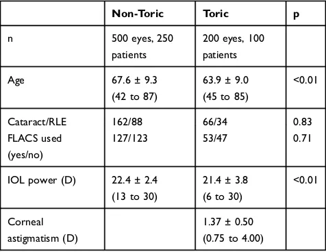
A review of existing clinical records identified 500 eyes of 250 patients implanted with the trifocal IOL of interest and 200 eyes of 100 patients implanted with the toric version of the IOL between July of 2015 and August of 2019 that met the inclusion criteria. Table 1 shows the available preoperative data for the two groups. Corneal astigmatism data were not recorded for non-toric eyes. As can be seen,
the groups are reasonably similar, though the patients with Toric IOLs were slightly younger and had a slightly lower spherical IOL power implanted. While these differences were statistically significant, they were not considered clinically significant.
Figure 1 shows the distribution of the postoperative
mean refraction spherical equivalent (MRSE). There was no statistically significant difference in the percentage of eyes within 0.25D of the intended correction (80% non-toric, 86% non-toric, Chi-squared test, p = 0.06). Ninety-six percent of all eyes were within 0.50D of the intended correction. The MRSE was not statistically significantly different between the toric and non-toric IOLs, nor was there any statistically significant difference between FLACS and standard surgery. There was a statistically significant difference in the MRSE between the cataract and RLE eyes, but the difference was clinically insignif-icant (−0.02D ± 0.02D for RLE eyes vs.−0.08D ± 0.01D for cataract eyes, p = 0.004).
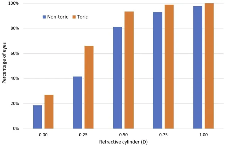
Figure 2 shows the cumulative distribution of
post-operative refractive cylinder by lens type. The percentage of eyes with residual cylinder of 0.0, or less than 0.25D, 0.50D and 0.75D was statistically significantly higher in the toric group vs. the non-toric group (p < 0.02 for all four comparisons). While not shown, the mean residual refractive cylinder in the non-toric group was 0.42 ± 0.29D and in the toric group was 0.28 ± 0.23D. In the toric group, 94% of eyes (187/200) had a residual refrac-tive cylinder less than or equal to 0.50D, compared to 81% of eyes (406/500) in the non-toric group.
The mean binocular uncorrected VAs by lens type at distance, intermediate and near were calculated. There was no statistically significant difference between the lens types, but the VA was statistically significantly different by test distance. Mean distance VA (−0.08) was about one line better than intermediate VA (+0.03) and about two letters better than near VA (−0.04). In all instances, the mean VA was better than 0.1 logMAR (20/25). While not shown, there was no statistically significant effect of sur-gery type (FLACS vs. standard) on any VA measures, but VA at all distances was statistically significantly better in the RLE group vs. the cataract group; the mean difference between the two groups at all distances was about 2 letters. Four out offive patients (80.6%, 282/350) had a binocular uncorrected VA of 0.1 logMAR (20/25) at all three dis-tances, with no significant difference in this percentage by lens type (p = 0.64).Figure 3shows the distribution of VA at each of the distances.
Table 1Patient Demographics
Non-Toric Toric p
n 500 eyes, 250
patients
200 eyes, 100 patients
Age 67.6 ± 9.3
(42 to 87)
63.9 ± 9.0 (45 to 85)
<0.01
Cataract/RLE 162/88 66/34 0.83
FLACS used (yes/no)
127/123 53/47 0.71
IOL power (D) 22.4 ± 2.4 (13 to 30)
21.4 ± 3.8 (6 to 30)
<0.01
Corneal astigmatism (D)
1.37 ± 0.50 (0.75 to 4.00)
Abbreviations:IOL, intraocular lens; RLE, refractive lens exchange; FLACS, fem-tosecond laser assisted cataract surgery; D, diopter.
Clinical Ophthalmology downloaded from https://www.dovepress.com/ by 118.70.13.36 on 24-Aug-2020
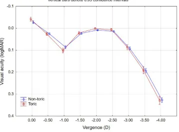
The binocular uncorrected defocus curve was measured using a decimal chart; decimal VA values were converted to logMAR; results by lens type are shown inFigure 4. The performance was similar for both lens types, with a mean VA difference of less than a letter at any given vergence. The
range of vision was calculated for each patient as the range of vergences, measured from 0.0D (distance) where the VA was 0.1 logMAR (Snellen 20/25) or better. Seventy-two percent of patients (251/350) had a range of vision of 2.5D or greater (from distance to −2.50 D, corresponding to Figure 1Distribution of the mean postoperative refraction spherical equivalent by lens type.
Abbreviation:D, diopter.
Figure 2Distribution of postoperative refractive cylinder by lens type. Abbreviation:D, diopter.
Clinical Ophthalmology downloaded from https://www.dovepress.com/ by 118.70.13.36 on 24-Aug-2020
a reading distance of 40 cm) using this 0.1 logMAR cutoff. Using a cutoff of 0.2 logMAR (Snellen 20/32 or better), 96% of patients (335/350) had a range of vision of 2.50 D or
greater, with 90% (315/350) having a range of vision of 3.0 D or better (from distance to −3.00 D, corresponding to a reading distance of 33 cm) at this acuity level.
Figure 3Binocular uncorrected visual acuity by distance.
Figure 4Binocular uncorrected defocus curve.
Clinical Ophthalmology downloaded from https://www.dovepress.com/ by 118.70.13.36 on 24-Aug-2020
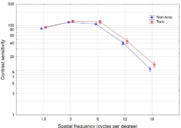
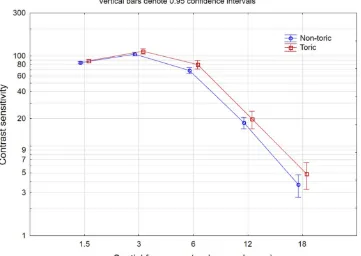
Binocular uncorrected contrast sensitivity results in both photopic and mesopic conditions are shown in
Figure 5 (photopic) and 6 (mesopic). Mean results for
the toric lens were slightly better than for the non-toric lens at all spatial frequencies in both photopic and mesopic conditions. There were several spatial frequencies where surgery type (refractive lens exchange or cataract) and/or lens type (non-toric or toric) were statistically significantly different, but the differences were not considered clinically significant (less than 5%). There were no statistically sig-nificant differences by surgery type (FLACS vs. standard) at any spatial frequency in either photopic or mesopic conditions.
Discussion
Previous results reported for this toric IOL have demon-strated good binocular visual acuity at various distances. Mean reported uncorrected distance VA (UDVA) varied from −0.148to 0.079logMAR; our results were closer to the former at −0.08 logMAR (the 0.07 value may be a monocular result, which may explain the lower value). Mean reported uncorrected intermediate VA (UIVA) at 60 cm varied from 0.0210,11to 0.085logMAR; our results were closer to the former at 0.03 logMAR. Mean reported uncorrected near VA (UNVA) at 40 cm varied from−0.0110
to 0.035 logMAR; again, our results were closer to the former at −0.04 logMAR. Some of the differences in the visual acuity results may relate to testing methods, residual refractive error and possibly length of follow-up.
In a previous study, the monocular uncorrected visual acuity results for those implanted with non-toric and toric versions of the same lens were noted to be 0.20 and 0.23 at intermediate (60 cm) and 0.05 and 0.07 at near (33 cm), respectively; the current study also showed no significant difference in VA by lens type.7
Previously reported defocus curve testing has consis-tently demonstrated a good range of visual acuity. Acuity of 0.3 logMAR or better from distance to near (−3.00D, equivalent to a reading distance of 33 cm) is common.5,9,11,12 Results appear consistent with these prior results, though perhaps not as good as reported by Gundersen and Potvin, possibly because they used best distance-corrected VA at 6 months to 2 years postop while the current study used uncorrected VA at 1-month postop.13 We found that 90% of our subjects had a 3.0D range of vision with an acuity of 0.2 logMAR or better. Similar tofindings in the current study, the defocus curve did not differ significantly between the toric and non-toric groups in a previous study by Rementería-Capelo et al, though they tested subjects in with their best
distance-Figure 5Photopic contrast sensitivity by lens type.
Clinical Ophthalmology downloaded from https://www.dovepress.com/ by 118.70.13.36 on 24-Aug-2020
correction.7Visual acuity at −1.0D (corresponding to a 1 meter viewing distance) was lower than for other ver-gences, while the acuity −1.5D (corresponding to a viewing distance of about 67 cm) was equivalent to the acuity at near. This is a function of the “closer intermedi-ate vision”feature of the lens design.11
Good contrast sensitivity outcomes have also been demonstrated in several studies with similar VA under different lighting conditions suggesting a level of pupil independence.8,9,14Results here appear within the normal range for subjects tested on the device.15 Contrast sensi-tivity results in the current study were better than the results reported by Ruiz-Mesa et al, especially at the higher spatial frequencies, despite the previous authors’ use of corrected VA for contrast sensitivity testing.12
There are limitations to the current study. It reports results from a single center. There were no specific inclusion or exclusion criteria besides the ones noted in the methods. Rather, this study was designed to capture a large number of patients in a routine clinical setting. In addition, visual acuity, defocus and contrast sensitivity were measured only in the binocular uncorrected state, which limits the ability to compare current data to other studies in the literature. Finally, the focus was on objective outcome measures such as visual acuity and contrast sensitivity; subjective measures such as
quality of vision and reported visual disturbances were not collected. Less importantly, refractions were limited to results from an autorefractor. However, autorefraction has been shown to provide only a nominally more minus refrac-tion relative to subjective methods.16
In conclusion, the current study demonstrates that both the non-toric and toric versions of the trifocal IOL eval-uated here provide excellent functional vision to patients, with good distance, intermediate and near uncorrected VA, a wide range of vision and good contrast sensitivity. We believe this is the largest study of clinical outcomes for this particular trifocal lens that has been reported to date.
Acknowledgments
Sarah Y. Makari, OD is a consultant to Science in Vision who received compensation for writing assistance in pre-paration of the manuscript.
Funding
This study was supported with an investigator-initiated study grant from Alcon (IIT #29955405), Fort Worth, TX.
Disclosure
Edgardo Carreño, Edgardo A. Carreño, Rodrigo Carreño, Maricarmen Carreño, and Veronica López received payment Figure 6Mesopic contrast sensitivity by lens type.
Clinical Ophthalmology downloaded from https://www.dovepress.com/ by 118.70.13.36 on 24-Aug-2020
for data analysis from Alcon. Richard Potvin reports personal fees from Centro Oftalmológico Carreño, during the conduct of the study; personal fees from Alcon, outside the submitted work. The authors report no other conflicts of interest in this work.
References
1. Sen HN, Sarikkola AU, Uusitalo RJ, Laatikainen L. Quality of vision after AMO array multifocal intraocular lens implantation.J Cataract Refract Surg.2004;30:2483–2493. doi:10.1016/j.jcrs.2004.04.049 2. Cillino S, Casuccio A, Di Pace F, et al. One-year outcomes with
new-generation multifocal intraocular lenses. Ophthalmology.
2008;115:1508–1516. doi:10.1016/j.ophtha.2008.04.017
3. Yoon CH, Shin IS, Kim MK. Trifocal versus bifocal diffractive intrao-cular lens implantation after cataract surgery or refractive lens exchange: a meta-analysis. J Korean Med Sci. 2018;33(44):e275. doi:10.3346/jkms.2018.33.e275
4. de Carneros-llorente AM, de Carneros AM, de Carneros-llorente PM, Jiménez-Alfaro I. Comparison of visual quality and subjective outcomes among 3 trifocal intraocular lenses and 1 bifocal intraocular lens.
J Cataract Refract Surg. 2019;45:587–594. doi:10.1016/j.jcrs. 2018.12.005
5. Sezgin Asena B. Visual and refractive outcomes, spectacle independence, and visual disturbances after cataract or refractive lens exchange surgery: comparison of 2 trifocal intraocular lenses.J Cataract Refract Surg.
2019;45:1539–1546. doi:10.1016/j.jcrs.2019.06.005
6. Monaco G, Gari M, Di Censo F, Poscia A, Ruggi G, Scialdone A. Visual performance after bilateral implantation of 2 new presbyopia-correcting intraocular lenses: trifocal versus extended range of vision. J Cataract Refract Surg. 2017;43:737–747. doi:10.1016/j.jcrs.2017.03.037
7. Rementería-Capelo LA, Contreras I, García-Pérez JL, Blázquez V, Ruiz-Alcocer J. Visual quality and patient satisfaction with a trifocal intraocular lens and its new toric version. J Cataract Refract Surg.
2019;45:1584–1590. doi:10.1016/j.jcrs.2019.06.014
8. Yesilirmak N, Akova YA, Donmez O. Comparison of mix-and-match implanted bifocal IOLs and bilateral implanted trifocal IOLs after femtosecond laser-assisted cataract surgery. J Refract Surg.
2019;35:559–564. doi:10.3928/1081597X-20190806-01
9. Alió JL, Plaza-Puche AB, Alió Del Barrio JL, et al. Clinical out-comes with a diffractive trifocal intraocular lens.Eur J Ophthalmol.
2018;28:419–424. doi:10.1177/1120672118762231
10. Böhm M, Hemkeppler E, Herzog M, et al. Comparison of a panfocal and trifocal diffractive intraocular lens after femtosecond laser-assisted lens surgery.J Cataract Refract Surg.2018;44:1454–1462. doi:10.1016/j. jcrs.2018.07.060
11. Kohnen T, Herzog M, Hemkeppler E, et al. Visual performance of a quadrifocal (trifocal) intraocular lens following removal of the crystalline lens. Am J Ophthalmol. 2017;184:52–62. doi:10.1016/j. ajo.2017.09.016
12. Ruiz-Mesa R, Abengózar-Vela A, Ruiz-Santos M. A comparative study of the visual outcomes between a new trifocal and an extended depth of focus intraocular lens.Eur J Ophthalmol.2018;28:182–187. doi:10.5301/ejo.5001029
13. Gundersen KG, Potvin R. Trifocal intraocular lenses: a comparison of the visual performance and quality of vision provided by two differ-ent lens designs.Clin Ophthalmol.2017;11:1081–1087. doi:10.2147/ OPTH
14. García-Pérez JL, Gros-Otero J, Sánchez-Ramos C, Blázquez V, Contreras I. Short term visual outcomes of a new trifocal intraocular lens.BMC Ophthalmol.2017;17(1):72. doi:10.1186/s12886-017-0462-y 15. Hohberger B, Laemmer R, Adler W, Juenemann AG, Horn FK.
Measuring contrast sensitivity in normal subjects with OPTEC 6500: influence of age and glare. Graefes Arch Clin Exp Ophthalmol.2007;245:1805–1814. doi:10.1007/s00417-007-0662-x 16. Garzón N, García-Montero M, López-Artero E, Poyales F,
Albarrán-Diego C. Influence of trifocal intraocular lenses on standard autorefraction and aberrometer-based autorefraction. J Cataract Refract Surg.2019;45(9):1265–1274. doi:10.1016/j.jcrs.2019.04.017
Clinical Ophthalmology
Dove
press
Publish your work in this journal
Clinical Ophthalmology is an international, peer-reviewed journal cover-ing all subspecialties within ophthalmology. Key topics include: Optometry; Visual science; Pharmacology and drug therapy in eye dis-eases; Basic Sciences; Primary and Secondary eye care; Patient Safety and Quality of Care Improvements. This journal is indexed on PubMed
Central and CAS, and is the official journal of The Society of Clinical Ophthalmology (SCO). The manuscript management system is completely online and includes a very quick and fair peer-review system, which is all easy to use. Visit http://www.dovepress.com/ testimonials.php to read real quotes from published authors.
Submit your manuscript here:https://www.dovepress.com/clinical-ophthalmology-journal
Clinical Ophthalmology downloaded from https://www.dovepress.com/ by 118.70.13.36 on 24-Aug-2020