O R I G I N A L R E S E A R C H
The Safety and Exploration of the
Pharmacokinetics of Intrapleural Liposomal
Curcumin
This article was published in the following Dove Press journal:
International Journal of Nanomedicine
Ashleigh Hocking1
Sara Tommasi 2
Peter Sordillo3
Sonja Klebe 1,4
1Department of Anatomical Pathology, Flinders University, Adelaide, SA, Australia;2Department of Clinical Pharmacology, Flinders University, Adelaide, SA, Australia;3SignPath Pharma Inc, New York, NY, USA;4Department of Surgical Pathology, SA Health, Flinders Medical Centre, Bedford Park, SA, Australia
Background: Malignant pleural effusion (MPE) is the accumulation offluid in the pleural
cavity as a result of malignancies affecting the lung, pleura and mediastinal lymph nodes. Curcumin, a compound found in turmeric, has anti-cancer properties that could not only treat MPE accumulation but also reduce cancer burden. To our knowledge, direct administration of curcumin into the pleural cavity has never been reported, neither in animals nor in humans.
Purpose: To explore the compartmental distribution, targeted pharmacokinetics and the safety
profile of liposomal curcumin following intrapleural and intravenous administration.
Methods: Liposomal curcumin (16 mg/kg) was administered into Fischer 344 rats by either
intrapleural injection or intravenous infusion. The concentration of curcumin in plasma and tissues (lung, liver and diaphragm) were measured using ultra-performance liquid chromatogra-phy-mass spectrometry (UPLC-MS). Blood and tissues were examined for pathological changes.
Results: No pleural or lung pathologies were observed following intrapleural liposomal
curcumin administration. Total curcumin concentration peaked 1.5 hrs after the administra-tion of intrapleural liposomal curcumin and red blood cell morphology appeared normal. A red blood cells abnormality (echinocytosis) was observed immediately and at 1.5 hrs after intravenous infusion of liposomal curcumin.
Conclusion: These results indicate that liposomal curcumin is safe when administered
directly into the pleural cavity and may represent a viable alternative to intravenous infusion in patients with pleural-based tumors.
Keywords:malignant pleural effusion, liposomal, curcumin, intrapleural, local administration
Introduction
A malignant pleural effusion (MPE) is the accumulation of fluid in the pleural
cavity as a result of malignancy. The most common causes of MPE are malignan-cies that have metastasized to the pleural or mediastinal lymph nodes; breast and lung cancer are the most prevalent causes in women and men, respectively. Malignant pleural mesothelioma, a tumor arising in the mesothelial cells lining the pleural cavity, also commonly results in MPE. Not only does MPE produce
significant discomfort and breathing difficulties in these patients, but it is also
a frequent cause of mortality.1 Controlling recurrent MPE is an integral part of
palliative care for these patients, which is achieved by either pleurodesis or
inser-tion of an indwelling pleural catheter for ongoing drainage.2
The use of anti-cancer agents, in conjunction with MPE management, could help
alleviate patient’s symptoms and reduce their cancer burden.3Curcumin−a polyphenol
Correspondence: Ashleigh Hocking Department of Anatomical Pathology, Flinders University, Flinders Medical Centre, Flinders Drive, Bedford Park, SA 5041, Australia
Email ash.hocking@flinders.edu.au
International Journal of Nanomedicine
Dove
press
open access to scientific and medical research
Open Access Full Text Article
International Journal of Nanomedicine downloaded from https://www.dovepress.com/ by 118.70.13.36 on 24-Aug-2020
derived from turmeric− can modulate numerous pathways involved in carcinogenesis, including those controlling
inflammation, cell cycle progression, and angiogenesis and
cell survival.4 Curcumin also has the potential to help to
reduce MPE since it can moderate numerous factors involved
influid accumulation, including vascular endothelial growth
factor-A (VEGF-A), interleukin-6 (IL-6) and tumor necrosis
factor-alpha, although this has not been verified.1,5–7
However, difficulties with clinical translation exist because
of curcumin’s low solubility in aqueous solution and oils,
instability at physiological pH, low bioavailability and rapid
molecular transformation and degradation.4
Liposomes are phospholipid vesicles that act as delivery systems for both hydrophobic and hydrophilic drugs. They are utilized to reduce early degradation and improve
stabi-lity, biodistribution and cellular uptake.8,9 In an effort to
overcome curcumin’s poor oral bioavailability and water
solubility, researchers have developed a liposomal curcumin formulation, which has been safely administered in humans
via intravenous infusion.10,11Liposomal curcumin could be
administered directly into the pleural cavity of patients with MPE through an existing intrapleural catheter or at the time of pleurodesis. Intrapleural drug delivery is an attractive alternative to intravenous therapies for pleural cancers because i) drugs reach higher concentrations at the site of the tumor ii) concentrations are sustained for longer periods due to a slower clearance rate and iii) there are reduced
systemic toxicities.12–15 Numerous drugs including
pacli-taxel, bevacizumab and cisplatin have been administered into the pleural space in clinical trial settings to control malignant pleural effusion, alleviate symptoms, or slow
dis-ease progression.12,15–25 To the best of our knowledge, the
direct administration of curcumin into the pleural cavity has never been reported. The purpose of this study was to evaluate the safety and bio-distribution of a pharmaceutical-grade liposomal curcumin formulation after intrapleural administration in healthy rats.
Materials and Methods
Chemicals and Reagents
Liposomal curcumin (Lipocurc™) was a kind gift from
SignPath Pharma Inc. (Sandy, Utah, United States).
Liposomal curcumin was synthesized at Polymun Scientific
GmbH, Vienna, Austria, according to the encapsulation
pro-tocol previously described.20,21 The formulation was
comprised of curcumin (6.0 mg/mL), DMPC (14:0–
-1,2-dimyristoyl-sn-glycero-3-phosphocholine) (72 mg/mL)
and DMPG (14:0–1,2-dimyristoyl-sn-glycero-3-
phosphoryl-glycerol) (8.0 mg/mL). Liposomal curcumin exhibited a zeta
potential of−36 mV at pH 5.0 and mean particle diameter of
117 nm. Aliquots were stored at−20°C in storage boxes that
were protected from light and aliquots were thawed immedi-ately before use to avoid degradation.
Animals
Male and female Fischer 344 rats (aged 12-weeks, Flinders University School of Medicine Animal Facility) were used for in vivo experiments. Rats were housed 3 per cage with Back-2-Nature Animal Bedding (Fibrecycle Pty Ltd, Queensland, Australia) in temperature-controlled (22±1°C), and humidity-controlled (60±5%) environment on a 12:12
light-dark cycle. Rats had free access to food (Gordon’s
Premium Rat and Mouse Pellets, Gordon’s Specialty Stock
Feed, New South Wales, Australia) and water. Approval for the use of animals was obtained from the Flinders University and Southern Adelaide Local Health Network Animal Welfare Committee (approval number 892/15) in accordance with the State Government of South Australia Animal Welfare Act, 1985 and the National Health and Medical Research Council Australian Code for the Care and Use of
Animals for Scientific Purposes, 2013.
Liposomal Curcumin Administration
Protocol
Intrapleural and Intravenous Administration of Liposomal Curcumin
Liposomal curcumin (16 mg/kg) was administered by intra-pleural or intravenous delivery. The dose of liposomal cur-cumin that was used in this study was based on doses previously administered intravenously in human and animal
studies.11,26–30 Liposomal curcumin was administered into
the pleural cavity of Fischer 344 rats (n=12, equal propor-tions of males and females) using an anterior sub-diaphragmatic approach, which has been validating in our laboratory using a talc model of pleurodesis. Rats were anaesthetized before intrapleural curcumin injections using
isoflurane (Veterinary Companies of Australia Pty Ltd, New
South Wales, Australia) in an isoflurane induction chamber
(Flinders University Biomedical Engineering, Adelaide,
Australia) set at 4% isoflurane and 2% oxygen. Once fully
anaesthetized rats were transferred to a nose mask with 2%
isoflurane and 2% oxygen for ongoing anesthesia. Rats were
given 0.3 mg/kg of buprenorphine for pain relief via
a subcutaneous injection. A small section of the rat’s chest
International Journal of Nanomedicine downloaded from https://www.dovepress.com/ by 118.70.13.36 on 24-Aug-2020
was shaved using an electric shaver to expose the bottom of the rib cage and xiphoid process. The injection point was positioned under the bottom of the right rib cage approxi-mately 0.5 cm away from the xiphoid process. Liposomal curcumin was slowly administered into the right lateral side of the pleural cavity using a 25-gauge 16 mm needle. Rats
were then taken off the isoflurane mask and were transferred
to a recovery cage for post-procedural monitoring of
respira-tory rate, righting reflex and temperature. Blood was taken
from the tail vein following intrapleural liposomal curcumin administration at 1.5 h, 24 h and 48 h, 1-week, 2-weeks and 3-weeks or until euthanasia (48 h (n=4), 1-week (n=4) and 3-weeks (n=4)). A separate group of male Fischer 344 rats (n=4) received liposomal curcumin via intravenous infusion. Prior to the infusion, rats were left in an incubator set to 35° C for at least 15 mins to allow vasodilation of the tail vein. Rats were anaesthetized before intravenous curcumin
infu-sions using isoflurane in an isoflurane induction chamber
with 3% isoflurane and 2% oxygen. Once fully
anaesthe-tized, rats were transferred to a nose mask (1–2% isoflurane
and 2% oxygen) for ongoing anesthesia on an insulated heat pad. Cannulation of the lateral tail vein was achieved using a 24G ¾ inch SURFLO I.V catheter set (Terumo
Corporation, Tokyo, Japan). The cannula wasflushed with
200 µL 10 IU of heparinized saline before it was immobi-lized. Liposomal curcumin was administered intravenously over 2 h at a dose rate of 3.4 mL/kg/h via a compact infusion pump (Harvard Apparatus, Holliston, Massachusetts, United States of America). Blood was taken immediately following cessation of the infusion and then at 1.5 h, 24 h and 48 h after the infusion. All rats in the intravenous infusion group were euthanized 48 h after the cessation of the infusion.
Tissue Collection
At euthanasia, approximately 100 mg each of lung, dia-phragm and liver tissue was washed in saline and then
snap-frozen in liquid nitrogen and stored at −80 °C until
curcumin concentrations could be measured. Sections of lungs, diaphragm, small intestine, chest wall, brain, heart,
liver and kidney were fixed in 4% formalin for
histolo-gical analysis. Sections were labeled with CONFIRM
Rabbit Anti-Human Ki-6730–39 monoclonal antibody on
a BenchMark ULTRA, automated immunohistochemistry slide staining system (Ventana Medical Systems, Oro Valley, Arizona, United States) using validated clinical procedures.
Blood Collection
Blood smears were performed at 0 h, 1.5 h, 24 h and 48 h, to evaluate the morphology of red blood cells after intra-pleural and intravenous liposomal curcumin administra-tion. Slides were then air-dried and Romanowsky-stained
(Diff-Quik). Approximately, 200 μL of blood was
col-lected into Lithium Heparin Microvette®(Sarstedt AG &
Co. Nümbrecht, Germany) and centrifuged for 5 mins at 2000 g. Plasma was transferred into a fresh tube and stored
at −80°C until ultra-performance liquid chromatography
mass-spectrometry analysis (UPLC-MS) was performed.
Quanti
fi
cation of Curcumin
Concentrations in Plasma via UPLC-MS
Sample PreparationEnzymatic hydrolysis of curcumin conjugates was
per-formed using β-glucuronidase, and sulfatase as previously
described.31,32Briefly, plasma samples (200μL) were diluted
in 70μL of water, 50μL ofβ-glucuronidase (446 units) in
0.1 M-phosphate buffer (pH 6.8) and 45 μL of sulfatase
(52 units) in 0.1 M sodium acetate buffer (pH 5.0) and incubated for 3.5 h at 37°C. Tissue samples were weighed and homogenized in 1 mL of human plasma. Calibrators and quality controls (QCs) were prepared using pooled human plasma from 5 healthy volunteers with no detectable
curcu-min. Plasma aliquots (190μL) were spiked with 10 μL of
a stock solution of curcumin in DMSO to yieldfinal
curcu-min concentrations of 0, 10, 20, 100, 200, 500, 900, and 1000 ng/mL for calibration standards and 40, 160, and 800 ng/mL
for QCs. The spiked plasma samples (200μL), or
homoge-nized tissue (200μL) were diluted in 70μL of water, 50μL of
0.1M-phosphate buffer (pH 6.8), and 45μL 0.1 M sodium
acetate buffer (pH 5.0). Rat plasma samples were diluted using pooled human plasma from 5 healthy volunteers to
make up a final volume of 200 μL when blood volumes
collected yielded less than 200μL. Curcumin-d6 (Toronto
Research Chemicals, C838502) was used as the internal
standard and 10 μL of an 8 µg/mL stock solution was
added to each sample, calibrator or QC prior to the curcumin extraction.
Extraction of Curcumin from Plasma
Samples
The extraction method was carried out as previously
described.32,33Briefly, samples were mixed with 1 mL of
extraction buffer (ethyl acetate: methanol, 95:5; v/v) and vortex mixed for 30 seconds. The upper solvent and lower
International Journal of Nanomedicine downloaded from https://www.dovepress.com/ by 118.70.13.36 on 24-Aug-2020
aqueous phases were left to separate for 10 mins at room temperature. The lower aqueous layer was frozen in an ethanol/dry-ice bath, and then the upper solvent layer was decanted into a clean 5 mL tube. The extraction was repeated twice more on the lower aqueous phase for a total of three extractions. The pooled solvent extracts were evaporated to dryness using a miVac Duo concen-trator for 30 mins at 40°C and the extracts were
reconsti-tuted in 100μL of methanol. A 5μL aliquot was analyzed
by UPLC-mass spectrometry.
Quantitation of Curcumin
Analysis was performed on a Waters Acquity ultra-performance liquid chromatography (UPLC) system coupled
to a Waters Premier quadrupole time offlight mass
spectro-meter (MS) with an electrospray ionization source operated
in negative ionization mode. Time-of-flight data were
col-lected in MS mode between 100 and 1000 Da with an instrument scan time of 1 second and inter-scan delay of 0.02 second. The experimental parameters were set as fol-lows: capillary voltage 3.0 kV, source temperature 100°C, desolvation temperature 300°C, sampling and extraction cone voltages were 30 and 5 eV respectively. The collision
gasflow was 0.5 mL per minute. Instrument control, data
acquisition, and data processing were performed using Waters MassLynx version 4.1 software. The
ultraviolet-visible chromatogram was recorded at 420 nm.
Chromatographic separation was performed at a flow rate
of 0.3 mL per minute on a Waters Acquity UPLC BEH C18 column (1.7 µm, 2.1 mm x 100 mm) held at 35 °C. The mobile phase composition was 10% v/v acetonitrile in water (mobile phase A) and acetonitrile (mobile phase B). Initial conditions were 70% mobile phase A and 30% mobile phase B. The proportion of mobile phase B was increased linearly to 60% over 5 mins and then returned to 30% for 2 mins to re-establish equilibrium before injection of the samples for analysis. Extracted ion chromatograms were obtained with a mass window of 0.02 Da from total ion chromatograms employing the m/z corresponding to the monoisotopic mass
of curcumin ([M-H]−= 367.13 amu) and for curcumin-d6 as
internal standard ([M-H]−= 373.16 amu). System suitability
testing and quality control assessments were conducted
according to quality management guidelines.34
Statistics
All results are expressed as mean ± standard deviation from
at least three separate animals. A Mann–Whitneyt-test was
used to determine the significance of variability of curcumin
concentrations between intrapleural and intravenous routes of administration.
Results
Macroscopic and Histological Observations
The visceral and parietal pleura appeared macroscopically normal following intrapleural administration of liposomal curcumin at all time points. There was no macroscopic or histological evidence of pleural adhesions following intra-pleural liposomal curcumin administration. Normal intra-pleural
fluid volumes were observed at post-mortem in rats at all
time points. No lung or pleural pathologies were observed in the histology sections 48 h, 1-week and 3-weeks
follow-ing instillation of liposomal curcumin (Figure 1). We did
not observe hemosiderin-laden macrophages, signs of hemorrhage or lung injury indicating that liposomal curcu-min was not erroneously injected into the lung tissue. Ki-67 (a protein present during the active stages of the cell cycle) immunolabelling revealed no active proliferation in these cells. Similarly, morphologically unremarkable mesothelial and lung histology was observed in all rats after the admin-istration of intravenous liposomal curcumin and Ki-67 immunolabelling revealed that there was no active prolif-eration in mesothelial cells. Heart, liver, kidney and chest wall from all time points displayed morphologically normal
histological appearances (Supplementary Figure 1).
Analytical Assessment, System Suitability
Testing and Quality Control
Curcumin and internal standards were resolved by UPLC-MS. The retention time of both curcumin and internal standard was
4.14 mins. The lower limit of quantification (LLOQ) of
cur-cumin based on this method was 10 ng/mL. A system suit-ability assessment was used as part of the assay validation
protocol. The assessment confirmed that: analyte peak area
accuracy at the LLOQ remained between 80–120% and blank
analyte peak area was less than 20% of LLOQ analyte peak area following detector saturation with six consecutive upper
limit of quantification (ULOQ, 1000 ng/mL) samples.
Additionally, variation in analyte and internal standard peak area at ULOQ was determined to be within the range
recom-mended by quality management guidelines (% coefficient of
variation (CV) <15%).34Four replicate quality control
sam-ples (40, 160, 800 ng/mL) were used to confirm that the assay
precision (%CV <15%) and accuracy (within 85–115%) were
compliant with quality management guidelines.
International Journal of Nanomedicine downloaded from https://www.dovepress.com/ by 118.70.13.36 on 24-Aug-2020
The Concentration of Curcumin in the
Blood Following Liposomal Curcumin
Injections
The concentration of total curcumin (free curcumin and theβ
-glucuronidase and sulfatase de-conjugation portion) was mea-sured at various time points following administration of either intrapleural or intravenous liposomal curcumin (16 mg/kg). Total curcumin was detected in the plasma of rats up to 48 h after administration of intrapleural liposomal curcumin with
concentrations peaking at 1.5 h (0.235 ± 0.0762 μg/mL).
Curcumin was not detected in any sample at 1-week, 2-weeks and 3-weeks following intrapleural injections. High total curcumin concentrations were measured in plasma sam-ples immediately after cessation of the intravenous infusion
(1.276 ± 0.505 μg/mL); however, they were considerably
reduced at 1.5 h (0.192 ± 0.06 μg/mL) (Table 1).
Comparable concentrations of total curcumin were observed in the plasma of rats at 1.5 h, 24 h and 48 h irrespective of the
delivery method (Figure 2).
The Concentration of Curcumin in Tissues
Following Liposomal Curcumin Injections
Free curcumin was detected at similar concentrations in the lung, diaphragm and liver tissues of rats 48 h following intrapleural and intravenous liposomal curcumin
adminis-tration (Table 2). No significant difference in tissue
con-centrations was detected amongst the rats in the
intrapleural injection and intravenous infusion groups.
Red Blood Cell Morphology
We observed changes in red blood cell morphology imme-diately and 1.5 h after intravenous liposomal curcumin
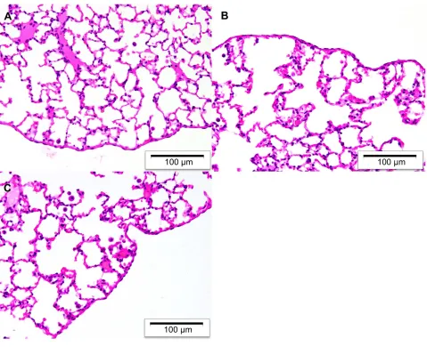
Figure 1Representative H&E stained sections of rat visceral pleura and underlying lung parenchyma after intrapleural administration of liposomal curcumin. Morphologically normal histology was observed at (A) 48 h (B) 1-week and (C) 3-weeks. A total of four rats were assessed at each time point.
International Journal of Nanomedicine downloaded from https://www.dovepress.com/ by 118.70.13.36 on 24-Aug-2020
infusion. Red blood cells showed marked echinocytosis, an abnormality wherein numerous, spikey projections are pre-sent on the cell membrane, indicating that red blood cells are
at risk of rupturing (Figure 3). Echinocytes were absent from
blood samples at 24 h and 48 h. Red blood cells displayed normal cell morphology at all time points following
intra-pleural liposomal curcumin administration (Figure 3).
Discussion
Curcumin is an attractive potential anti-cancer agent as it can act on a wide range of molecular pathways to
stimulate tumour cell death and decrease tumour cell pro-liferation including phosphatidylinositol-3-kinase (PI3K)/
Akt signaling,35–39 Nuclear Factor (NF)-κB, and JAK/
STAT3 signaling.40 It has also been shown to reduce
chemotherapy-induced toxic side effects.41,42 To the best
of our knowledge, curcumin has never been administered directly into the pleural cavity of animals, or humans. Therefore, the safety and compartmental distribution of intrapleural liposomal curcumin following intrapleural needs to be evaluated.
We detected peak total curcumin plasma concentrations in the plasma 1.5 h after intrapleural delivery of liposomal curcumin, indicating that a proportion of curcumin had entered the systemic circulation. These peak plasma concen-trations are comparable to the systemic levels of total curcu-min that we have previously measured in rats after consumption of an oral, bioavailable curcumin formulation, which can be purchased over-the-counter for human use and
therefore is considered safe.43We detected little to no total
curcumin in the plasma of rats at 24 h, and 48 h after intrapleural administration of liposomal curcumin, suggest-ing that liposomal curcumin is mostly metabolized or
dis-tributed to blood cells or tissues within thefirst 24 h after
administration. We detected high levels of total curcumin in the plasma immediately following intravenous infusion of
liposomal curcumin (1.276 ± 0.505μg/mL). Total curcumin
plasma concentration rapidly dropped 1.5 h after cessation of
the infusion (0.192 ± 0.06μg/mL), which was consistent with
other studies conducted in animals and humans that assessed
the pharmacokinetics of intravenous liposomal
curcumin.11,26 In humans, plasma concentrations of free
curcumin were not detected above the limit of detection (25
ng/mL) at times greater than 1 hr post-infusion.11Bolger and
colleagues recently established that liposomal curcumin rapidly diffuses into peripheral blood mononuclear cells and red blood cells; therefore, curcumin may be present
Table 1Total Curcumin Plasma Concentrations (Mean ± Standard
Deviation) Following Intrapleural and Intravenous Administration of Liposomal Curcumin (16 mg/kg). Values That Were Below the Detection Limit of the Assay Were Assigned a Value of 0μg/Ml. No Significant Difference in Plasma Concentrations Was Detected Between the Rats in the Intrapleural Injection and Intravenous Infusion Group (p=0.287, p=0.2545, p=0.6476, for 1.5 h, 24 h and 48 h Respectively)
Time (Hours) Intrapleural
Administration (μg/mL)a
Intravenous Infusion (μg/mL)a
0 h Not measured 1.276 ± 0.505 (n=4)b
1.5 h 0.235 ± 0.0762 (n=10)b
0.192 ± 0.06 (n=3)b
24 h 0.025 ± 0.022 (n=9)b
0.007 ± 0.01 (n=3)b
48 h 0.006 ± 0.009 (n=9)b
0.011 ± 0.03 (n=4)b
168 h (1-week) Not detected (n=6)b
Not measured 336 h (2-weeks) Not detected (n=3)b
Not measured 504 h (3-weeks) Not detected (n=3)b
Not measured
Notes: a
Values are presented as the mean ± standard deviation of at least 3 separate animals.bNumber of animals.
0 20 40 60
0.0 0.1 0.2 0.3 0.405 10 15 20
Time (hours)
Tot
al c
ur
cum
in
(μ
g/m
l)
Intravenous group Intrapleural group
Figure 2The concentration of total curcumin in the plasma of rats following intravenous and intrapleural administration of liposomal curcumin (16 mg/kg). Each data point represents the mean total curcumin concentration in at least three separate animals and error bars represent the standard deviation. Values that were below the detection limit of the assay were assigned a value of 0μg/mL.
Table 2 Curcumin tissue Concentrations (Mean ± Standard
Deviation) Following Intrapleural and Intravenous Administration of Liposomal Curcumin (16 mg/kg). No Significant Difference in Tissue Concentrations Was Detected Between the Rats in the Intrapleural Injection and Intravenous Infusion Group (p=0.4857, p=0.3429, p=0.6857, for Diaphragm, Lung and Liver Respectively)
Delivery Method Concentration of Curcumin (μg/g)a
Diaphragm Lung Liver
Intrapleural 0.1281 ± 0.076 0.17585 ± 0.193 0.02995 ± 0.029 Intravenous 0.1737 ± 0. 0.06515 ± 0.017 0.03487 ± 0.012
Notes:a
Values are presented as the mean ± standard deviation of 4 separate animals.
International Journal of Nanomedicine downloaded from https://www.dovepress.com/ by 118.70.13.36 on 24-Aug-2020
with circulating blood cells and subsequently distributed to
tissues.44,45
We observed transient echinocytosis and possible hemoly-sis in the blood of rats following a 2 h intravenous infusion of liposomal curcumin. Our results are in agreement with data from other studies investigating the safety of intravenous
lipo-somal curcumin administration.10,11,26,27,46Several factors can
trigger echinocytosis, which include, but are not limited to, uremia, chronic renal disease, liver disease and
hyperlipide-mia. To confirm that the observed echinocytosis was real and
not an artifact of the processes of drying or staining of the blood sample on the slide, control and test blood smears were run alongside and the former showed normal morphology. Storka and colleagues demonstrated that both empty liposomes and curcumin itself could contribute to echinocytosis in vivo, and
may indicate dose-limiting toxicity.46 In advanced cancer
patients, researchers observed a significant increase in
hema-tological adverse events in patients receiving intravenous
lipo-somal curcumin (300 mg/m2), including one case of
dose-limiting hemolysis.10 Here, we observed normal red blood
cell morphology in rats after the administration of intrapleural liposomal curcumin at all time points. This was not surprising since we observed lower peak concentrations in the systemic circulation after intrapleural delivery. From these data, we conclude that liposomal curcumin can be administered at higher concentrations in the pleural cavity without causing red blood cell abnormalities. Importantly, we also showed that intrapleural delivery of liposomal curcumin was not asso-ciated with pleural or lung toxicity in healthy rats, indicating that this mode of delivery is a feasible alternative to
intravenous infusion, which may achieve higher drug concen-trations within a pleural-based tumour. We utilized both male and female rats to investigate the effects of intrapleural liposo-mal curcumin in accordance with The National Health and
Medical Research Council (NHMRC) ‘guidelines for best
practice methodology for the use of animals for scientific
purposes’as sex-specific variation in angiogenesis, infl
amma-tion and wound healing exist.47
We measured free curcumin in diaphragm and lungs to estimate the amount of curcumin that diffused from the pleural cavity into surrounding tissues after intrapleural administration and compared the values to those found after intravenous administration. We also measured the concentration of free curcumin in the liver, as this is where curcumin is predomi-nately metabolized. We detected free curcumin in the dia-phragm, lungs and liver at similar concentrations in both the intrapleural and intravenous administration groups. This was expected since little to no total curcumin was detected in the plasma at the 48 h time point. Measuring curcumin tissue concentrations at earlier time points in an MPE tumor model, would be valuable to ascertain if intrapleural delivery is, in fact, superior to intravenous delivery when targeting pleural tumors.
Liposomal-drug release rates will impact a drug’s
ability to elicit a therapeutic response. Ando and col-leagues investigated intrapleural delivery of two
lipo-somal formulations of pemetrexed:
cholesterol-containing, and cholesterol-free liposomes, in an
ortho-topic mouse model of mesothelioma.48 The authors
found that only the cholesterol-free liposomes reduced tumor growth. This was thought to be dependent on the
Figure 3Representative Romanowski stained blood smears collected following intravenous infusion and intrapleural injection of liposomal curcumin (16 mg/kg) (A) Echinocyte formation was observed in the blood 1.5 h after intravenous liposomal curcumin infusions (B) Normal erythrocyte morphology was observed at 1.5 h following the administration of intrapleural liposomal curcumin. A total of four rats were assessed in each group.
International Journal of Nanomedicine downloaded from https://www.dovepress.com/ by 118.70.13.36 on 24-Aug-2020
higher release rate of pemetrexed from the cholesterol-free liposomes since the incorporation of cholesterol in liposomes can increase membrane rigidity, thereby delaying the drug-release. The liposomal curcumin used in this study does not contain cholesterol and has an average particle size of 117 nm, (a comparable size to both liposomes utilized in these studies (cho-lesterol-liposomes; 117.8 nm, cholesterol-free lipo-somes; 103.8 nm)). Additionally, our results indicate that liposomal curcumin is not retained within the pleural cavity past 48 h, suggesting that liposomal curcumin is a suitable liposome formulation to deliver high doses of curcumin to a pleural tumor by intra-pleural administration.
These experiments were conducted in healthy animals
and therefore, may not reflect the situation in patients
suffer-ing from MPE. It is important to note that pleural pharmaco-kinetics may be altered in patients with an MPE; For example, the propensity of a drug to enter systemic circula-tion may be limited if a tumor obstructs the lymphatic sto-mata or if the lymphatic vessels become saturated due to the presence of an MPE. Consequently, drugs are more likely to diffuse into the visceral and parietal pleura, thereby max-imizing drug exposure to the tumor. An obstructing tumor may slow the redistribution of drugs into blood, reducing systemic toxicities, but as a consequence, may also increase
the risk of local toxicities, such as pleural adhesions.49,50
Pleural adhesions are induced in patients undergoing talc pleurodesis as a way to prevent recurrent pleural effusion and are not considered life-threatening, and are indeed
desired in this circumstance.51Nevertheless, Marazioti and
colleagues recently demonstrated that there was no differ-ence in liposome retention time between healthy mice and
mice with pleural adenocarcinoma.52
Intrapleural liposomal curcumin therapy offers several potential advantages over intravenous therapy in patients with primary and secondary malignancies of the pleura. Few studies have directly compared intrapleural and intra-venous delivery of drugs; but these studies have consis-tently shown that intrapleural administration reduces peak plasma levels, reduces systemic toxicity and yields a higher drug concentration at the pleura compared with
intravenous administration.12–14,50,53 The position of the
tumour cells adjacent to the pleural cavity provides a unique opportunity to administer therapeutics directly to the tumour site. Therapeutic delivery via an existing pleura catheter or at pleurodesis means patients could be given tumour-site targeted therapies while also avoiding
any additional needling of the pleura. The efficacy of
intrapleural liposomal curcumin may be restricted in areas of the tumor that does not have a direct connection to the pleural cavity. Areas of locculated pleural effusion, chest wall invasion and the mediastinum showed no tumor
response towards intrapleural liposomal-entrapped
chemotherapy.25 Intravenous liposomal curcumin could
be used in combination with intrapleural administration as these may target regions of tumors that are not in direct contact with the pleural cavity.
Conclusion
No local or systemic toxicity was observed following intra-pleural administration liposomal curcumin, indicating that it is a safe alternative to intravenous administration. We hypothesize that intrapleural liposomal curcumin could pro-vide patients with MPE an alternative approach to che-motherapy, which could help to alleviate their symptoms, and reduce their cancer burden. Additionally, curcumin could be used as an adjunct therapy to improve outcomes and reduce toxic side effects. From a practice standpoint, liposo-mal curcumin could be delivered via patients existing indwelling pleural catheter, which is placed to manage
pleuralfluid drainage, or at the time of pleurodesis. Further
investigations are required to determine the efficacy of
intra-pleural liposomal curcumin in patients with MPE.
Acknowledgments
This research was funded by the Tour de Cure Pioneering Cancer Research Grant. Liposomal curcumin was a kind gift from SignPath Pharma In.
Disclosure
Dr. Sordillo is the Chief Scientific Officer at SignPath Pharma,
Inc. Dr Sordillo has a patent“Numerous”issued to SignPath
Pharma, Inc. Professor Sonja Klebe prepares medicolegal reports for the courts of Australia on the diagnosis of lung disease, outside the submitted work. The authors report no
other conflicts of interest in this work.
References
1. Psallidas I, Kalomenidis I, Porcel JM, Robinson BW, Stathopoulos GT. Malignant pleural effusion: from bench to bedside.Eur Respir Rev.
2016;25(140):189–198. doi:10.1183/16000617.0019-2016
2. Thomas R, Fysh ETH, Smith NA, et al. Effect of an indwelling pleural catheter vs talc pleurodesis on hospitalization days in patients with malignant pleural effusion: the AMPLE randomized clinical trial. JAMA.2017;318(19):1903–1912. doi:10.1001/jama.2017.17426
International Journal of Nanomedicine downloaded from https://www.dovepress.com/ by 118.70.13.36 on 24-Aug-2020
3. Bibby AC, Dorn P, Psallidas I, et al. ERS/EACTS statement on the management of malignant pleural effusions.Eur Respir J.2018;52 (1):1800349. doi:10.1183/13993003.00349-2018
4. Heger M, van Golen RF, Broekgaarden M, Michel MC. The molecular basis for the pharmacokinetics and pharmacodynamics of curcumin and its metabolites in relation to cancer.Pharmacol Rev.2014;66(1):222–307. 5. Yeh HH, Lai WW, Chen HH, Liu HS, Su WC. Autocrine IL-6-induced Stat3 activation contributes to the pathogenesis of lung adenocarcinoma and malignant pleural effusion. Oncogene.
2006;25(31):4300–4309. doi:10.1038/sj.onc.1209464
6. Stathopoulos GT, Kollintza A, Moschos C, et al. Tumor necrosis factor-alpha promotes malignant pleural effusion. Cancer Res.
2007;67(20):9825–9834. doi:10.1158/0008-5472.CAN-07-1064 7. Shanmugam MK, Rane G, Kanchi MM, et al. The multifaceted role
of curcumin in cancer prevention and treatment.Molecules.2015;20 (2):2728–2769. doi:10.3390/molecules20022728
8. Feng T, Wei Y, Lee RJ, Zhao L. Liposomal curcumin and its application in cancer.Int J Nanomedicine.2017;12:6027–6044. doi:10.2147/IJN 9. Sercombe L, Veerati T, Moheimani F, Wu SY, Sood AK, Hua S.
Advances and challenges of liposome assisted drug delivery. Front Pharmacol.2015;6:286. doi:10.3389/fphar.2015.00286
10. Greil R, Greil-Ressler S, Weiss L, et al. A Phase 1 dose-escalation study on the safety, tolerability and activity of liposomal curcumin (Lipocurc™) in patients with locally advanced or metastatic cancer. Cancer Chemother Pharmacol. 2018;82(4):695–706. doi:10.1007/ s00280-018-3654-0
11. Storka A, Vcelar B, Klickovic U, et al. Safety, tolerability and phar-macokinetics of liposomal curcumin in healthy humans. Int J Clin Pharmacol Ther.2015;53(1):54–65. doi:10.5414/CP202076 12. Sakaguchi H, Ishida H, Nitanda H, Yamazaki N, Kaneko K,
Kobayashi K. Pharmacokinetic evaluation of intrapleural perfusion with hyperthermic chemotherapy using cisplatin in patients with malignant pleural effusion. Lung Cancer. 2017;104:70–74. doi:10.1016/j.lungcan.2016.12.015
13. Froudarakis ME, Greillier L, Monjanel-Mouterde S, et al. Intrapleural administration of lipoplatin in an animal model. Lung Cancer.
2011;72(1):78–83. doi:10.1016/j.lungcan.2010.07.010
14. Li J, Tang J, Li Y, Yu J, Zhang B, Yu C. Pharmacokinetic profile of paclitaxel in the plasma, lung, and diaphragm following intravenous or intrapleural administration in rats. Thorac Cancer. 2015;6 (1):43–48. doi:10.1111/1759-7714.12139
15. Perng RP, Chen YM, Wu MF, et al. Phase II trial of intrapleural paclitaxel injection for non-small-cell lung cancer patients with malignant pleural effusions. Respir Med. 1998;92(3):473–479. doi:10.1016/S0954-6111(98)90294-3
16. Biaoxue R, Hui P, Wenlong G, Shuanying Y. Evaluation of efficacy and safety for recombinant human adenovirus-p53 in the control of the malignant pleural effusions via thoracic perfusion. Sci Rep.
2016;6:39355. doi:10.1038/srep39355
17. Biaoxue R, Xiguang C, Hua L, Wenlong G, Shuanying Y. Thoracic perfusion of recombinant human endostatin (Endostar) combined with chemotherapeutic agents versus chemotherapeutic agents alone for treat-ing malignant pleural effusions: a systematic evaluation and meta-analysis.BMC Cancer.2016;16(1):888. doi:10.1186/s12885-016-2935-4
18. Hu R, Jiang H, Li H, Wei D, Wang G, Ma S. Intrapleural perfusion thermo-chemotherapy for pleural effusion caused by lung carcinoma under VATS.J Thorac Dis.2017;9(5):1317–1321. doi:10.21037/jtd 19. Ishida A, Miyazawa T, Miyazu Y, et al. Intrapleural cisplatin and
OK432 therapy for malignant pleural effusion caused by non-small cell lung cancer. Respirology. 2006;11(1):90–97. doi:10.1111/ res.2006.11.issue-1
20. Lombardi G, Nicoletto MO, Gusella M, et al. Intrapleural paclitaxel for malignant pleural effusion from ovarian and breast cancer: a phase II study with pharmacokinetic analysis.Cancer Chemother Pharmacol.2012;69(3):781–787. doi:10.1007/s00280-011-1765-y
21. Sterman DH, Alley E, Stevenson JP, et al. Pilot and feasibility trial evaluating immuno-gene therapy of malignant mesothelioma using intrapleural delivery of adenovirus-IFNalpha combined with chemotherapy.Clin Cancer Res.2016;22(15):3791–3800. doi:10.1158/ 1078-0432.CCR-15-2133
22. Sterman DH, Recio A, Carroll RG, et al. A Phase I clinical trial of single-dose intrapleural IFN-beta gene transfer for malignant pleural mesothelioma and metastatic pleural effusions: high rate of antitumor immune responses.Clin Cancer Res.2007;13(15 Pt 1):4456–4466. doi:10.1158/1078-0432.CCR-07-0403
23. Perez-Soler R, Shin DM, Siddik ZH, et al. Phase I clinical and pharmacological study of liposome-entrapped NDDP administered intrapleurally in patients with malignant pleural effusions. Clin Cancer Res.1997;3(3):373–379.
24. Perng RP, Wu MF, Lin SY, Chen YM, Lin JY, Whang-Peng J. A phase I feasibility and pharmacokinetic study of intrapleural pacli-taxel in patients with malignant pleural effusions.Anticancer Drugs.
1997;8(6):565–573. doi:10.1097/00001813-199707000-00003 25. Lu C, Perez-Soler R, Piperdi B, et al. Phase II study of a
liposome-entrapped cisplatin analog (L-NDDP) administered intrapleu-rally and pathologic response rates in patients with malignant pleural mesothelioma. J Clin Oncol. 2005;23(15):3495–3501. doi:10.1200/ JCO.2005.00.802
26. Helson L, Bolger G, Majeed M, Vcelar B, Pucaj K, Matabudul D. Infusion pharmacokinetics of Lipocurc (liposomal curcumin) and its metabolite tetrahydrocurcumin in beagle dogs. Anticancer Res.
2012;32(10):4365–4370.
27. Matabudul D, Pucaj K, Bolger G, Vcelar B, Majeed M, Helson L. Tissue distribution of (Lipocurc) liposomal curcumin and tetrahydro-curcumin following two- and eight-hour infusions in beagle dogs. Anticancer Res.2012;32(10):4359–4364.
28. Kanai M, Yoshimura K, Asada M, et al. A phase I/II study of gemcitabine-based chemotherapy plus curcumin for patients with gemcitabine-resistant pancreatic cancer. Cancer Chemother Pharmacol.2011;68(1):157–164. doi:10.1007/s00280-010-1470-2 29. Bayet-Robert M, Kwiatkowski F, Leheurteur M, et al. Phase I dose
escalation trial of docetaxel plus curcumin in patients with advanced and metastatic breast cancer. Cancer Biol Ther. 2010;9(1):8–14. doi:10.4161/cbt.9.1.10392
30. Dhillon N, Aggarwal BB, Newman RA, et al. Phase II trial of curcumin in patients with advanced pancreatic cancer.Clin Cancer Res.2008;14(14):4491–4499. doi:10.1158/1078-0432.CCR-08-0024 31. Asai A, Miyazawa T. Occurrence of orally administered curcuminoid as
glucuronide and glucuronide/sulfate conjugates in rat plasma.Life Sci.
2000;67(23):2785–2793. doi:10.1016/S0024-3205(00)00868-7 32. Vareed SK, Kakarala M, Ruffin MT, et al. Pharmacokinetics of
curcumin conjugate metabolites in healthy human subjects.Cancer Epidemiol Biomarkers Prev. 2008;17(6):1411–1417. doi:10.1158/ 1055-9965.EPI-07-2693
33. Heath DD, Pruitt MA, Brenner DE, Rock CL. Curcumin in plasma and urine: quantitation by high-performance liquid chromatography. J Chromatogr B Analyt Technol Biomed Life Sci. 2003;783 (1):287–295. doi:10.1016/S1570-0232(02)00714-6
34. U.S Food and Drug Administration. Bioanalytical method validation. Guidance for industry.2018. Available from:https://www.fda.gov/ fi les/drugs/published/Bioanalytical-Method-Validation-Guidance-for-Industry.pdf. Accessed February 03, 2020.
35. Xu X, Qin J, Liu W. Curcumin inhibits the invasion of thyroid cancer cells via down-regulation of PI3K/Akt signaling pat hway. Gene. 2014;546(2):226–232. doi:10.1016/j.gene.2014.06. 006
36. Yu Z, Wan Y, Liu Y, Yang J, Li L, Zhang W. Curcumin induced apoptosis via PI3K/Akt-signalling pathways in SKOV3 cells. Pharm Biol. 2016;54(10):2026–2032. doi:10.3109/13880 209.2016.1139601
International Journal of Nanomedicine downloaded from https://www.dovepress.com/ by 118.70.13.36 on 24-Aug-2020
37. Qiao Q, Jiang Y, Li G. Inhibition of the PI3K/AKT-NF-kappaB pathway with curcumin enhanced radiation-induced apoptosis in human burkitt’s lymphoma.J Pharmacol Sci.2013;121(4):247–256. doi:10.1254/jphs.12149FP
38. Akkoc Y, Berrak O, Arisan ED, Obakan P, Coker-Gurkan A, Palavan-Unsal N. Inhibition of PI3K signaling triggered apoptotic potential of curcumin which is hindered by Bcl-2 through activation of autophagy in MCF-7 cells.Biomed Pharmacother.2015;71:161–171. doi:10.1016/j. biopha.2015.02.029
39. Fu H, Wang C, Yang D, et al. Curcumin regulates proliferation, autophagy and apoptosis in gastric cancer cells by affecting PI3K and P53 signaling.J Cell Physiol.2017.
40. Zhang C, Li B, Zhang X, Hazarika P, Aggarwal BB, Duvic M. Curcumin selectively induces apoptosis in cutaneous T-cell lymphoma cell lines and patients’PBMCs: potential role for STAT-3 and NF-kappaB signaling. J Invest Dermatol. 2010;130(8):2110–2119. doi:10.1038/jid.2010.86
41. Liu Z, Huang P, Law S, Tian H, Leung W, Xu C. Preventive effect of curcumin against chemotherapy-induced side-effects. Front Pharmacol.2018;9:1374. doi:10.3389/fphar.2018.01374
42. Onen HI, Yilmaz A, Alp E, et al. EF24 and RAD001 potentiates the anticancer effect of platinum-based agents in human malignant pleural mesothelioma (MSTO-211H) cells and protects nonmalignant mesothelial (MET-5A) cells.Hum Exp Toxicol.2015;34(2):117–126. doi:10.1177/0960327114542965
43. Hocking AJ, Elliot D, Hua J, Klebe S. Administeringfixed oral doses of curcumin to rats through voluntary consumption.J Am Assoc Lab Anim Sci.2018;57(5):508–512. doi:10.30802/AALAS-JAALAS-17-000143 44. Bolger GT, Licollari A, Tan A, et al. Distribution of curcumin and
THC in peripheral blood mononuclear cells isolated from healthy individuals and patients with chronic lymphocytic leukemia. Anticancer Res.2018;38(1):121–130. doi:10.21873/anticanres.12199
45. Bolger GT, Licollari A, Bagshaw R, et al. Intense uptake of liposomal curcumin by multiple myeloma cell lines: comparison to normal lym-phocytes, red blood cells and chronic lymphocytic leukemia cells. Anticancer Res.2019;39(3):1161–1168. doi:10.21873/anticanres.13225 46. Storka A, Vcelar B, Klickovic U, et al. Effect of liposomal curcumin on red blood cells in vitro.Anticancer Res.2013;33(9):3629–3634. 47. Guidelines for best practice methodology for the use of animals for
scientific purposes 2017.2018. Contract No.: ISBN: 9781925129977. 48. Ando H, Kobayashi S, Abu Lila AS, Eldin NE, Kato C, Shimizu T, et al. Advanced therapeutic approach for the treatment of malignant pleural mesothelioma via the intrapleural administration of liposomal pemetrexed.J Control Release. 2015;220(Pt A):29–36. doi:10.1016/j. jconrel.2015.10.019
49. Popowicz N, Sparling B. Pleural phamacokinetics. In: Lee YCG, Light RW, editors. Textbook of Pleural Diseases. 3rd ed. London, UK: CRC Press;2016:104–121.
50. Tada Y, Hiroshima K, Shimada H, et al. An intrapleural administra-tion of zoledronic acid for inoperable malignant mesothelioma patients: a phase I clinical study protocol.Springerplus.2016;5:195. doi:10.1186/s40064-016-1893-2
51. Penz E, Watt KN, Hergott CA, Rahman NM, Psallidas I. Management of malignant pleural effusion: challenges and solutions.Cancer Manag Res.2017;9:229–241. doi:10.2147/CMAR
52. Marazioti A, Papadia K, Giannou A, Stathopoulos GT, Antimisiaris SG. Prolonged retention of liposomes in the pleural cavity of normal mice and high tumor distribution in mice with malignant pleural effusion, after intrapleural injection.Int J Nanomedicine.2019;14:3773–3784. doi:10.2147/IJN.S202568
53. Bogliolo GV, Lerza R, Bottino GB, et al. Regional pharmacokinetic selectivity of intrapleural cisplatin. Eur J Cancer. 1991;27 (7):839–842. doi:10.1016/0277-5379(91)90129-2
International Journal of Nanomedicine
Dove
press
Publish your work in this journal
The International Journal of Nanomedicine is an international, peer-reviewed journal focusing on the application of nanotechnology in diagnostics, therapeutics, and drug delivery systems throughout the biomedical field. This journal is indexed on PubMed Central, MedLine, CAS, SciSearch®, Current Contents®/Clinical Medicine,
Journal Citation Reports/Science Edition, EMBase, Scopus and the Elsevier Bibliographic databases. The manuscript management system is completely online and includes a very quick and fair peer-review system, which is all easy to use. Visit http://www.dovepress.com/ testimonials.php to read real quotes from published authors.
Submit your manuscript here:https://www.dovepress.com/international-journal-of-nanomedicine-journal
International Journal of Nanomedicine downloaded from https://www.dovepress.com/ by 118.70.13.36 on 24-Aug-2020