University of South Carolina
Scholar Commons
Theses and Dissertations
6-30-2016
Enabling Studies to Optimize Biomaterials for the
Treatment of Myocardial Infarction
Eva Adriana Romito
University of South Carolina
Follow this and additional works at:https://scholarcommons.sc.edu/etd Part of theBiomedical Engineering and Bioengineering Commons
This Open Access Dissertation is brought to you by Scholar Commons. It has been accepted for inclusion in Theses and Dissertations by an authorized administrator of Scholar Commons. For more information, please [email protected].
Recommended Citation
Enabling Studies to Optimize Biomaterials for the Treatment of Myocardial
Infarction
by
Eva Adriana Romito
Bachelor of Science
University of South Carolina, 2012
Submitted in Partial Fulfillment of the Requirements
For the Degree of Doctor of Philosophy in
Biomedical Engineering
College of Engineering and Computing
University of South Carolina
2016
Accepted by:
Francis Spinale, Major Professor
Tarek Shazly, Major Professor
Edie Goldsmith, Committee Member
Michael Sutton, Committee Member
Jason Burdick, Committee Member
DEDICATION
To my husband who has borne all of my stress, frustrations, stubbornness, and
overwhelming joys with impressive exuberance for all the years we’ve been together.
To my family that fiendishly encouraged inquiry, and developed my argumentative
nature with great affection.
To my friends always responsive to moments of insanity with creative sources of
ACKNOWLEDGEMENTS
My sincere acknowledgment and gratitude go to my advisors Drs. Francis Spinale
and Tarek Shazly. I have received invaluable assistance, advice and innumerable
suggestions that have furthered my knowledge base as well as impacted my individual
growth as a researcher. They have both devoted an enormous amount of time into
fostering my scientific capabilities in thought as well as writing and presentation. I would
like to thank my dissertation committee members Dr. Jason Burdick, Dr. Edie Goldsmith
and Dr. Michael Sutton for their guidance and support. I express my sincere thanks to all
my lab-mates, colleagues, and friends who are always with me for support and assistance.
Lastly, and with utmost importance I would like to thank my family, my husband and my
friends for their unconditional love, understanding and unfaltering support of my
ABSTRACT
The canonical mechanism of wound healing is disrupted following a myocardial
infarction (MI), manifesting as an unregulated response that negatively impacts left
ventricular (LV) function. This mechanism, termed post-MI remodeling, culminates in an
outcome that favors progression to a systolic heart failure state and death for the patient.
Therapeutic approaches following the occurrence of a MI are designed to modulate the
natural remodeling process and mitigate the loss of cardiac function. The mechanics and
structure of the healing infarct have been the focus of numerous pre-clinical and clinical
investigations, leading to the impending clinical introduction of material injections as a
means to favorably alter remodeling outcomes. However, to date there is no body of
work that provides a coherent framework for evaluation of targeted material therapies. To
form a basis for optimization of material-based MI treatments, we have integrated
measurements of MI regional mechanics, the morphology of the local extracellular
matrix, and the biophysical impact of material injections into the MI region in a porcine
model of MI. The combined findings of this study have enhanced a mechanistic
understanding of material-based post-MI interventions, elucidated the relationship
between MI regional mechanics and LV function throughout the natural and attenuated
history of LV remodeling, and has developed mechanical metrics of value to move forth
towards future developments of a generalizable computational tools for screening and
TABLE OF CONTENTS
DEDICATION ... iii
ACKNOWLEDGEMENTS ... iv
ABSTRACT ...v
LIST OF TABLES ... ix
LIST OF FIGURES ...x
LIST OF ABBREVIATIONS ... xii
CHAPTER 1:INTRODUCTION ...1
1.1OVERVIEW ...1
1.2 PROJECT SCOPE ...9
1.3 SPECIFIC AIMS ...10
CHAPTER 2:ASSESSMENT OF IN-VIVO POST-MYOCARDIAL INFARCTION;A FOCUS ON REGIONAL MECHANICS AND THE ROAD AHEAD ...13
2.1 ABSTRACT ...14
2.2INTRODUCTION ...14
2.3POST-ACUTE TIME PHASE (3-7 DAYS) ...17
2.4INTERMEDIATE TIME PHASE (7-14 DAYS) ...24
2.5LATE TIME PHASE (14 DAYS AND BEYOND) ...31
2.6SUMMARY ...39
CHAPTER 3:SONOMICROMETRY-BASED ANALYSIS OF POST-MYOCARDIAL INFARCTION REGIONAL MECHANICS ...52
3.2INTRODUCTION ...53
3.3 MATERIALS AND METHODS ...56
3.4RESULTS ...61
3.5DISCUSSION ...62
3.6CONCLUSION ...65
3.7LIMITATIONS ...65
CHAPTER 4:BIOPHYSICAL EFFECTS OF A HYALURONIC ACID BASED HYDROGEL ON AVERAGE PEAK WALL STRESS OF THE LEFT VENTRICULAR WALL POST-MYOCARDIAL INFARCTION ...72
4.1 INTRODUCTION ...73
4.2MATERIALS AND METHODS ...73
4.3RESULTS ...75
4.4LIMITATIONS ...75
4.5CONCLUSIONS ...76
4.6PROPOSED NEXT STEPS ...76
CHAPTER 5:HARPANALYSIS OF CHANGES IN STRAIN POST-MI AS A RESULT OF A HYALURONIC ACID GEL INTERVENTION ...85
5.1INTRODUCTION...86
5.2MATERIALS AND METHODS ...86
5.3RESULTS ...88
5.4LIMITATIONS ...88
5.5CONCLUSIONS ...89
CHAPTER 6:FUTURE WORK AND THESIS CONCLUSION ...98
6.1FUTURE WORK ...98
REFERENCES ...100
LIST OF TABLES
Table 2.1 Summary of techniques ...43
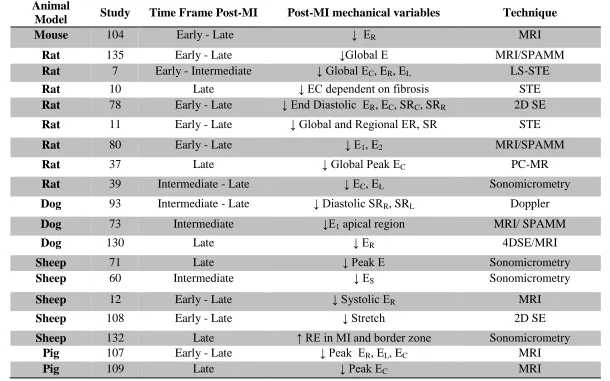
Table 2.2 Tabulated Studies ...44
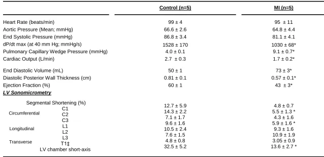
Table 3.1 Left Ventricular global and regional function in referent control and
post-myocardial infarction ...67
Table 5.1 Tabulated Strain values of the MI region by treatment and layer for the basal region of the LV ...92
Table 5.2 Tabulated Strain values of the MI region by treatment and layer for the mid-ventricular region of the LV ...93
LIST OF FIGURES
Figure 1.1 3-D rendering of LV with MI ...12
Figure 1.2 Representative MI and incidence ...12
Figure 2.1 Species Differences ...46
Figure 2.2 Strain Heterogeneity ...47
Figure 2.3 Extent of infarction across myocardial wall ...48
Figure 2.4 Directional dependence ...49
Figure 2.5 Summary recommendations ...45
Figure 2.6 In-vivo chamber stiffness ...51
Figure 3.1 Sonomicrometry array ...68
Figure 3.2 Regional Stiffness ...69
Figure 3.3 Relative anisotropy and heterogeneity ...70
Figure 3.4 Stylized relative anisotropy and heterogeneity ...71
Figure 4.1 HA structure and injection pattern ...77
Figure 4.2 Echocardiography ...78
Figure 4.3 Schematic of gel time line ...79
Figure 4.4 Isochronal plot of LV stress and gel modulus ...80
Figure 4.5 Change in MI thinning...81
Figure 4.6 Change in LV stress...82
Figure 4.7 Representative data HA compression ...83
Figure 5.1 SPAMM image filtering for HARP ...90
Figure 5.2 HARP analysis toolbox ...91
Figure 5.3 Radial thickening ...95
Figure 5.4 Circumferential shortening ...96
LIST OF ABBREVIATIONS
CVD ... Cardiovascular Disease
ECM ... Extra cellular matrix
EF ... Ejection Fraction
HF ... Heart Failure
LG-MRI ... Late Gadolinium Enhanced
LV ... Left Ventricle
LS-STE ... Layer Specific Speckle Tracking Echocardiography
LVEDP ... Left Ventricular End Diastolic Pressure
LVEDV ... Left Ventricular End Diastolic Volume
MI ... Myocardial Infarction
MRI ... Magnetic Resonance Imaging
SPAMM ... Spatial Modulation Magnetization
CHAPTER
1INTRODUCTION
1.1 Overview
Clinical Motivation and Natural History of MI
The prevalence of heart disease across the United States and the western world
presents an ever-increasing problem that has severe health implications with regards to
longevity and quality of life. Currently, cardiovascular disease (CVD) accounts for the
largest mortality rate in the U.S. every year 61. In its broadest definition, CVD refers to
many different etiologies affecting the circulatory system, with its central component
being the heart muscle 61,62. Acute coronary events directly affect the heart and ultimately
compromise cardiac function to an extent beyond repair with current clinical practice
111,154
. Every year three-quarters of a million of Americans suffer a new coronary event 62.
One such event is that of a myocardial infarction (MI) in the left ventricle (LV). A MI is
an ischemic event, meaning there is a lack of oxygen delivered to a specific portion of the
myocardium. Once the oxygen supply is lost the tissue in the affected region quickly
looses viability (Figure 1.1).
As the contractile units of the myocardium (cardiomyocytes) die, the portion of
affected tissue becomes necrotic and loses functionality 33,111,117. Survival of a MI is
possible, but patient quality of life is invariably compromised. The reduction of life
as a major contributor to the development of systolic heart failure 33,71,111. Once a patient
enters a state of HF the rate of survival to a normal life expectancy decreases for both
sexes. A study carried out over a decade in Scotland comprehensively observed patients
of both sexes that were suffering from common cancers (ovarian, lung, stomach, prostate,
breast) as well as MI and HF with regards to their survival rates 161. The resulting data
demonstrated markedly low survival rates for patients suffering from HF with a trend that
was significantly lower than most cancers affecting both sexes (with the exception of
stomach cancer) 161. Such a threatening trend gave verisimilitude to an extreme
conclusion from the authors of the study; as they deemed HF to perhaps be “more
malignant than cancer”. Although MI showed high probability of survival in this study,
the likelihood of transgressing to HF after the initial acute occurrence presents patients
and healthcare professionals with a delicate situation for treatment in patients. A primary
and decisive factor in the pathway to HF from MI is the effect of the structural
remodeling that takes place post-MI 9,33,111. Since MI and post-MI remodeling are major
contributors to the etiology of the HF state, it is imperative to rapidly curtail the
otherwise progressive loss of cardiac function.
In a normal setting, the most common treatment of MI involves reperfusion
therapy, but it is only achievable for a short period of time after its onset, where a supply
of oxygenated blood can be reestablished at the affected site 33,111,117.Studies have shown
there is a notable benefit from reperfusion therapy on cardiac function and post
remodeling markers 33. It is the remodeling aspect post-MI that becomes the largest
driving force in the decline of cardiac function 33,111,133. Affecting the progression of
Post-MI remodeling is a multifactorial process that overtakes the canonical wound
healing response that would occur after injury. Following the ischemic event, a series of
pathways are triggered by the natural inflammatory response to injury. Various
biochemical cascades contribute to the readsorption of necrotic tissue post-MI, as the
proliferation of fibroblasts and fibrillar collagen deposition within the affected region
fundamentally change the material composition of the extra cellular matrix (ECM) at the
site of injury 51,111,145,154. Drastic changes in geometry and structure combine to create
adverse effects on cardiac function and the likely inception of HF. The chief attributes of
remodeling include changes in LV geometry evidenced by the thinning of the infarct
region (MI thinning) and hypertrophy of the remote/opposing myocardium. The thinning
of the MI region is in part due to the scarification that occurs as necrotic tissue is
removed, and a proliferative phase of fibroblasts takes place. During this infiltration there
is a marked increase in the collagen content in the ischemic region as the non-viable
injured myocardium is transformed into scar tissue 111,145. This MI scar becomes thin and
expands the LV in volume (Figure 1.2 top) such that the difference in geometry has deep
mechanical repercussions.
The dispersion of mechanical loads across the LV through the cardiac cycle is
severely affected when there is a geometric change. The peak wall stresses and strains
experienced by the LV wall are a function of material characteristics and geometry. The
behavior of the material (viable myocardium and MI scar) is vastly changed after injury.
The scar lacks the contractile forces required to adequately contribute to the main
pumping action of the LV and eject blood during the systolic part of the cardiac cycle. In
functional stroke volumes and reduce insufficiencies. Such an increase in tension exerts a
higher mechanical demand on viable tissue 49,154,193. The structural integrity of the MI
scar is so changed from the normal myocardium that its inherent stiffness is notably
reduced and therefore cannot adequately generate the contractile force required for
ejection 72,73,117. The dense network of collagen that forms the scar is more susceptible to
deformation induced by changes in pressure (mechanical load) 49,72,73,154. The change in
geometry from expansion of the thinned MI region and hypertrophy of the remote region
also impinges on the mechanical dispersion of loads across the wall. The peak stresses
and deformation observed in the MI region only serve to contribute to the
forward-feeding mechanism of remodeling, where expansion begets more expansion 133. In the
presence of an aberrant mechanical environment the remodeling is unbridled and will
continue until catastrophic failure occurs. Although an initially compensatory and
adaptive process, the changes in tissue architecture that occur as a result of remodeling
also create an unsuitable environment to retain LV function.
Among the most important determinants of cardiac function that are indicative of
increased risk towards HF are those dealing with volume, pressure and pumping
efficiency. The clinical parameters that directly describe these functions are: ejection
fraction (EF %), left ventricular end diastolic volume and pressure (LVEDV and LVEDP,
respectively), heart rate, cardiac output and other associated pressures (dP/dt, Ao
pressures). As the consequences of remodeling become more pronounced the behavior of
the LV is deeply affected, which in turn will worsen cardiac function.
Increasing the understanding of the mechanical behavior and composition of the
remodeling. Forward strides have been made in the hopes of reaching a more
comprehensive solution; among these steps are classic mechanical studies and
interventional approaches utilizing biocompatible materials delivered directly to the MI
region.
State of the art investigations on Post-MI mechanics
Classical studies of normal myocardium are well established, and utilize a range
of in vitro and in silico techniques. However, understanding the totality of change
induced by remodeling requires a combination of not only these techniques, but also
complementary animal models to identify key remodeling outcomes of high clinical
significance 72,92,155.
The bulk of investigations on the mechanics of scar tissue formed following MI
have taken place under ex vivo conditions with hearts isolated from their respective
animal models, arrested then passively inflated for study 49,68,73,145,173. Canonical studies
on the mechanics of scar tissue include those done by Fomosky, Holmes and Covell.
Their investigations centered on the mechanics of the developing scar post-MI in porcine,
and rat models, but have limitations in their respective testing parameters and
experimental set up. There are three major limitations to the study of myocardial scar
mechanics: the employed animal model, discernment of passive vs. active mechanics, and
maintaining the integrity of the tissue. A clear focus on the impact of collagen deposition
has dominated these studies. Collagen architecture is thought to be the largest contributor
to scar mechanics throughout the healing process and is therefore of main concern in
have shown opposing outcomes in the organization of collagen in the developing scar 51.
In considering a rat model, studies have found scar tissue to be highly disorganized in
collagen structure and mechanically isotropic 49. In ovine, canine and porcine models
however, it has been found that collagen fibers can be highly organized and can even
display a preferred alignment through their thickness when compared to the surrounding
viable tissue 73,133,181. Though creation of MI in various animal models is well
established, the mechanical testing of the heart itself is often flawed in the basic
simulation of the in vivo environment and has been deprived of the contractile forces that
control the major function of the muscle—rendering mechanics to be of a passive nature
alone. The lack of an active component in mechanical testing deprives the full description
and understanding of the underlying regional mechanics of the scar. Scar tissue from the
myocardium has also been subjected to mechanical testing with biaxial experiments, but
has suffered from the limitations and introduction of artifact associated with such. Planar
biaxial testing is appropriate for specifying constitutive relations of mechanically
orthotropic tissues (seen in MI scar of large animal models), but cutting the tissue to
necessary dimensions to facilitate testing often is difficult and compromises the specimen
integrity55,87,115.
Studies that provide a direct study of mechanics of the heart in an in vivo state
commonly utilize tools such as MRI imaging, and more directly sonomicrometry array
Sonomicrometry Array Localization
The use of highly specialized sonomicrometry crystals has garnered a lot of
interest in studying mechanics and function of the heart. Sonomicrometry crystals
transmit and receive sound energy. Each crystal is a digital sonomicrometer that takes
some electrical signal (voltage) to emit an ultra-sound burst that can ping multiple
receiving crystals. The voltages are converted into sound energy through the tissue
medium the crystals are placed in. The frequencies that transmitting crystals output are in
the 1 MHz range. This allows for a calculation of distance between crystals as the
information gathered from transmission-reception time lapse is recorded in Herz (number
of cycles/second) and the known speed of sound through biological materials is 1540
meters/ second 34,38,181. The size of the crystals can be modified for implantation directly
on the epicardial and endocardial surfaces of the heart or embedded within the
myocardial wall.
The capabilities of these crystals to transmit information as well as their size
provide an advantageous tool with which to study mechanics in an in vivo context.
Investigations of MI scar mechanics have utilized this tool in a limited capacity with its
primary focus being a means to measure global LV function or strain alone instead of a
comprehensive regional mechanical characterization of scar behavior during the cardiac
cycle (scar evolution, extension of border zone, local mechanical characterization, etc.)
38,49,51,73,193 .
State of the art biomaterial therapies
In the pursuit of viable therapies to impede negative remodeling effects,
the relationship between regional mechanics and their impact on collagen deposition have
demonstrated a correlation between the two in a rat model 51,54,102,155,175. Such studies
present the foundation for the idea of impacting the regional scar mechanics via
interventional therapies to modulate collagen architecture and altering the natural
remodeling process.
Approaches to modify the mechanics of the scar include the use of passive
restraints, biomaterial injectates and cellularized versions of each 92,102,114,155. Further
categorizing these approaches is the distinction of synthetics vs. natural materials. In the
passive restraints avenue of treatment cardiac patches and meshes have been widely
studied. The idea behind mesh strategies is to impede the outward dilation of the infarct
region by physically restraining it in the area of coverage 45,89,90,121,155. Materials that have
been investigated include a wide range of synthetic and natural polymers such as
polypropelene (merselene, synthetic) and fibrin based (natural) options 121. The range of
coverage has also been tested with meshes that cover the infarct alone to cardiac support
devices that cover both ventricles of the heart with positive outcomes for both 45. Aside
from acting as purely passive mechanical supports, meshes and patches have also
incorporated cellular and bioactive deliverables with some successful impact on
remodeling outcomes 28,89,114,171. The largest detractor from these solutions in a clinical
setting is the invasive nature of applying the devices to the epicardial surface of the heart
and the associated risks with such a procedure in patients that have already incurred
increased health risks from acute coronary events.
The promise of biomaterial polymeric injectates provides a possible, thought
targeted approach that has the potential to translate into a catheter style delivery for
injectable hydrogels 22,54,56,70,77,114. Polymers that are synthetic or natural in origin can be
tailored for specific material characteristics including: mechanical stiffness, degradation
mechanics, bioactive delivery kinetics and biocompatibility. Principal studies in the field
of biomaterial injections have shown similar promise in altering remodeling outcomes
post-MI in various animal models 77,102,171 with a multitude of material choices 155.
Naturally derived materials such as fibrin, collagen, alginates and hyaluronic acid
derivatives are attractive options due to their tuneability for specific mechanics as well as
biocompatibility of their cross-linked forms and degradation byproducts 29,54,155.
1.2 Project Scope
Studies in the mechanics of myocardial scar have explored experimental avenues
that have severe limitations in representing the physiological environment and bypassing
active components of mechanical behavior of the MI scar. The direct use of
sonomicrometry crystals to obtain measures of regional scar mechanics outside of global
LV responses demonstrates a gap in the basic understanding and knowledge in the field.
Various combinations of experimental techniques have been used in assessing plausible
therapeutic approaches, yet no single comprehensive model exists to directly correlate
parameters of material characteristics to regional remodeling outcomes. The creation of
such a predictive framework could further the optimization of material parameters and
define the regional dependence necessary to optimize material delivery strategies. To
this effect a comprehensive understanding of biomaterial-tissue interactions needs to be
1. Can we characterize the regional behavior of the developing MI scar in terms of classical mechanistic variables of stress, strain, material stiffness and
compliance via a sonomicrometry approach?
2. Can we parameterize material characteristics and their imparted effects on the
MI tissue in terms of global and regional mechanics and LV geometric
outcomes?
3. Can we characterize mechanical effects of the HA treatment in a regional
manner to aid the creation of a predictive computational framework relating
mechanical characteristics of the initial MI region, biomaterial-MI tissue
hybrid and remodeling outcomes for optimization of injectable therapies?
1.3 Specific Aims
The specific aims of this dissertation are focused on further investigation MI
regional mechanics as well as developing a comprehensive strategy for optimization of a
hyaluronic acid based hydrogel injectate. The goals of this work are divided into three
primary specific aims:
Specific Aim 1: Characterize the mechanics of the MI region throughout early post-MI remodeling following injection of an HA gel primarily via sonomicrometry in a pig
model and supported by echocardiography and ex-vivo mechanical testing of a MI-HA
gel hybrid.
Specific Aim 2: Generate in-vitro metrics of HA gel degradation, erosion, and mechanical stiffness and seek correlation to in-vivo metrics of remodeling outcomes
Specific Aim 3: Generate a robust data set of mechanical metrics with a regional focus in the LV that incorporates measurements of MI region geometry and material mechanics
(with and without HA gel injection) to better characterize the effects of an HA gel
injectate at a relevant time point post-MI with methods applicable to other possible
therapeutics as a framework for understanding the mechanical effects of material
Figure 1.1 3-D rendering of LV with MI. Three-dimensional rendering of porcine heart depicting an acute coronary event. Marked region demonstrates a blockage in oxygenated blood delivery causing an ischemic region to form (within dotted line).
CHAPTER 2
ASSESSMENT OF IN-VIVO POST-MYOCARDIAL INFARCTION; A
FOCUS ON REGIONAL MECHANICS AND THE ROAD AHEAD
1
1
2.1 Abstract
Cardiovascular disease, particularly the occurrence of myocardial infarction (MI),
remains a leading cause of morbidity and mortality.61,62 There is growing recognition that
a key factor for post-MI outcomes is adverse remodeling and changes in the regional
structure, composition, and mechanical properties of the MI region itself. However,
in-vivo assessment of regional mechanics post-MI can be confounded by the species,
temporal aspects of MI healing, as well as size, location, and extent of infarction across
myocardial wall of the MI. Moreover, MI regional mechanics have been assessed over
varying phases of the cardiac cycle and thus uniform conclusions regarding the material
properties of the MI region can be difficult. This review assesses past studies that have
performed in-vivo measures of MI mechanics and attempts to provide coalescence on key
points from these studies as well as put forth potential recommendations for unifying
approaches in terms of regional post-MI mechanics. A uniform approach to biophysical
measures of import will allow comparisons across studies as well as provide a basis for
potential therapeutic markers.
2.2 Introduction
Despite improvements in reperfusion therapy, prolonged periods of
ischemia often culminate in permanent injury to the myocardial parenchyma, otherwise
known as myocardial infarction (MI).20,61,62,118,129,134,150,161,170 The resultant MI can lead to
left ventricular (LV) pump dysfunction due to both a direct loss of contractile units and
by placing the viable myocardium at a mechanical disadvantage.133,164 The MI region
itself is not static but rather undergoes significant changes in composition and structure
healing response: post-acute/inflammation, proliferation, and maturation.
31,32,39,40,46,52,53,142,163,177
However, the complete resolution of the wound into a fully
formed and contracted scar, which is the end result of a prototypical wound healing
response, does not occur in this context. Instead, the MI region undergoes continuous
remodeling due to persistent proliferation of fibroblasts and turnover of newly formed
extracellular matrix, and as a consequence, is structurally unstable.50,120,130,163 This
deviation from prototypical wound healing can be partially attributed to the continuous
cyclical loading of the myocardium with resultant stresses and strains governing the
activity of resident fibroblast cells. 23,25,100 Thus, while the remodeling process is certainly
complex and multifactorial, it is likely that local alterations in stress and strain patterns
that occur within the MI region contribute significantly to structural instability, thinning
of the MI region, and increased MI area as a function of time.85,113,179,180 Moreover, the
continuous changes in the material properties of the MI coupled with abnormalities in
stress and strain result in a “feed forward” mechanism for progressive MI expansion, LV
dilation, and ultimately LV failure - termed adverse post-MI remodeling.33,164 Therefore,
the continued development of therapeutic strategies to interrupt/attenuate adverse
post-MI remodeling requires consideration of the mechanical properties and behavior of the
MI region itself. 29,69,77,90,179 For example, mechanical restraint devices that surround part
or all of the LV have shown potential to limit MI region dilation up to 8 weeks post-MI.
90
Accordingly, the overall goal of this review is to evaluate past studies that have
examined biophysical properties of the MI region and to coalesce these findings into
unifying concepts regarding the relation of these properties to the post-MI remodeling
Past studies have utilized different post-MI models, measurement systems, and
time points to quantify MI region mechanics, all of which must be taken into
consideration for meaningful interpretation of reported results. To that end, this review
places past studies in temporal context to the post-MI phases of remodeling identified as
post-acute (3-7 days), intermediate (7-14 day), and late remodeling (>14 days). While
these temporal phases are rather arbitrary and overlapping, they serve to demark relevant
cellular/extracellular changes that likely impact biophysical properties of the MI
region.133,140 However, it is important to note that for clinical observations, some
exceptions have been made for inclusion within the post-acute phase, as most studies
approximate the onset of MI and only report the elapsed time since reperfusion. In this
review, the acute phase of ischemia will not be considered, because biophysical
measurements in this time period are confounded by the effects of reperfusion,
differences in myocardial viability and stunning, and significant variances in overall
tissue viability. In general, these events have dissipated by ~72 hours following the index
event.19,41,48,89,164 A number of landmark studies have been performed, whereby the
myocardial mechanical properties have been examined from excised specimens taken
from normal and infarcted LV regions.66,88,116,117,120,123,187 These studies have provided
critical insight into the development of models that characterize key aspects of cardiac
muscle biophysical behavior. However, the translation of these in-vitro studies to the
intact cardiac preparation, specifically in terms of the mechanical properties of the MI
region, is much less clear. Accordingly, this review will focus upon studies that have
2.3Post-Acute Time Phase (3-7 Days)
Significant cardiac myocyte cell loss via necrosis/apoptosis occurs in the
post-acute phase, which is accompanied by the elaboration of cytokines and an egress of
inflammatory cells, such as neutrophils and macrophages.40,52,53,145,166 These
biological/cellular events are accompanied by an increased release of a number of
proteases that degrade cellular and extracellular components and thus contribute to
structural remodeling of the MI region.40,52,145,162 Notable physiological results of these
cellular and extracellular events are increased vascular permeability and changes in
interstitial oncotic pressure, which in turn promote localized edema within the MI
region.4,50 Together, these changes in composition, structure, and turgor pressure will
significantly affect mechanics within the MI region.
While the generalized inflammatory pathways and cell types can be similar across
species in this post-acute phase, there are temporal differences in the pattern of
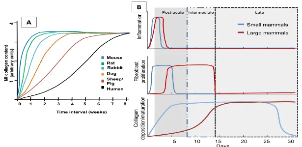
inflammation between small and large mammals post-MI.39 For example, a robust
increase in the cytokine tumor necrosis factor alpha is observed early in rodent MI
models with a tendency to plateau and resolve by one week post-MI.40,52,162 In contrast, a
more progressive and sustained increase in tumor necrosis factor alpha within the MI
region is reported in large animal models.39,40,52 The temporal cytokine profile observed
in large animal models in this post-MI period is similar to the cytokine profile observed
in plasma samples obtained in humans.18,33,46,52,53,85 Similarly, temporal differences in the
release of matrix proteases and development of local tissue edema have been observed
between small and large animal models post-MI. A summary of the temporal changes in
outcome of interspecies differences is the occurrence of myocardial rupture. Specifically,
myocardial rupture is a rare event clinically post-MI and exceedingly uncommon in large
animal models.17,108,119 In contrast, rodent models of MI, particularly those of mice, have
reported rupture rates between 29-53%.26,162,166 These observations would suggest
significant differences in the cellular and extracellular architecture and composition
within the MI region of rodents as opposed to large mammals, which in turn would
directly influence regional mechanical properties. Accordingly, the type of animal model
used will have significant implications for measurements of MI region mechanics in the
post-acute phase.
Imaging studies
A large number of studies have utilized various imaging modalities to study the
function and mechanics of the LV and specifically the MI region. The most common
techniques include magnetic resonance imaging (MRI) as well as ultrasound in both
humans and animals. 2,6,57,59,128,143,172,176,186 The primary goal of most studies has been to
identify correlations among MI size, mechanics, and functional measures. Imaging
techniques present an attractive opportunity for less invasive and clinically translatable
approaches. 30,64
Ultrasound
Traditional 2-D ultrasound measurements have been utilized extensively to
examine LV volumetric and pump function in the post-MI period. For the purposes of
focus, studies that have utilized ultrasound to examine aspects of myocardial strain, and
in particular strain during the filling phase of the cardiac cycle, will be examined to
Doppler Imaging (TDI), a form of ultrasound, is a noninvasive method by which regional
indices of myocardial function, such as contraction (from endocardial and epicardial
velocities) and wall motion, may be assessed. 8,60,172,182 Strain rate imaging (SRI) can be
acquired and has proven to be an effective tool in the evaluation of both MI and viable
surrounding myocardium (Figure 2.2).43,60,168,189 While the precise post-MI period was
not specified, Edvardsen et al reported that following the acute post-MI period in
patients, abnormal deformation patterns exist in the post-MI LV with longitudinal
stretching in the MI region compared to shortening (in the same direction) in the remote
myocardium. 43 Moreover, radial cycle dependent strains within the MI region were
indicative of localized thinning rather than thickening (Figure 2.2A 43,78,189). The study by
Zhang et al used strain rate imaging to assess various markers of LV function relevant to
the mechanics of the region, which included early diastolic strain rate and peak systolic
strain rate (reference length at end diastole) in patients with varying extents of infarction
across myocardial wall at 2-6 days post-MI. 189 This study determined that the peak
systolic strain rate and early diastolic strain rate decreased most markedly in patients
where a completely transmural MI was present (Figure 2.2B 43,78,189). In those patients
with a transmural MI, the greatest decline in both peak systolic strain rate and early
diastolic strain rates occurred. From the results of this study, the influence of infarction
across myocardial wall as a factor affecting the mechanics of the MI region can be
Magnetic Resonance Imaging
MRI has long been heralded as the gold standard technique for assessment of LV
mechanics due to its high spatial resolution and non-invasive nature. 3,58,149 A common
use of MRI involves the tagging of the LV and MI region with a set of horizontal and
vertical lines (by saturation) to track points or segments during multiple cardiac cycles,
termed spatial modulation of magnetization (SPAMM, or MR tagging). In general, MR
tagging strain measurements are derived from radiofrequency pulses that produce
magnetic saturation, enabling the tracking of segment or line lengths that can be analyzed
over cardiac cycles.3,30 Measurements of tracked points/segments through the cardiac
cycle can then be converted to linear dimensions, hence an index of mechanical strain.
Using this approach, it has been identified, for example, that a reduction (from referent
control values) in peak systolic strain (absolute values) within the MI region is related to
the reduction in LV pump performance, whether strain is measured along a longitudinal,
circumferential, or radial direction.174 With consideration of the relatively accelerated
remodeling process in rodents, Ross et al utilized MRI to assess the extent of wall
thickening (among other regional functional measures) at 1 day post-MI.143 The authors
noted that the percentage of wall thickening (presented as % of end diastolic wall
thickness) showed an approximate 60% decline from baseline to roughly 10% after only
1 day post-MI. While wall thickening clearly declined, radial strain at different times
post-MI varied significantly, with differences attributed to edema-related changes in the
end diastolic wall thickness. This study also reported the disappearance of such edema at
day seven post-MI, which was accompanied by a further decrease in wall thickening.
change in strain, likely in the radial direction (here seemingly affected by edema). These
changes in MI thickness and motion over a 3-7 day time period are consistent with the
inflammatory timeline for rodents (Figure 2.1 39,40,46,52,53,79,80,86,98,183). Young et al
reported circumferential and longitudinal cycle dependent strains in a rodent MI model to
exhibit spatial variation throughout the LV, although there is a persistent decrease in
magnitude (compared to referent normal strains) at all locations.188 Since there is a
relatively collapsed time window for the acute post-MI period and the spatial resolution
of MRI in rodents can be limited, this technique is primarily relegated to large animal
models to examine regional mechanics post-MI. Using a pig model, Saeed et al found
similar decreases in longitudinal and circumferential cycle dependent strains at 3 days
post-MI (compared to remote myocardium) and also reported significantly decreased
peak systolic strain in all directions (radial, circumferential, longitudinal) in the analyzed
segments contained within the MI region.149 Another study in pigs quantified the
differences in peak circumferential cycle-dependent strain between the MI region and
peri-infarct zone. 152 This study used late-gadolinium enhancement-cardiac MR to
quantify regional strains, finding the 3 day time point to show a significant decrease in
both MI region and peri-infarct zone strain. Such findings of variances in strain in the
different regions (MI region, peri-infarct zone, remote, etc) underscore that variations in
strain occur non-uniformly over time.
Various studies have been carried out in a clinical setting with MRI techniques as
a basis for the study of the post-MI myocardium. Using MR tagging in patients, Bogaert
et al found that normal and shear strains in the MI region decreased significantly by 5
thickness, which has previously been shown to modulate wall stress and strain patterns
and lead to an increase in structural stiffness.4 Garot et al used tagged MRI coupled with
harmonic phase imaging (HARP).57 This dual approach isolates segmental motion of
tagged myocardium (MRI tagging), which is then analyzed using a HARP algorithm that
produces phase images (based on the spectral peaks in the Fourier domain). In these
images, the motion of points can then be tracked along with the accompanying direction
of movement.126 In this past clinical study, MRI using HARP was performed
approximately 3 days post-MI while arguably, prior to the time points of interest in this
review, holding relevance in terms of regional mechanics within both normally perfused
and MI segments. Specifically, this study identified that regional systolic strain pattern
changes in both perfused and non-perfused segments occurred at this early post-MI time
point. Gerber et al also utilized tagged MRI in conjunction with contrast enhanced MRI
at 4 days post-MI in patients to analyze the peak circumferential shortening strain
(fractional change of length between end diastole and end systole) recorded at end
systole. The authors found that circumferential strain significantly decreased in segments
that displayed early hypo-enhancement (lower retention of contrast indicative of viable
tissue) as well as delayed hyper-enhancement (increased retention of contrast indicative
of MI) when compared to remote segments. These findings demonstrate that in addition
to quantifying the extent of an infarction, hypo- and hyper-enhancement can be used to
develop MRI-based indices of myocardial function. In this study, the dysfunctional
segments contained within the MI region demonstrated a significant relationship of
hyper/hypo-enhancement that represented the extent of infarction across myocardial wall
of the MI. 58
Mechanical implications
The post-acute phase is temporally offset across animal models with rodents
exhibiting collagen deposition and scar maturation twice as fast as large mammals. In all
animal models, the strain in the MI region decreases as compared to controls, albeit to
different extents. It is important to note that the remote myocardial region in treatment
groups serves as a referent control in some studies. Although this is a common study
design, it is well-known that the remote region geometry and properties change
throughout the remodeling process.16 Therefore, sham animals should be used for control
groups when possible. The MI region nominally exhibits a high degree of mechanical
anisotropy with most studies reporting changes in the absolute values of strain that differ
along the principal directions (where coordinate directions correspond to zero shear) and
over time. The local decrease in strain is often interpreted as a change in the material
properties of the MI region and has been described in terms of dyskinetic wall motion
with respect to the remote myocardium.165 Studies that utilized various imaging
modalities have identified progressive wall thinning and decreased curvature in the LV,
both of which contribute to an observed increase in global wall stress. Some studies have
focused on the mechanics of the regions adjacent to the MI and likewise found a decrease
in recorded strain magnitudes. Post-MI remodeling results in changes to both material
local change in myocardial composition (increased collagen content, presence of edema,
etc) and can only be identified based on stress-strain relations. Post-MI changes in
structural properties of the LV depend on the evolution of both material properties and
geometry and can be quantified from pressure-volume relations obtained at the chamber
level (i.e. chamber stiffness). Measurements of both material and structural properties
are affected by the loss of contractile units in the MI region and the forces applied on this
region by the surrounding viable myocardium. As a result, quantification of MI region
material properties are better assessed during diastole, with the myocardium considered
as a passively deforming material. Moreover, structural properties of the LV should
always be reported along with a corresponding point in the cardiac cycle. Together,
studies suggest that changes in regional strain and stress do correlate with concurrent
changes in functional indices.
2.4 Intermediate Time Phase (7-14 Days)
This post-MI phase is associated with increased expression of anti-inflammatory
cytokines and pro-fibrotic molecules, where one of the most notable is transforming
growth factor beta. 21,37,44,99,106,158 Studies involving a range of animal models consistently
report increased fibroblast proliferation within the MI region in concurrence with changes
in intra- and intercellular protein expression profiles.39,40,46,69 Fibrillar collagen content
within the MI region is elevated in comparison to the border or remote regions, and
changes in LV geometry consistent with the MI expansion process occur. 178–180 Since
this post-MI phase is accompanied by significant transitions in inflammation, signaling
molecules, and matrix formation, very dynamic changes also occur in biophysical
that must be calculated). As stated previously, the temporal changes in
biochemical/cellular events that occur in this post-MI phase are also species dependent
(Figure 2.1a 39,40,46,52,53,79,80,86,98,183), whereby temporal events in inflammation and matrix
formation/maturation are shifted to the left (as per the figure) with respect to small and
large animals. For example, in mouse MI models, a relatively mature MI scar (defined as
a thinned, collagen rich region) has been reported to occur within two weeks post-MI.31,32
Conversely, the MI region in larger animal models remains highly cellular and
structurally dynamic throughout this phase and up through the late phase (28 days
post-MI and beyond). Biomechanical response variables (which include stress and strain) are
directly affected by the loss of contractile forces in the MI and active viable surrounding
myocardium (affecting calculated stress) with architectural changes also impacting strain.
11,31,49,109,110,125,130,138
Thus, the temporal difference in scar maturation within the MI
region with respect to different animal species constitutes an important consideration
when evaluating stiffness in this post-MI time period.
Ultrasound
Modifications and expanded use of ultrasound, such as speckle tracking
echocardiography (STE), layer-specific STE (LS-STE), and Tissue Doppler Imaging
(TDI), have been used to examine changes in the length of segments and direction of
strain within the MI, often at time points beyond this intermediate period. More
specifically, these methods allow for computations of strain and strain rates of targeted
chords within the MI region based on tracking the relative positions of individual
section will focus upon the admittedly few studies that have examined relative strain
values within the MI region using these techniques in this time frame.
While the use of high resolution/frequency transducers (ultrasound probes) has
improved spatial resolution and thus applications in rodent post-MI models, the majority
of studies (primarily at acute or later time points) have been performed in large animals
and humans. However, a study utilizing LS-STE in a rodent model at two weeks post-MI
reported a depth-dependent decrease in the peak circumferential strain compared to
baseline values. In transmural infarcts, a significant decrease in peak circumferential
strain was found only in the layers spanning the endocardium and midwall. Conversely,
non-transmural infarcts exhibited an increase in peak circumferential strain in the
endocardial layer. These findings highlight the dependence of strain on extent of infarct
across the myocardial wall, a model-specific factor which therefore should be considered
and reported along with mechanical measurements of the MI region. (Figure 2.3 10)
Using a dog model, Park et al reported a decrease in radial and longitudinal strain
rate at both early and late diastole in concurrence with a reduction (from control values)
in strain magnitudes. 127 The authors reported stiffness indices in the MI region (derived
from wall thickness and hemodynamic pressure measurements) to increase in correlation
with a decrease in diastolic strain rate and changes in composition. The end diastolic
stress in the circumferential direction of the MI region was also noted to have a more
drastic decrement compared to longitudinal direction. Here, the authors conclude with
the suggestion that diastolic deformation indexes and functional measurements (rather
than systolic) of the MI region could be significant metrics of study in future clinical
In patients, it has been established that the extent of infarction across the
myocardial wall affects the magnitude of strain changes post-MI.12,189 To this effect,
studies in patient populations using STE and 2D-echocardiography at a time point
described as early, but beyond acute occurrence of MI, are here categorized as falling in
the intermediate phase. In these studies, the segments with a completely transmural MI
resulted in decreased peak systolic strains in both circumferential and radial directions at
both short and long axis. A significant decrease compared to referent normal values in
strain rates (systolic, same directions) was also associated with extent of infarction across
the myocardial wall.12,27
Magnetic Resonance Imaging (MRI)
MRI studies using rat and mouse models in the intermediate time frame have
shown a continued decrease in strain (in all directions) compared to the acute time period.
This decrease is consistent with the progress toward maturation of the MI (Figure 2.1
39,40,46,52,53,79,80,86,98,183
). In rats, Young et al utilized MRI tagging and late gadolinium
enhancement-MRI (LGE-MRI) techniques with a finite element model (FE) to map LV
wall strain. The results at the 7 day time point found the longitudinal, circumferential,
and 3D principal strains to be most affected (compared to the control) in the apical
region and midventricular regions.188 Here, the 3D principal strain was defined as the
maximal contraction at a given point in a direction not usually aligned with a traditional
image plane, highlighting regional differences of strain post-MI.
In a dog model, SPAMM tagged MRI in conjunction with a finite element (FE)
circumferential strain at the apex of the LV most affected.97 Studies on pig MI models
have sought to define regional strains and regional mechanical dyssynchrony using
cardiac MRI and LGE-MRI, with results that are in general agreement with previous
findings in large mammals (decreased strain in all directions).1 Similarly, a pig model
studied at 11 days post-MI with LGE-MRI in conjunction with HARP analysis showed
MI region strains that were significantly less than baseline healthy segments. The
circumferential strain displayed the greatest reduction in the MI region, although adjacent
regions also exhibited significant alteration of principal strains. Here, principal strains are
defined along coordinate directions that correspond to zero shear. It is interesting to note
that this study eliminated segments that were not completely transmural, thus anticipating
and accounting for the possibility of mechanical heterogeneity in the radial direction.159
Physical Approaches: Sonomicrometry and Markers
Sonomicrometry arrays are formed with ultrasonic crystals that each emit and
receive high-frequency pulse waves, enabling the measure of relative distances between
crystal pairs when implanted within the myocardium.186 Tissue-embedded arrays can
provide high spatial resolution of local deformation fields with some freedom in the
creation of a reference coordinate system. A study carried out in a rat model by
Fomovsky et al used seven sonomicrometry crystals to study MI region mechanics from
1-6 weeks post-MI. The authors found the circumferential and longitudinal strains were
significantly decreased from referent normal values at both 1 week and 2 week time
points. Critically, no collagen fiber alignment was seen in the MI region of the rats and
categorization of MI region mechanical properties as isotropic may not be suitable for
larger animal models that exhibit a high degree of fiber alignment. 49
In larger animal models, such as sheep, the implantation of 9 sonomicrometry
crystals along the MI region showed significant regional differences in strain with a two
week time point. In this study, remodeling strain was defined as the change in end
diastolic dimensions from baseline configuration of the crystals on the LV free wall to a
deformed configuration at a specific time post-MI. In essence, this remodeling strain is
representative of transient changes in geometry rather than cycle dependent ones. A
significant increase in remodeling strain was observed at the 14 day time point, with a
concomitant reduction in MI region end-systolic cyclical strain also promoting akinesis.
The authors also found a correlation between markers of apoptosis and measures of
remodeling strain and concluded that this is a promising mechanics-based target for
interruption of post-MI remodeling.93 Jackson et al tracked the areas of the MI region,
border zone, and remote regions from baseline to 8 weeks post-MI and reported nearly
40% expansion (increase in total area) within the MI region after two weeks compared to
the baseline. At the same time point, the border zone experienced a 10% expansion, and
the remote showed <5% expansion. (Figure 2.2C 43,78,189) Interestingly, the authors note
that the rate of expansion immediately following the onset of infarction was similar in all
regions.78 Although not reported in this study, a measure of remodeling strain (change
from baseline length) would have likely shown an increase as the expansion of the MI
region took place.
A combinatorial approach entails implantation of physical markers within the MI
technique to track their relative positions. Using a sheep model, Gorman et al used MRI
tagging and titanium markers along the border zone of the MI region. Obtained results
show that systolic border zone radial strain had significantly decreased by one week
post-MI and would continue to decline over the next 12 weeks, while the post-MI region area
gradually increased in size. 15 Another study by Holmes et al used gold beads implanted
in the LV free wall of pigs and biplane cineradiography to track deformations at 1 and 3
weeks post-MI. This study used a plexiglass phantom with embedded beads to calibrate a
coordinated system for calculation of principal and remodeling strains. Reference lengths
for strain calculations were obtained at end diastole in a control animal. At one week, the
authors report a marked circumferential LV expansion, an increase in remodeling strains,
and a decrease in cycle-dependent radial and in-plane strains, with the plane of interest
defined by the longitudinal and circumferential directions (tangent to the epicardium).74
Mechanical implications
Post-MI healing throughout the intermediate phase significantly differs among
animal models, with rodents entering a period of scar maturation while larger mammals
continue to exhibit fibroblast proliferation and collagen deposition. Numerous factors
impact recorded strain patterns, including the presence of edema, the MI size, the extent
of infarction across myocardial wall, and its location with respect to the anatomical long
axis of the LV. Consistent mechanical observations include a decrease in MI region
cyclic strain and an increase in remodeling strain. Direct comparisons among studies
show quantitatively different strain magnitudes due to the use of different reference
lengths, cardiac axis definition, extent of MI, location, and cardiac points of reference
trends coupled with related qualitative descriptors of wall motion facilitate comparison
among studies, although a comprehensive structure-function relationship of in-vivo
mechanics of the MI region has not yet been reached. Increased remodeling strains were
noted to be most prominent in the circumferential direction, with segment lengths
changing as the MI region dilates post-MI. Along with changing strain, reports indicate
that wall stress is generally increased, with the most significant changes observed in the
diastolic pressure range.
2.5 Late Time Phase (14 days and beyond)
The late remodeling phase is characterized by lowered or absent cellular
inflammatory infiltrate in the MI region.39,40,46,52 The scar maturation process is underway
across species in the MI region, whereby an index of MI/matrix maturation includes
increased expression of lysyl oxidase, interleukin-10, and TIMP-1.40,50,163 While total
collagen content is uniformly elevated within the MI region, the relative stability and
maturation of the matrix is species dependent as described previously and summarized in
Figure 2.1. Moreover, the increased proliferation of fibroblasts, particularly fibroblasts
with a secretive phenotype, occurs in this phase of post-MI remodeling. Thus, this phase
can be associated with continued/persistent release of matrix proteases, which in turn will
affect collagen maturation and turnover within the MI region. As a result, species
dependent as well as fibroblast/protease dependent processes will directly affect the
structural composition and integrity of the MI region, and in turn, measurements of
Ultrasound
Benejam et al used a rat model and STE to characterize segmental dysfunction
throughout the LV at a time point of 4-10 weeks post-MI. Their results demonstrated
lowered absolute values for midwall circumferential and radial strains and strain rates.
The authors also report a marked difference in strain amongst segments, with highly
fibrotic segments displaying the lowest strain in the MI region. Peak systolic and early
diastolic circumferential and radial strains showed lowered absolute values and strain
variations in the MI region.13 Liao et al used 2D strain echocardiography at this same
time point and animal model in order to assess the development of the MI region. The
authors determined strain and strain rate were decreased at different magnitudes
dependent on location within the MI region, which was divided into anteroseptal,
anterior, and anterolateral sections.105 The results of these studies are generally consistent
in rat models at the 4 week time point.13,14,105
A variation of STE was used in a dog model by Wong et al, where speckle
tracking displacement estimates in the mid-wall were combined with shape tracking
using compactly supported radial basis functions to improve quantitative deformation
analyses. At a six week post-MI time point, acquired radial strain maps were consistent
with previous reports of reduced MI region strains.182
Sakamoto et al utilized 2-D echocardiography and a sheep model to compare
reperfused versus non-reperfused MIs at 8 weeks post-MI. The authors extend their
measurements of remodeling strain (defined as the ratio of a baseline reference length to
reperfusion impacted MI region stiffness. The authors found that although thinning still
occurred, the animals in the reperfusion group had MI regions that trended toward
akinesis, signifying greater stiffening in contrast to the quantified dyskenesis in the
non-reperfused group. 150 Comparably, a previously mentioned study in a dog model by Park
et al at a similar time point reported the radial diastolic strain rates significantly
decreased (by more than half in the case of radial late diastolic strain) compared to
baseline values.127 The decrease in strain rate prompted the authors to suggest an increase
in MI region stiffness in the presented sheep study, although it is important to note
contractility also inherently plays a role in the calculated strain rate.
In clinical observations, Thorstensen et al used 3-D echocardiography to compare
global function in patients with small and large MI volume fractions (large >12% infarct
volume fraction). They report decreased 3D global strain rate values in all directions
(longitudinal, circumferential, and radial) that were significant in patients with large MI
as compared to healthy controls.169 Helle-valle et al used STE at 6 months post-MI to
analyze strain, rotation, and wall motion in the segments within the MI region, border
zone, and remote myocardium. 68 This study found that the center of the MI region
displayed the greatest dyskenesis with a concomitant reduction (from referent normal
values) in strain, i.e. abnormal motion is most extreme at the center of the infarct with a
tendency to normal motion toward the edges. The authors noted an interesting pattern in
rotation, with the counter-clockwise border (assuming a clockface orientation) zone
showing hyper-rotation shifting to intermediate rotation towards the center of the MI
region and trending towards hypo-rotation at the clockwise MI border. The authors
portion of the cardiac phase. Experimental findings from this study were combined with a
finite element model to resolve the apparent inconsistencies between regional apical
rotation and strain. The authors conclude from simulation results that an imbalance in
active contractile forces from the interaction of the MI region and surrounding viable
myocardium is the cause for the oppositely directed rotations at the two MI border
regions and low strain in the center of the MI region.68
Magnetic Resonance Imaging
A mouse model study by Ross et al used MRI and reported that the percent wall
thickness (interpreted as increased remodeling radial strain) at 4 weeks post-MI was
negative in the MI region, indicative of local thinning. This study also reported systolic
wall bulging, which is representative of dyskinetic motion in the MI region.143 Liu et al
found post-MI heterogeneous changes in strain (presented as maximal stretch and
maximal shortening independent of any coordinate system) in rats at a 4 week time point.
This study utilized SPAMM tagging along with HARP analysis and found a reduction
(compared to control values) in strain at the apex, while the strain at the LV base
increased (Figure 2.4C-D 107,191). These results suggest regionally dependent changes in
strain occur in this late time period, as the apex and base of the LV were noted to have
opposing changes post-MI in a model with an anteroapical infarct. 107 Espe et al studied
the LV motion in a rat model at 6-weeks post-MI by using phase contrast-MRI
(PC-MRI).47 This study calculated regional circumferential strain from the acquired PC-MRI
velocity data and found the peak circumferential strain to decrease both globally and in
the MI region with significant heterogeneity throughout the LV. This study focused its