Medical Image Analysis on Software and
Hardware System
Divya1, Sunitha Lasrado2
PG Student, Dept. of Electronics and Communication Engineering, NMAMIT, Nitte, Udupi District, Karnataka, India1 Asst. Professor, Dept. of Electronics and Communication Engineering, NMAMIT, Nitte, Udupi District, Karnataka,
India2
ABSTRACT: Biomedical Engineering is one of the main areas where in the image processing plays a wide role. Medical imaging and its processing is a topic of interest which helps for easier diagnosis and analysis of the medical issues. Detection of the areas affected is the basic step in the processing. In this work, Prewitt edge detection and Sobel edge detection of a Computed Tomography image of brain with tumour is considered. The process is carried out on Software and Hardware system for different sizes of the image and the time taken are analysed.
KEYWORDS: Biomedical Engineering, Image Processing, Medical Imaging, Edge Detection, Computed Tomography, Software and Hardware system
I. INTRODUCTION
Biomedical Engineering is the field which bridges the medical field to many other fields like engineering, applied mathematics, physics and so on [1]. The advancements brought in the technology is now being effectively used in medicine. One such area which brings improvement in the medical diagnosis is the medical imaging and its processing. Images of various parts of the body are obtained from various techniques like Computed Tomography (CT), X-Rays, Magnetic Resonance Imaging (MRI), endoscopy, ultrasonic imaging etc. These obtained images are to be processed for better identification of the problems. In that stage, image processing comes into picture. Image features detection, extraction, segmentation, colour enhancement and many more criterions are achieved through image processing. Brain is the central system of a human body. It controls all the activities. Growth of abnormal cells in the tissues of brain are the tumours. These tumours could be either cancerous (malignant) or non-cancerous (benign) [2]. Detection of these tumours and immediate treatments are essential. The size of the tumour and its location are to be identified [3]. To achieve this segmentation of the image is to be carried out. Segmentation is mainly achieved by techniques based on edge detection or based on thresholding or region based [4].
Edge detection is the fundamental step in most of the image analysis methods. Changes in the intensity is obtained through edge detection. Derivatives helps in the finding the changes in intensity of the image and thus help in detection of edges. Edge detection are basically classified as Classical or Gradient based edge detectors (first derivative), Zero crossing (second derivative), Laplacian of Gaussian (LoG), Gaussian edge detectors, Coloured edge detectors [5]. The classical or gradient based methods include Roberts, Prewitt, Sobel and Canny edge detections.
First derivative is defined by the magnitude of gradient. The gradient of an image at is given,
The magnitude of the gradient is given by,
where is the value of rate of change in the gradient vector direction.
Edge detection is done by convolution of original image with the coefficient of gradient along and direction.
PREWITT EDGE DETECTION
In Prewitt edge detection method, equal weights are assigned to the neighbourhood pixels of the pixel whose edge strength to be detected. The mathematical equation given in equation (5) is used to design the kernel.
where is the coefficient of gradient operator. is the coefficient place value of mask pixels. is the gradient operator. Prewitt operator convolution mask is given in Figure 1.
Figure 1: Convolution mask of Prewitt edge detection [6]
SOBEL EDGE DETECTION
In Sobel edge detection algorithm, pixel weights assigned to the pixel in neighbourhood of the candidate pixel are not equal like in the case of Prewitt edge detection. Operations are similar to that practised in case of Prewitt edge detection. The mathematical equation defining the kernel is given in equation (6).
where is the coefficient of gradient operator. is the coefficient place value of mask pixels. is the gradient operator. The convolution mask of Sobel edge detection is given in Figure 2.
Figure 2: Convolution mask of Sobel edge detection [6]
II. RELATED WORK
Segmentation is one of the significant step in these image processing application. Various methods of segmentation is discussed in [4]. Edge detection is one of the methods for segmentation of image. Various methods of edge detection for segmentation is given in [5], [11], [12] and [13]. In [6], [14] and [15] edge detection techniques are implemented on various platforms like Xilinx System Generator (XSG), Vivado HLS and on FPGA boards.
III.METHODOLOGY
SOFTWAREIMPLEMENTATION
Edge detection is implemented on Matrix Laboratory (MATLAB) software by writing a MATLAB code. The Prewitt and Sobel edge detected images are obtained. The time taken for the execution of the code and to obtain the results is got by the 'tic' and 'toc' functions.
Edge detection is also carried out on MATLAB Simulink platform. The block sets present in the Simulink library are used to detect the Prewitt and Sobel edge detected images. The time taken is measured by the 'tic' and 'toc' functions and execution of the Simulink model is performed through the MATLAB command prompt.
The above procedures were carried out on a Intel core i7 processor and in MATLAB 2012a platform.
HARDWAREIMPLEMENTATION
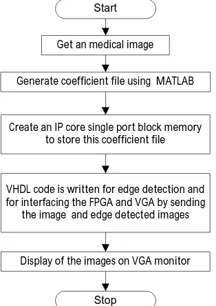
Field Programmable Gate Array (FPGA) is used to implement Prewitt edge detection and Sobel edge detection on hardware. Very High Speed Hardware Description Language (VHDL) code is written for edge detection. The image details are converted to coefficient file (.coe) using MATLAB. This .coe is used to generate an Intellectual Property (IP) core of a Single Port Block Random Access Memory (RAM). From the RAM, the image pixel details are buffered and the edge detection is carried out using VHDL code. The image and the edge detected images are then displayed on the Video Graphics Array (VGA) by interfacing FPGA with VGA monitor. The flowchart given in Figure 3 explains the steps involved in edge detection of image in FPGA and display of it in the VGA monitor.
Start
Get an medical image
Generate coefficient file using MATLAB
Create an IP core single port block memory to store this coefficient file
Display of the images on VGA monitor VHDL code is written for edge detection and for interfacing the FPGA and VGA by sending
the image and edge detected images
Stop
Figure 3: Flowchart showing the steps involved in edge detection in FPGA and display of images on VGA monitor
IV.RESULTS
A brain with tumour CT image [7] is considered for the work which is shown in Figure 4. The white patch in the figure is the tumour. This is to be detected.
Figure 4: CT image of brain with tumour [7]
SOFTWAREIMPLEMENTATION
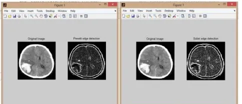
The output obtained from the Prewitt edge detection and Sobel edge detection MATLAB codes is given in Figure 5. Prewitt edge detection implementation output is to the left side. Sobel edge detection implementation output is to the right side. In both of them the original image and the edge detected images are present.
Figure 5: Output of Prewitt and Sobel edge detection in MATLAB
Figure 6 shows the output obtained for Prewitt and Sobel edge detections in MATLAB Simulink. Left part is the Prewitt edge detection output and the right part is the output obtained from Sobel edge detection.
HARDWAREIMPLEMENTATION
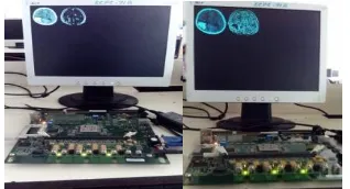
Figure 7 shows the Prewitt and Sobel edge detection techniques implemented on FPGA and its result displayed on the VGA monitor.
Figure 7: Output of Hardware implementation of Prewitt and Sobel edge detection
Table 1 shows the logic utilization for Prewitt and Sobel edge detection implementation on FPGA Virtex 2 Pro for the image sizes 128*128, 160*160 and 200*200.
Table 1: Logic utilization for Prewitt and Sobel edge detection implementation on FPGA
Logic Utilization Available Prewitt edge detection Sobel edge detection
128*128 160*160 200*200 128*128 160*160 200*200
Total No. of slice registers 27392 265 272 282 265 272 282
Number of slice flipflops 201 208 218 201 208 218
Number of latches 64 64 64 64 64 64
Number of 4 input LUTs 378 455 457 374 451 453
Number of occupied slices 355 412 451 353 410 449
Total number of 4 input LUTs 27392 607 718 802 605 716 800
Number used as logic 378 455 457 374 451 453
Number used as route thru 93 95 81 95 97 83
Number used for 32x1 RAMs 128 160 256 128 160 256
Number used as shift registers 8 8 8 8 8 8
Number of bonded IOBs 556 30 30 30 30 30 30
IOB latches 8 8 8 8 8 8
Number of block RAMs 136 8 13 20 8 13 20
Number of GCLKs 16 3 3 3 3 3 3
Total equivalent gate count for design
546999 879541 1350829 546983 879505 1350793
Additional JTAG gate count for IOBs
1440 1440 1440 1440 1440 1440
Table 2 shows the time taken for executing the Prewitt edge detection on different platform - MATLAB, MATLAB Simulink and on FPGA Virtex 2 Pro. Image sizes 128*128, 160*160 and 200*200 are considered.
Table 2: Time taken for the execution of Prewitt edge detection on various platform for different image sizes
Image Sizes MATLAB MATLAB Simulink FPGA-Virtex 2 Pro
128*128 0.16s 0.30s 6.14ns
160*160 0.19s 0.34s 6.62ns
Table 3 shows the time taken for executing the Sobel edge detection on different platform - MATLAB, MATLAB Simulink and on FPGA Virtex 2 Pro. Image sizes 128*128, 160*160 and 200*200 are considered.
Table 3: Time taken for the execution of Sobel edge detection on various platform for different image sizes
V. CONCLUSION
Prewitt edge detection and Sobel edge detection algorithms were implemented on Software and Hardware system for a CT image of brain with tumour. The time taken for the execution of edge detection on these platforms for different image sizes (128*128, 160*160 and 200*200) is noted. It is observed that the hardware implementation is faster in this case compared to the software implementation.
The edge detection can be further used for segmentation, feature extraction, measuring size or volume of the tumour etc. The methods can be implemented for any medical images or videos.
REFERENCES
[1] Zhu, H., "Medical Image Processing Overview", University of Calgary, Summer School Program-Introduction to Mathematical Medicine, held at the University of Waterloo, 2003.
[2] Brain tumour-Patient Version, http://www.cancer.gov/types/brain
[3] Mustaqeem, A., Javed, A. and Fatima, T., "An efficient brain tumor detection algorithm using watershed & thresholding based segmentation", International Journal of Image, Graphics and Signal Processing, Vol.4, No.10, pp. 34-39, 2012.
[4] Matta, S., "Review: Various image segmentation techniques", International Journal of Computer Science and Information Technologies ( IJCSIT), Vol.5, No.6, pp.7536-7539, 2014.
[5] Lakshmi, S. and Sankaranarayanan, D.V., "A study of edge detection techniques for segmentation computing approaches", IJCA Special Issue on “Computer Aided Soft Computing Techniques for Imaging and Biomedical Applications” CASCT, pp.35-40, 2010.
[6] Paikrao, P.L. and Swapnil, G.K., "FPGA based image feature extraction using xilinx system generator", International Journal of Computer Science and Information Technologies, Vol.5, No.3, pp. 3743-3747, 2014.
[7] Al-Ashwal, R.H., Supriyanto, E., Rani, N.A.B., Abdullah, N.A.B., Illani, N., Aziz, B. and Mahfooz, R.B., "Digital Processing for Computed Tomography Images: Brain Tumor Extraction and Histogram Analysis", Mathematics and Computers in Contemporary Science, pp.119-128.
[8] Ge, R. and Shen, H., "Researching on Feature Extraction of Brain CT Image", International Journal of Signal Processing, Image Processing and Pattern Recognition, Vol.6, No.5, pp. 39-48, 2013.
[9] Uemura, T., Koutaki, G. and Uchimura, K., "Image segmentation based on edge detection using boundary code", International Journal of Innovative Computing, Information and Control, Vol.7, No.10, pp.6073-6083, 2011.
[10] SELEÞCHI, E. and Duliu, O.G., "Image Processing and Data Analysis in Computed Tomography", Rom. Journ. Phys., Vol. 52, No. 5–7, pp. 667–675, 2007 .
[11] Senthilkumaran, N. and Rajesh, R., "Edge detection techniques for image segmentation–a survey of soft computing approaches", International journal of recent trends in engineering, Vol.1, No.2, pp.250-254, 2009.
[12] Maini, R. and Aggarwal, H., "Study and comparison of various image edge detection techniques", International journal of image processing (IJIP), Vol.3, No.1, pp.1-11, 2009.
[13] Nadernejad, E., Sharifzadeh, S. and Hassanpour, H., "Edge detection techniques: Evaluations and comparison", Applied Mathematical Sciences Vol.2, No.31, pp.1507-1520, 2008.
[14] TK, H.N., Belleudy, C. and Pham, T.V., "Performance and Evaluation Sobel Edge Detection on Various Methodologies", International Journal of research in Engineering and Technology, Vol.3, No.5 pp.549-553, 2014.
[15] Said, Y., Saidani, T., Smach, F. and Atri, M., "Real time hardware co-simulation of edge detection for video processing system", 16th IEEE Mediterranean Electrotechnical Conference (MELECON), pp.852-855, 2012.
[16] Volnei A. Pedroni, "Circuit Design and Simulation with VHDL", 2nd edition, MIT Press, 2010.
[17] R. C. Gonzalez and R. E. Woods, “Digital Image Processing”, 2nd edition, Pearson Education (Asia) Pte. Ltd/Prentice Hall of India, 2004.
[18] Virtex 2 Pro Hardware Refernce Manual, http://www.xilinx.com/univ/XUPV2P/Documentation/XUPV2P_User_Guide.pdf [19] Virtex 2 Pro User Guide, www.xilinx.com/support/documentation/user_guides/ug012.pdf
[20] VGA Controller, https://eewiki.net/pages/viewpage.action?pageId=15925278
Image Sizes MATLAB MATLAB Simulink FPGA-Virtex 2 Pro
128*128 0.16s 0.36s 6.07ns
160*160 0.18s 0.36s 6.63ns