| WORMBOOK CELL FATE, SIGNALING AND DEVELOPMENT
Programmed Cell Death During
Caenorhabditis elegans
Development
Barbara Conradt,*,1Yi-Chun Wu,†,‡,1and Ding Xue§,1 *Department Biology II, Center for Integrated Protein Science Munich, Ludwig Maximilian-University Munich, Planegg, 82152, Germany,†Institute of Molecular and Cellular Biology, National Taiwan University and‡Institute of Atomic and Molecular Sciences,
Academia Sinica, Taipei, 10617, Taiwan,§Department of Molecular, Cellular and Developmental Biology, University of Colorado, Boulder, Colorado 80309 ORCID ID: 0000-0002-8429-8136 (D.X.)
ABSTRACTProgrammed cell death is an integral component ofCaenorhabditis elegansdevelopment. Genetic and reverse genetic studies in C. elegans have led to the identification of many genes and conserved cell death pathways that are important for the specification of which cells should live or die, the activation of the suicide program, and the dismantling and removal of dying cells. Molecular, cell biological, and biochemical studies have revealed the underlying mechanisms that control these three phases of programmed cell death. In particular, the interplay of transcriptional regulatory cascades and networks involving multiple transcrip-tional regulators is crucial in activating the expression of the key death-inducing geneegl-1and, in some cases, theced-3gene in cells destined to die. A protein interaction cascade involvingEGL-1,CED-9,CED-4, andCED-3results in the activation of the key cell death proteaseCED-3, which is tightly controlled by multiple positive and negative regulators. The activation of theCED-3caspase then initiates the cell disassembly process by cleaving and activating or inactivating crucialCED-3substrates; leading to activation of multiple cell death execution events, including nuclear DNA fragmentation, mitochondrial elimination, phosphatidylserine externalization, inactivation of survival signals, and clearance of apoptotic cells. Further studies of programmed cell death inC. eleganswill continue to advance our understanding of how programmed cell death is regulated, activated, and executed in general.
KEYWORDSCaenorhabditis elegans; activation phase; execution phase; programmed cell death; specification phase; WormBook
TABLE OF CONTENTS
Abstract 1533
Cell Death Activation 1537
The core machinery involved in the activation of the apoptotic program 1537 Biochemical and structural analyses of the activation of the core apoptotic program 1537
Regulation of cell death activation in C. elegans 1538
Cell Death Specification 1539
Role of egl-1transcriptional control 1539
RID lineage: 1539
The deaths of the hermaphrodite-specific neurons and cephalic companion neurons: 1540
Continued
Copyright © 2016 by the Genetics Society of America doi: 10.1534/genetics.115.186247
Manuscript received February 3, 2016; accepted for publication April 22, 2016.
CONTENTS,continued
Role of asymmetric cell division 1541
The Q lineage: 1541
The NSM sister cell death: 1542
The M4 sister cell death: 1542
Noncanonical apoptotic cell death 1543
Cell Death Execution 1544
CED-3-activated cell death execution events 1544
Nuclear DNA fragmentation: 1544
PS externalization: 1544
Mitochondrial elimination: 1545
Inactivation of survival signals: 1545
Clearance of Apoptotic Cells 1546
Presentation of eat me signals 1546
Surface PS expression on phagocytes 1546
Engulfment receptors and signaling pathways 1547
The CED-1, CED-6, and CED-7 pathway: 1547
The CED-5 and CED-12 pathway: 1548
The PAT-2 and PAT-3 pathway: 1549
Negative regulators of the engulfment process 1549
MTM-1, SRGP-1, and PDR-1: 1549
PGRN-1: 1550
ABL-1, SLI-1, and SWAN-1: 1550
Engulfment promotes apoptosis 1550
Formation and maturation of phagosomes 1551
Sealing of phagosomes: 1551
Rab small GTPases in phagosome maturation: 1551
Homotypic fusion and protein sorting complex—a potential RAB-7 effector in phagosome maturation: 1552 Lipid second messenger PtdIns(3)P and its effector proteins: 1552
Autophagy genes: 1553
Acidification of phagosomal lumen 1553
Digestion of apoptotic cells 1554
Degradation of proteins of apoptotic cells: 1554
Degradation of DNA of apoptotic cells: 1554
Nonapoptotic Cell Death 1554
Conclusions 1555
G
ENETIC studies of programmed cell death in Caenorhab-ditis elegansled to the identification of key players involved in this important physiological process, whose functions are conserved fromC. elegansto humans (Adams 2003; Horvitz 2003; Danial and Korsmeyer 2004; Fuchs and Steller 2011). These pioneering studies were made possible by the follow-ing biology ofC. elegans: (1) unlike in many other organ-isms, programmed cell death is not essential forC. elegansviability, at least under laboratory conditions (Ellis and Horvitz 1986); (2) cells undergoing programmed cell death inC. eleganschange their morphology and refractivity and can be observed in living animals using differential in-terference contrast (DIC) microscopy (also referred to as Nomarski optics; Figure 1) (Robertson and Thomson 1982);
(3) programmed cell death that occurs during the devel-opment of somatic tissues ofC. elegansis determined by the essentially invariant cell lineage, therefore, it is known not only which cells undergo programmed cell death but also when and where they die (Sulston and Horvitz 1977; Sulston
et al.1983). These unique features made it possible to geneti-cally dissect the process of programmed cell death inC. elegans
at single-cell resolution. The resulting groundbreaking work was recognized with the Nobel Prize for Medicine in 2002, which was awarded to Sydney Brenner, John E. Sulston, and H. Robert Horvitz for their leading roles in deciphering the
Programmed cell death occurs during two stages of
C. elegans life and in two different types of tissues: during embryonic and postembryonic development of the soma (re-ferred to as developmental cell death) (Sulston and Horvitz 1977; Sulstonet al. 1983), and in the gonad of adult her-maphrodites (germ cell death) (Sulston 1988; White 1988; Gumienny et al. 1999). Developmental cell death is deter-mined by the essentially invariant somatic cell lineage: out of the 1090 cells generated during the development of the hermaphrodite soma, exactly 131 reproducibly undergo pro-grammed cell death (113 of these cells die during embryonic and 18 during postembryonic development) (Sulston and Horvitz 1977; Sulstonet al.1983). Germ cell death affects the majority of all developing germ cells (possibly to provide resources for surviving germ cells) and occurs in a manner that is not determined by cell lineage (Gumiennyet al.1999; Hansen and Schedl 2013). Furthermore, various types of in-sults such as, for example, exposure to DNA damage-inducing treatments cause additional germ cells to die (Gartneret al.
2000). Since germ cell death has been reviewed recently (Gartneret al.2008; Bailly and Gartner 2013), in this review we will focus on developmental cell death.
A combination of morphological observations and genetic analyses led to the finding that developmental cell death proceeds in three phases: during the“specification phase”, it is determined which cells will undergo programmed cell death and which cells will survive; during the “activation phase”, the cell death program is activated in those cells that are programmed to die; during the“execution phase”, cells are dismantled, killed, and subsequently engulfed and de-graded by neighboring cells (Figure 2) (Horvitz 1999). What happens when one of these phases is disrupted? Mutations that affect the specification phase alter the highly reproduc-ible pattern of developmental cell death and result in the inappropriate survival or death of one or a small number of cells (for example Ellis and Horvitz 1991). Mutations that
affect the activation phase can cause a general block in pro-grammed cell death (resulting in the inappropriate survival of the majority of the 131 cells that are programmed to die) or result in the inappropriate deaths of many cells that nor-mally live (leading to the loss of viability) (Ellis and Horvitz 1986; Hengartner et al.1992; Conradt and Horvitz 1998). Finally, mutations that disrupt the execution phase block cel-lular disassembly (Nakagawa et al.2010) and result in the accumulation of dead cells (referred to as cell corpses) that fail to be engulfed and/or degraded (Sulston 1976; Hedgecock
et al.1983; Elliset al.1991).
Thefirstcelldeath abnormality (ced) genes identified were ced-1andced-2(Hedgecocket al.1983). Loss-of-function (lf) mutations inced-1orced-2partially block the engulfment of cell corpses. A block or delay in cell corpse engulfment results in the accumulation of cell corpses in embryos and young larvae, which can easily be detected using DIC mi-croscopy. Indeed, subsequent genetic screens for mutants with similar phenotypes resulted in the identification of additional genes required for cell corpse engulfment and other aspects of the execution phase (Ellis et al. 1991; Gumienny et al.2001; Wuet al.2001; Zhouet al.2001a). Mutations inced-1andced-2were also instrumental in the identification of genes involved in the activation phase. The persistent cell corpse defect in ced-1 mutants was used to screen for mutations that suppress this phenotype with the rationale that some of the suppressors should suppress this phenotype by blocking the upstream activation phase, and hence cause a general block in programmed cell death. This resulted in the identification of lf mutations in theced-3gene, which (as discussed in more detail below) is required for most programmed cell deaths inC. elegans(Ellis and Horvitz 1986). Genes involved in thefirst phase of programmed cell death, the specification phase, were identified in genetic screens with goals to identify mutations that do not cause a general block in programmed cell death, but are able to block only specific cell deaths such as, for example, the deaths of theneurosecretorymotorneuron (NSM) sister cells (Ellis and Horvitz 1991; Thellmannet al.2003; Hatzold and Conradt 2008) or the sexually dimorphic neuronscephalic com
pan-ion neurons (CEMs) (Peden et al. 2007; Schwartz and
Horvitz 2007). These cell deathspecification (ces) screens not only resulted in the identification of genes important for the specification phase, but also another crucial compo-nent of the activation phase,ced-9(Hengartneret al.1992). In contrast toced-3, the function ofced-9is to protect against programmed cell death in the 959 somatic cells that are pro-grammed to live, and lf mutations inced-9cause many cells to inappropriately die; thereby leading to embryonic lethality. Doesced-3negatively controlced-9to allow the 131 cell deaths to occur, or doesced-9negatively controlced-3to allow the 959 somatic cells to survive? The activation phase of pro-grammed cell death can be considered a regulatory pathway that controls a“life-death”switch. In such a regulatory path-way, lf mutations in genes that are closer (downstream) to the switch generally suppress the phenotype caused by lf
mutations in genes that are further away (upstream) from the switch. In the case ofced-3andced-9, double mutant analysis revealed that the loss ofced-3suppresses the inappropriate cell death phenotype and the embryonic lethal phenotype caused by ced-9(lf) mutations. Hence, in the regulatory pathway of the activation phase,ced-3is epistatic to or acts downstream ofced-9(Hengartneret al.1992). Similar double mutant analyses have been used to place additional compo-nents of the activation phase into this life-death regulatory pathway. In addition, they have allowed analyses of genetic pathways that underlie the specification and execution phase, as described below.
Of note, rather than strictly in a sequential and linear fashion; certain aspects of the specification, activation, and execution phases may occur in parallel. Furthermore, feed-back exists between the execution phase and the activation phase. This is demonstrated by the fact that in genetic back-grounds in which the activation phase is functionally com-promised, a block in engulfment (execution phase) reduces
the likelihood of a cell that is programmed to die to actually die (Hoeppneret al.2001; Reddienet al.2001; Chakrabortyet al.
2015). This“killing”function of engulfment appears to act on the activation phase and promotes its swift induction and completion (Chakrabortyet al.2015).
For most of the 131 somatic cells programmed to die, the activation phase is mediated by a core apoptotic machinery that is conserved from C. elegans to mammals [egg laying defective-1 (EGL-1) homologous to the BH3-only proteins,
CED-9homologous to Bcl-2,CED-4homologous to apoptotic
protease activating factor 1 (Apaf-1), andCED-3homologous to caspases] (Horvitz 2003; Lettre and Hengartner 2006; Conradt 2009). However, for at least one programmed cell death, the death of the“linker cell”in males, the activation phase occurs through a nonapoptotic machinery (Abraham
et al.2007; Blumet al.2012). In the following, we will review our current understanding of the specification, activation, and execution phases of apoptotic developmental cell deaths. Furthermore, we will summarize recent advances in our
understanding of the nonapoptotic death of the linker cell. Genetic perturbations and/or various treatments can also lead to various forms of nonapoptotic cell death inC. elegans
(pathological cell death). Since this type of cell death does not occur during normalC. elegansdevelopment and since it has recently been reviewed (Vlachos and Tavernarakis 2010; Kinet and Shaham 2014), it will not be covered here.
Cell Death Activation
The core machinery involved in the activation of the apoptotic program
Three death-promoting genes,egl-1,ced-3, andced-4, are re-quired for most, if not all, developmental cell death in
C. elegans. Strong lf mutations in any of these genes result in the survival of most somatic cells that normally undergo pro-grammed cell death during development (Ellis and Horvitz 1986; Conradt and Horvitz 1998). Furthermore, these three genes act within dying cells to promote apoptosis, indicating that cells die by an intrinsic suicide mechanism (Yuan and Horvitz 1990; Shaham and Horvitz 1996b; Conradt and Horvitz 1998). By contrast, the activity of theced-9gene pro-tects cells from undergoing programmed cell death during
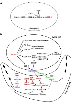
C. elegansdevelopment (Hengartneret al.1992). lf mutations inced-9cause embryonic lethality as a consequence of ectopic deaths of cells that normally live.ced-3,ced-4,egl-1, andced-9 appear to act in a simple genetic pathway in whichegl-1acts upstream ofced-9to induce cell death,ced-9acts upstream of ced-4to inhibit cell death, andced-4acts upstream ofced-3to kill cells (Figure 2A) (Hengartner et al.1992; Shaham and Horvitz 1996b; Conradt and Horvitz 1998).
ced-9 encodes a protein similar to the gene product of the human proto-oncogenebcl-2(Hengartner and Horvitz 1994b), which plays a similar role in preventing apoptosis
in mammals (Adams and Cory 2001). ced-9 and bcl-2 are
members of a gene family that plays important roles in reg-ulating apoptosis in diverse organisms (Reed 1997; Adams and Cory 2001). Members of the Bcl-2 protein family contain one or several characteristic Bcl-2 homology (BH) domains, BH1, BH2, BH3, and BH4, which are domains important for mediating interactions among different members of the Bcl-2 family (Adams and Cory 2001).egl-1encodes a small protein of 91 amino acids with a BH3 motif, which has been found in all proapoptotic members of the Bcl-2 gene family and medi-ates direct binding of these proteins to antipoptotic Bcl-2 members (Conradt and Horvitz 1998; Bouillet and Strasser 2002). ced-3 encodes the founding member of a family of aspartate-specific cysteine proteases named caspases (Yuan
et al.1993; Alnemriet al.1996). Like other caspases,CED-3is synthesized as a proenzyme and is proteolytically activated to generate an active protease containing a large subunit of 17 kDa (p17) and a small subunit of 15 kDa (p15) or 13 kDa (p13) (Alnemri et al. 1996; Xue et al. 1996). The protease activity of CED-3appears to be essential forced-3 to cause programmed cell death inC. elegans(Xueet al.1996;
Shaham et al. 1999). However, a deletion mutation that removes the ced-3 region encoding the entire protease do-main, including the p17 and p15 domains, causes a weaker cell death defect than those observed in multiple ced-3(lf)
mutants carrying missense mutations (Shahamet al.1999), suggesting that some of the developmental cell death can occur in the absence of the CED-3 protease activity. ced-4 encodes a protein similar to human Apaf-1, an activator of human caspase-9 (Yuan and Horvitz 1992; Zouet al.1997).
Both CED-4 and Apaf-1 contain a caspase-recruitment
do-main and nucleotide-binding motifs that are critical for the function of these proteins (Seshagiri and Miller 1997; Zou
et al.1999). Likewise,CED-4plays a critical role in activating
CED-3during apoptosis. Interestingly,ced-4may also produce an alternatively spliced transcript, ced-4L, which encodes a slightly larger protein (CED-4L) with a 24-amino acid insertion between its two nucleotide-binding motifs and which might protect against programmed cell death (Shaham and Horvitz 1996a). The serine/arginine-rich (SR) protein kinase 1 (spk-1) gene, which encodes a homolog of SR protein kinases impli-cated in regulating splicing, has been proposed to inhibit cell death inC. elegansby promoting the generation of the
ced-4Lsplice variant (Galvinet al.2011). Consistently, loss ofspk-1preferentially suppresses the cell death defects of some partialced-4(lf)mutants but not those of strongced-4(lf)
mutants.
Biochemical and structural analyses of the activation of the core apoptotic program
Biochemical, cell biological, and structural analyses ofEGL-1,
CED-9,CED-4, andCED-3have provided important insights
into how these proteins function to regulate the activation of programmed cell death during C. elegans development (Horvitz 2003).CED-4has been shown to physically interact with CED-9in vitro and in cultured cells (Chinnaiyanet al.
1997; Spector et al.1997; Wu et al. 1997), forming a 2:1
CED-4/CED-9 protein complex (Yan et al. 2005). In vivo,
endogenousCED-9andCED-4proteins have been shown to colocalize at mitochondria in C. elegans embryos, and the mitochondrial localization ofCED-4appears to be dependent onCED-9(Chenet al.2000). In addition toCED-9,CED-4has been shown to interact withCED-3in vitroand in mammalian cells (Chinnaiyanet al.1997; Yanget al.1998).
Interestingly, ectopic egl-1 expression in C. elegans em-bryos results in the translocation of CED-4 to perinuclear membranes and ectopic programmed cell death (Chenet al.
2000). CED-4 translocation from mitochondria to perinu-clear membranes appears to be triggered by the binding of
EGL-1 to CED-9, which induces a major conformational
change in theCED-9protein (Yanet al.2004), resulting in the disassociation of theCED-4dimer from theCED-4/CED-9
complex (Conradt and Horvitz 1998; del Peso et al.1998; Parrish et al. 2000; Yan et al. 2005). The released CED-4
dimers then oligomerize to form a funnel-shaped CED-4
(Qiet al.2010; W. Huanget al.2013). Moreover, this series of events can be recapitulatedin vitrousing recombinantEGL-1,
CED-4, andCED-9protein; leading to the proteolytic activa-tion of theCED-3zymogen (Yanet al.2005). Therefore, these four proteins are necessary and sufficient to activateCED-3
in vitro.
A gain-of-function mutation in ced-9 (n1950) results in the substitution of glycine 169 with glutamate, blocks most somatic cell death during development (Hengartner and Horvitz 1994a), and impairs the binding ofEGL-1to
CED-9 and EGL-1-induced release of the CED-4 dimers
(Parrish et al. 2000; Chen et al. 2000; Yan et al. 2004,
2005). EGL-1-induced CED-4 disassociation from CED-9
and its translocation to perinuclear membranes are thought to be important for the activation of CED-4 and the
sub-sequent activation of the CED-3 zymogen (Chen et al.
2000; Yanet al.2005).CED-4translocation to perinuclear membranes may help stabilizeCED-4octamers or help fa-cilitate the interaction between CED-4 octamers and the
CED-3 zymogens. However, the subcellular localization
pattern of the CED-3 zymogen and the mechanism that
relocatesCED-4to perinuclear membranes have not been determined and are critical for understanding cell death activation inC. elegans. A recent study proposes thatCED-4
predominantly localizes to perinuclear membranes in living cells and further accumulates on perinuclear membranes in response to apoptotic stimuli in a manner dependent on
EGL-1(Pourkarimiet al.2012). It is unclear why different
CED-4antibodies used in these two studies exhibited
dras-tically different CED-4 localization patterns (Chenet al.
2000).
The mechanism by whichCED-3is activated appears to differ somewhat from the mechanisms that activate mamma-lian caspases, which involve either release of cytochrome c from mitochondria and assembly of an oligomerized Apaf-1/ caspase-9 apoptosome (caspase-9 activation), the formation of caspase-8 trimers induced by activation of death receptors (caspases-8 activation), or direct proteolytic activation of downstream executor caspases (such as caspase-3, caspase-6, and caspase 7) by upstream initiator caspases (such as caspase-8 and caspase-9) (Liu et al. 1996; Budihardjo et al. 1999; Jiang and Wang 2004).
Although CED-9 clearly serves as a cell death inhibi-tor, some genetic evidence suggests that ced-9also has a proapoptotic activity (Hengartner and Horvitz 1994a). In a partial ced-3(lf) mutant background, loss of ced-9 can significantly enhance the cell death defect of the ced-3 mutant. It is unclear if ced-9 generates a different tran-script that encodes a proapoptotic protein. Alternatively, the proapoptotic activity of CED-9 could be due to its ability to act as a chaperone to assemble an asymmetric
CED-4dimmer, which is required for the formation of the
proapoptotic CED-4 octamers (Yan et al. 2005; Qi et al.
2010). It has also been suggested thatCED-9may promote cell killing by promoting mitochondrial fragmentation (Jagasiaet al.2005).
Regulation of cell death activation in C. elegans
Given the cell killing function ofCED-3, it is critical that the killing activity ofCED-3be tightly regulated. The control of caspase activity can be achieved at two different levels: the activation of the caspase zymogens and the catalytic activity of activated caspases. In mammals, inhibitors of apoptosis (IAPs) directly suppress both the activation of caspase zy-mogens and the catalytic activity of activated caspases (Budihardjoet al.1999; Riedl and Shi 2004). Intriguingly, no IAP homolog has been identified inC. elegans, suggesting that different caspase inhibitors are employed to negatively regulate the activation or the activity of theCED-3caspase.
There are three genes inC. elegansencoding caspase-like proteins:caspase (csp) 1,csp-2, andcsp-3(Shaham 1998). Two of the caspase-like proteins,CSP-2andCSP-3, appear to lack a caspase activityin vitro.CSP-3is a smaller protein that shares sequence similarity with the small subunit of theCED-3 cas-pase and is not expected to act as a functional cascas-pase. Al-though CSP-2 has an overall sequence similarity to the protease domain of theCED-3caspase, it lacks the invariant catalytic pentapeptide QACXG (C is the active site and X could be R, Q, or G) that is found in all active caspases (VCCRG in
CSP-2) (Cohen 1997; Genget al. 2008; Genget al. 2009).
CSP-2andCSP-3may thus act dominant-negatively to
inter-fere with the activation or the activity ofCED-3. Indeed, both
CSP-2andCSP-3can associate with theCED-3zymogen and
inhibit CED-3 autocatalytic activation in vitro (Geng et al.
2008, 2009). However, CED-4 oligomers can overcome the inhibitory effects of CSP-2andCSP-3 to activate theCED-3
zymogen, providing a mechanism by whichCED-3is only ac-tivated in dying cells whereCED-4is activated and is inhibited in cells that are not programmed to die. Consistent with these
in vitroobservations, inactivation of thecsp-2andcsp-3gene in
C. eleganscauses ectopic cell death in germ cells and somatic cells, respectively (Genget al.2008, 2009; Huanget al.2012). Therefore,CSP-2andCSP-3employ the same mechanism to prevent undesired caspase zymogen autoactivation and apo-ptosis in different tissues ofC. elegansand define a new class of caspase inhibitors that act at the level of preventing caspase zymogen autoactivation. The cell death inhibitory effects of
CSP-2andCSP-3appear to be quite weak, since the effect of csp-3 on cell death was not observed in another study (Denninget al.2013). There are probably additional caspase inhibitors acting in parallel. One potential caspase inhibitor
isCED-9, which is an excellentCED-3substrate (Xue and
Horvitz 1997).CED-9has been shown to act as a competi-tive inhibitor ofCED-3(Xue and Horvitz 1997), as alterations of twoCED-3cleavage sites inCED-9markedly impair its cell death inhibitory activity. UnlikeCSP-2andCSP-3, the third caspase homolog,CSP-1, does show caspase activityin vitro, which has a different substrate specificity from that ofCED-3
(Shaham 1998), and has been shown to have a weak pro-apoptotic activity in some specific cells; acting independently of the core apoptotic pathway, includingCED-4andCED-9
In addition toegl-1,ced-3,ced-4, andced-9, several other genes have been implicated in regulating the activation of
the apoptotic program during C. elegans development.
These include the defender against apoptotic death 1
(dad-1) gene (Sugimotoet al.1995), which encodes a pro-tein similar to the mammalian apoptosis inhibitor DAD1 (Nakashima et al. 1993); the inhibitor of cell death 1
(icd-1) gene, which encodes a protein similar to theb sub-unit of the nascent polypeptide-associated complex (Bloss
et al. 2003); thedynamin-related protein 1(drp-1) gene, which encodes a dynamin GTPase related protein that mediates mitochondrial fission (Jagasia et al.2005); the
adenine nucleotide translocator 1.1 (ant-1.1) gene (also
called wan-1), which encodes a homolog of the human
adenine nucleotide translocator (Shen et al. 2009); and the eukaryotic initiation factor 3 subunit K(eif-3.K) gene, which encodes a homolog of eif-3.k (Huang et al.2012).
BothDRP-1andANT-1.1localize to mitochondria and are
thought to interact withCED-9and/orCED-4(forANT-1.1) to affect apoptosis (Jagasiaet al.2005; Shenet al.2009; Y. Lu
et al.2011).eif-3.Kappears to act upstream ofced-3to pro-mote apoptosis (Huang et al. 2012). Howdad-1 and icd-1 might interact with the core killing machinery is currently unclear.
Cell Death Specification
The observation that cell fate-altering mutations, such as lf mutations of the genes uncoordinated 86 (unc-86)POU or
pattern of reportergene expression abnormal 3(pag-3) Gfi-1, can affect the pattern of developmental cell death suggests that programmed cell death which occurs during the devel-opment of theC. eleganssoma can be regarded as a cell fate (Chalfieet al.1981; Sulston and Horvitz 1981; Finneyet al.
1988; Cameron et al. 2002). Furthermore, most of the
131 cells that die are generated through a cell division that is asymmetric with respect to both cell fate and cell size (with the smaller daughter being the cell that is programmed to die) and die in a cell-autonomous manner (Sulston and White 1980; Yuan and Horvitz 1990). This suggests that these cells“know”at the time of their birth that their fate is to die and, hence, are indeed programmed to die. Finally, many of the 131 cells that die are sisters of cells that differ-entiate into neurons and adopt a neuronal fate, if prevented from dying (Ellis and Horvitz 1986; Ellis and Horvitz 1991; Whiteet al.1991). At least some of these“undead”neurons appear to be fully functional (Avery and Horvitz 1987).
egl-1is the key activator of the activation phase of apopto-tic cell death. The current model for what determines which cells will live and which cells will die during development is that in the 959 cells programmed to live, the activity ofegl-1 is low or absent and that in the 131 cells that are programmed to die,egl-1activity is high. Highegl-1activity inhibitsced-9 activity, resulting in the activation ofced-4andced-3and the induction of the execution phase of apoptotic cell death (Horvitz 2003). Therefore, during the specification phase of
apoptotic cell death, the activity ofegl-1has to be increased specifically in those cells that are programmed to die.
Role of egl-1 transcriptional control
egl-1activity is regulated at the level of transcription. The egl-1gene is expressed at a detectable level predominantly in cells programmed to die (Conradt and Horvitz 1999;
Thellmann et al. 2003; Liu et al. 2006; Hatzold and
Conradt 2008; Pottset al.2009; Hiroseet al.2010; Winn
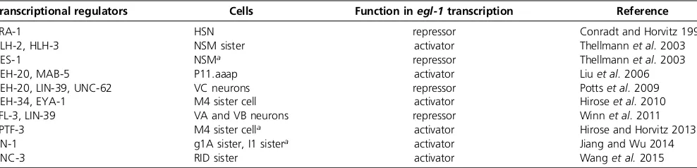
et al.2011; Hirose and Horvitz 2013; Jiang and Wu 2014; Wang et al.2015). Furthermore, mutations incis-regulatory elements of theegl-1locus not only cause changes inegl-1 expression but also in the pattern of programmed cell death (Conradt and Horvitz 1999; Hiroseet al.2010). Thesecis -regulatory elements are located either downstream or up-stream of theegl-1transcription unit and are conserved in related Caenorhabditis species (Figure 3A). A number of direct transcriptional regulators (trans-acting factors) of theegl-1gene that act through these elements have been identified (Table 1) (Conradt and Horvitz 1999; Thellmann
et al.2003; Liuet al.2006; Hatzold and Conradt 2008; Potts
et al. 2009; Hirose et al. 2010; Winn et al. 2011; Hirose and Horvitz 2013; Jiang and Wu 2014; Wang et al.2015). Their genetic analyses revealed that most control egl-1 transcription (and, hence, programmed cell death) only in one type of cell lineage, or a limited number of different types of cell lineages. Furthermore, most of these transcrip-tional regulators act through one specific cis-regulatory element of theegl-1locus (Figure 3A). Hence,egl-1 tran-scriptional control appears to be mediated by a composite of lineage-specific modules. In support of this notion, anal-yses of known regulators of egl-1 transcription suggest that many of them have additional, nonapoptotic functions, including nonapoptotic functions in the particular lineage or lineages in which they controlegl-1transcription. Neverthe-less, the functions of these transcriptional regulators in the regulation of apoptotic cell death appear to be conserved since most of them have human homologs that have been implicated in the regulation of apoptotic cell death and/or tumorigenesis (Potts and Cameron 2011).
RID lineage: The Collier/Olf1/EBF1 (COE) transcription factor UNC-3 plays such a dual role in the RID lineage (Wang et al. 2015). The RID progenitor gives rise to the RID, which differentiates into a neuron; and the RID sister cell, which dies (Sulstonet al.1983).unc-3expression is de-tected in all three cells, the RID progenitor, the RID neuron, and the RID sister cell. In the RID neuron, the loss ofunc-3 function results in a defect in certain aspects of neurite growth; in the RID sister cell, however, the loss of unc-3
prevents its programmed death (Wang et al. 2015). The
directly activating egl-1 transcription in the RID sister cell (Figure 3A) (Wanget al.2015). What remains to be deter-mined is through what mechanismUNC-3-dependent activa-tion of egl-1transcription is prevented in the RID neuron, which is programmed to survive.
The deaths of the hermaphrodite-specific neurons and cephalic companion neurons:Two types of programmed cell deaths occur in a sexually dimorphic manner duringC. elegans
development: the deaths of the two hermaphrodite-specific neurons (HSNs) in males and the deaths of the four CEMs in hermaphrodites (Sulston et al.1983). The Zn finger DNA-binding protein and transcriptional repressor transformer 1 (TRA-1) Gli, which was originally identified because of its role in sex determination (Hodgkin 1987; Zarkower and Hodgkin 1992), plays a critical role in the specification of
the HSN and CEM death. TRA-1functions as the terminal, global regulator of somatic sexual fate and, hence, specifies the development of sexually dimorphic features, including the sexually dimorphic presence of the HSNs and CEMs. In hermaphrodites, in whichTRA-1activity is high,TRA-1binds to another downstream cis-regulatory element of theegl-1 locus; thereby directly repressing egl-1 transcription in the HSNs to allow HSN survival in hermaphrodites (Figure 3A) (Conradt and Horvitz 1999). However, by directly repressing the transcription of the geneC. elegans homeobox 30(ceh-30), which encodes a BarH homeodomain transcription factor that may directly repress egl-1 transcription in the CEMs,
TRA-1indirectly activatesegl-1transcription in the CEMs to
cause CEM death in hermaphrodites (Peden et al. 2007;
Schwartz and Horvitz 2007). In males, in whichTRA-1 activ-ity is low,TRA-1is unable to repressegl-1transcription in the
HSNs andceh-30transcription in the CEMs; consequently, the HSNs die and the CEMs survive.
Role of asymmetric cell division
As mentioned above, most of the 131 cells that die during development are generated through a cell division that is asymmetric with respect to both cell fate and cell size (Sulston and Horvitz 1977; Sulstonet al.1983). Furthermore, in those lineages in whichegl-1transcription has been analyzed,egl-1 transcription can specifically be detected in the daughter that is programmed to die (Conradt and Horvitz 1999; Thellmann
et al.2003; Liuet al.2006; Hatzold and Conradt 2008; Potts
et al.2009; Hiroseet al.2010; Winnet al.2011; Hirose and Horvitz 2013; Jiang and Wu 2014; Wanget al.2015). How is this asymmetry inegl-1transcriptional activation and, hence, egl-1 activity achieved? It has been proposed that this is achieved through the asymmetric presence or activity in the two daughter cells of activators and/or repressors of the cell death fate and egl-1 transcription (Guenther and Garriga 1996; Franket al.2005; Hatzold and Conradt 2008; Chien
et al. 2013). Indeed, mutations that affect the abilities of mothers of cells programmed to die to divide asymmetrically can affect the cell death fate of their daughters (Guenther and Garriga 1996; Cordeset al.2006; Hatzold and Conradt 2008; Ouet al.2010; Singhviet al.2011; Chienet al.2013; Gurlinget al.2014; Teuliereet al.2014). Therefore, events that lead to the polarization of mothers of cells programmed to die and that are required for their abilities to divide asymmetrically are fundamentally important for cell death specification. These events most probably include cell non-autonomous signaling events. One signaling pathway that has recently been implicated in the regulation of cell death is theC. elegansLET-60Ras and MAP kinase 1 (MPK-1) ERK MAPK pathway. It was shown that the EGF-like ligand abnor-mal celllineage 3 (LIN-3) can promote a number of devel-opmental cell deaths, such as the deaths of the g1A sister cell and the I1 sister cell (Jiang and Wu 2014). In this context,
LIN-3 activates the LET-60 Ras and MPK-1 ERK MAPK
pathway, which results in the binding of the ETS-like tran-scription factorLIN-1to a downstreamcis-regulatory region of the egl-1 locus and LIN-1-dependent activation ofegl-1
transcription (Figure 3A) (Jiang and Wu 2014). Based on these results it has been proposed that LIN-3-dependent signaling contributes to the activation of the core apopto-tic machinery in a cell-nonautonomous manner by promot-ingegl-1transcriptional upregulation in cells programmed to die. An alternative explanation could be that LIN-3 -dependent signaling may promote the polarization of mothers of cells programmed to die (and their abilities to divide asymmetrically) and prime the egl-1 locus for transcrip-tional upregulation in the smaller daughter after cell division.
The Q lineage:A number of genes have been identified that are required for the asymmetric divisions of the left and right posterior daughter of the Q cell (Q.p) during thefirst larval stage (L1 stage) and the apoptotic deaths of their smaller daughters (Cordeset al.2006; Ouet al.2010; Singhviet al.
2011; Chienet al.2013; Gurlinget al.2014; Teuliereet al.
2014). [Q.pL and Q.pR divide asymmetrically and each gives rise to a larger daughter, Q.pa, which further divides to gen-erate two neurons (AVM/PVM and SDQL/SDQR, respec-tively), and a smaller daughter, Q.pp, which dies (Sulston and Horvitz 1977).] These genes define three pathways that contribute to the ability of Q.p to divide asymmetrically by size and/or fate: the Par-1-like gene 1 (pig-1) pathway (Cordeset al.2006; Chienet al.2013), theADP-ribosylation factor (arf) pathway (Singhvi et al. 2011; Teuliere et al.
2014), and atargetofERK kinaseMPK-1(toe-2)-dependent pathway (Gurling et al. 2014). The pig-1 pathway is com-prised of the genesabnormal embryonicpartitioning of cyto-plasma 4 (par-4) LKB1, pig-1 MELK, yeast STE20 related
adaptor protein homolog 1 (strd-1) STRAD, and themouse
embryo scaffolding protein homolog mop-25.2 MOP25.
Thearfpathway includes the genesarf-1.2Arf,arf-6Arf,
centaurin 2(cnt-2) Arf GTPase-activating protein (GAP),g en-eralreceptor forphosphoinositides 1(grp-1) Arfguanine nu-cleotideexchangefactor (GEF),exchangefactor forArf 6(efa-6) Arf GEF, BRag/Iqsec/Schizo related Arf GEF family member
1 (bris-1) Arf GEF,RAB family 5(rab-5) Rab5, anddynamin related 1(dyn-1) Dynamin. Many of these genes affect addi-tional asymmetric cell divisions that give rise to cells that are
Table 1 Direct regulators ofegl-1BH3-only transcription
Transcriptional regulators Cells Function inegl-1transcription Reference
TRA-1 HSN repressor Conradt and Horvitz 1999
HLH-2, HLH-3 NSM sister activator Thellmannet al.2003
CES-1 NSMa repressor Thellmannet al.2003
CEH-20, MAB-5 P11.aaap activator Liuet al.2006
CEH-20, LIN-39, UNC-62 VC neurons repressor Pottset al.2009
CEH-34, EYA-1 M4 sister cell activator Hiroseet al.2010
EFL-3, LIN-39 VA and VB neurons repressor Winnet al.2011
SPTF-3 M4 sister cella activator Hirose and Horvitz 2013
LIN-1 g1A sister, I1 sistera activator Jiang and Wu 2014
UNC-3 RID sister activator Wanget al.2015
Direct regulators ofegl-1transcription identified that either function as repressors or activators ofegl-1transcription in specific cells.
programmed to die, such as the division of Q.a, and divisions in the PLM/ALN and HSN/PHB lineages. Thepig-1pathway potentially also affects the asymmetric divisions of cells that give rise to some of thefirst 14 cell deaths that occur during development as well as the M4 mother cell that gives rise to the M4 sister cell, which is programmed to die (see below:
The M4 sister cell death) (Denning et al.2012; Hirose and Horvitz 2013). The identification of these pathways suggests that events at the plasma membrane as well as membrane trafficking events (potentially in an endocytic compartment) play an important role in the polarization of Q.p and its ability to divide asymmetrically. What remains to be determined is how these three pathways interact, what signal or signals they help to transduce, and how their functions relate to the programming of the cell death fate and the activation ofegl-1transcription in the smaller daughter cell, in particu-lar (Guenther and Garriga 1996; Frank et al.2005; Cordes
et al. 2006; Hatzold and Conradt 2008; Ou et al. 2010; Singhviet al.2011; Chien et al.2013; Gurlinget al.2014; Teuliereet al.2014).
The NSM sister cell death:In the NSM lineage, transcription factors have been identified that functionally connect the asymmetric division of a mother cell with asymmetricegl-1 transcription in the daughters (Figure 3B). The NSM neuro-blast divides asymmetrically to give rise to a larger daughter, the NSM, which differentiates into a serotonergic neuron; and a smaller daughter, the NSM sister cell, which dies (Sulstonet al.1983; Hatzold and Conradt 2008). It has been proposed thategl-1transcription can potentially be activated in the NSM and the NSM sister cell through a heterodimer of helix-loop-helix 2 (HLH-2) Daughterless andHLH-3Achaete scute (HLH-2/HLH-3), which is present in both daughter cells and which can bind to four E boxes/Snail binding sites located in a downstreamcis-regulatory element of theegl-1 locus (Figure 3A) (Thellmannet al.2003). However,HLH-2/
HLH-3-dependent activation ofegl-1transcription appears to be prevented in the larger daughter, the NSM, by the Snail-like Zn-finger transcription factorCES-1, which can only be detected in the NSM but not the NSM sister cell (Ellis and Horvitz 1991; Metzstein and Horvitz 1999; Thellmannet al.
2003; Hatzold and Conradt 2008).CES-1preventsegl-1 tran-scription by competing withHLH-2/HLH-3for binding to the same E boxes/Snail binding sites. The mechanism or
mecha-nisms through which the asymmetric presence of CES-1
protein in the daughter cells is achieved remains to be de-termined; however,ces-1function itself appears to contribute to the ability of the NSM neuroblast to divide asymmetrically. Mutations that result in the mis- or overexpression of theces-1 gene in the NSM neuroblast cause the NSM neuroblast to divide symmetrically giving rise to two daughter cells of sim-ilar sizes, both containing detectable levels ofCES-1(Hatzold and Conradt 2008). Mutations that causeces-1mis- or over-expression in the NSM neuroblast are lf mutations of the genesces-2andDNaJdomain 11(dnj-11), which encode a hepatic leukemia factor (HLF)-like bZIP transcription factor
and a Mida1/ZRF1-like chaperone, respectively; and a gain-of-function mutation of ces-1, which is located in a
cis-regulatory region of the ces-1locus (Ellis and Horvitz 1991; Metzsteinet al.1996; Metzstein and Horvitz 1999; Hatzold and Conradt 2008). Consequently,egl-1transcription is repressed in both daughters and both daughters survive. Mis- or overexpression of theces-1gene in the NSM neuroblast also compromises cell cycle progression in this cell, which suggests that the correct level ofCES-1in the NSM neuroblast is not only critical for the ability of this cell to divide asym-metrically but also for its ability to divide at the correct time (Yanet al.2013). It has been proposed thatCES-1exerts its effect on cell cycle progression in the NSM lineage by directly controlling the transcription of the genecell division cycle 25.2
(cdc25.2), which encodes a CDC25-like phosphatase that pro-motes cell cycle progression (Yan et al.2013). HowCES-1
affects the polarization of the NSM neuroblast is still unclear (Figure 3B). Interestingly, the cell death regulatory function of theCES-2HLF,CES-1Snail, andEGL-1BH3-only pathway appears to be conserved in mammals. HLF and the Snail-related Zn-finger transcription factor SLUG act in the hema-topoietic lineage in mammals to regulate the transcription of the BH3-only gene Puma (Inaba et al. 1996; Inukaiet al.
1999; Inoue et al. 2002; Wu et al.2005). Finally, as men-tioned in the Introduction, the pathways that mediate cell corpse engulfment also have a killing function (Hoeppner
et al.2001; Reddien et al.2001). This killing function was recently investigated in the NSM lineage. Based on this study it has been proposed that the engulfment pathways promote the death of the NSM sister cell by contributing to the polar-ization of the NSM neuroblast; the generation of a gradient of
“apoptotic potential”(including activeCED-3caspase and the potential to synthesize additional CED-3 protein) in the NSM neuroblast; and the asymmetric segregation of apo-ptotic potential into the smaller daughter, which is
pro-grammed to die (Figure 3C) (Chakraborty et al. 2015).
The study also revealed that the activation of the engulf-ment pathways in this context is induced through the core apoptotic machinery, which appears to already be active to a certain degree in the NSM neuroblast (Figure 3C). However, many questions remain, including the mechanism through which the core apoptotic machinery is tightly controlled in the NSM neuroblast and the nature of the signaling path-ways involved. Furthermore, it is currently unclear whether the engulfment pathways play a similar role in other cell lineages.
the M4 sister cell (Hiroseet al.2010). The death of the M4 sister cell and the upregulation ofegl-1transcription in the M4 sister cell are also at least partially dependent on the specificity protein 1 (SP1)-like transcription factorSPTF-3, which binds to another upstreamcis-regulatory element of theegl-1locus (Figure 3A) (Hirose and Horvitz 2013). In-terestingly, SPTF-3 is a direct transcriptional activator of both theegl-1gene and thepig-1gene, which encodes an AMPK-like protein kinase most similar to the mammalian
kinase MELK and which, as discussed above (see The Q
lineage), is a component of a genetic pathway that has been implicated in asymmetric cell division (by size and fate) in a number of cell lineages. Consistent with the notion that
SPTF-3 activates pig-1 transcription in the M4 lineage to
cause the death of the M4 sister cell; like the loss of sptf-3, the loss ofpig-1blocks the death of50% of the M4 sister cells (Hirose and Horvitz 2013). Based on thesefindings it was proposed that the death of the M4 sister cell is con-trolled by two parallel pathways that are both induced by sptf-3function: the core apoptotic cell death pathway that is activated by the sptf-3-, ceh-34-, and eya-1-dependent transcriptional upregulation of egl-1 (the sptf-3, ceh-34, eya-1,egl-1pathway) and a pathway that is independent of the core apoptotic pathway and that is activated by sptf-3-dependent transcriptional activation ofpig-1(thesptf-3, pig-1pathway). Finally, the death of the M4 sister cell (as well as a number of other programmed cell deaths, includ-ing the death of the NSM sister cells) is also at least partially dependent on the yeastgeneralcontrolnondepressible ho-mologgcn-1gene and theABC transporter,class F 1(abcf-1) gene, whose gene products physically interact and, based on sequence homologies, may function in the regulation of messenger RNA translation (Hirose and Horvitz 2014). This pathway (gcn-1,abcf-1pathway) has been proposed to act in parallel to the twosptf-3-dependent pathways to contribute to the death of the M4 sister cell as well (Hirose and Horvitz 2014). However, based on the known function ofpig-1 in asymmetric cell division and recentfindings in the NSM lineage (Cordes et al. 2006; Chien et al. 2013; Chakrabortyet al.2015), it is also possible that, rather than acting in parallel to thesptf-3,ceh-34,eya-1,egl-1pathway; thesptf-3,pig-1pathway and thegcn-1,abcf-1pathway may act at different time points in the M4 lineage to promote the asymmetric division of the M4 mother cell and the segre-gation of the apoptotic potential into the M4 sister cell (sptf-3, pig-1pathway) as well as the synthesis of CED-3
protein in the M4 sister cell after cell division (gcn-1,abcf-1 pathway).
Noncanonical apoptotic cell death
Most of the apoptotic cell deaths that occur duringC. elegans
development occur very rapidly: the cells are generated and within30 min they have been killed and turned into a cell corpse (Sulston and Horvitz 1977; Sulston et al. 1983). There is evidence in support of the notion that the transcrip-tional activation of egl-1 is not only necessary but also
sufficient for this type of apoptotic cell deaths (referred to as “canonical” apoptotic death) and that egl-1 transcrip-tional upregulation marks their onset (Conradt and Horvitz 1999; Thellmannet al.2003; Liuet al.2006; Hatzold and Conradt 2008; Pottset al. 2009; Hiroseet al.2010; Winn
et al. 2011; Hirose and Horvitz 2013; Jiang and Wu 2014; Wanget al.2015). However, there are at least two types of apoptotic cell deaths that occur duringC. elegans develop-ment for which the transcriptional upregulation ofegl-1does not seem to be sufficient: the death of the four CEMs and the death of the tail-spike cell. Furthermore, the death of the CEMs is still dependent onegl-1function (96% of the CEMs survive in hermaphrodites lackingegl-1function); however, the death of the tail-spike cell is only partially dependent on egl-1(30% of the tail-spike cells survive in animals lacking egl-1function) (Maureret al.2007; Nehmeet al.2010). In-terestingly, rather than dying within30 min, the CEMs and the tail-spike cell die 150 min or 300 min after being generated, respectively (Sulston and Horvitz 1977; Sulston
et al. 1983). It has been proposed that this delay of their deaths could be the reason whyegl-1transcriptional upre-gulation is not sufficient for them (Nehmeet al.2010). The answer might lie in the amount of theCED-3Caspase zymo-gen present in cells programmed to die. The ced-3 gene is strongly expressed in mothers of cells programmed to die and inactive CED-3 zymogens produced in the mothers are inherited to the daughters, where it presumably gets processed and activated in the daughter that is programmed to die once the onset of death has been triggered by egl-1 transcriptional upregulation (Maureret al.2007; Chakraborty
et al.2015). Interestingly, in both the CEMs and the tail-spike cell, transcriptional upregulation ofced-3is observed just prior to their deaths (Maurer et al. 2007; Nehme et al. 2010). Based on these observations it has been proposed that at the time the CEMs and the tail-spike cell die, the level of
theCED-3zymogen in these cells might have decreased
be-low a threshold (due to protein turnover) that is necessary to generate enough activeCED-3to trigger the execution phase of programmed cell death, and upregulation ofced-3 becomes necessary (Nehmeet al.2010). Based on antibody staining,CED-9Bcl-2 andCED-4Apaf-1 appear to be pre-sent in most if not all cells, at least during most of embry-onic development (Chenet al.2000). The transcriptional activation ofced-3would induce newCED-3zymogen syn-thesis, raising the level of theCED-3zymogens above the necessary threshold. Interestingly, in the CEMs, the tran-scriptional upregulation ofced-3occurs after the transcrip-tional upregulation of egl-1, indicating that the onset of CEM death is regulated byced-3transcriptional activation rather thanegl-1 transcriptional activation (Nehmeet al.
Cell Death Execution
Once the cell death program is activated, it initiates the highly regulated cell disassembly process, which includes nuclear DNA fragmentation, cytoplasm shrinkage, mitochondria elim-ination, and exposure of an“eat me”signal(s) such as phos-phatidylserine (PS) on the surface of the dying cell to induce phagocytosis by neighboring cells or macrophages (Steller 1995). The activated caspases play crucial roles in coordinat-ing the execution of different cell disassembly events by cleaving and activating proapoptotic protease targets and by cleaving and inactivating prosurvival protease substrates. The activated protease targets then initiate different cell kill-ing events that contribute to the demise of the cell.
CED-3-activated cell death execution events
Nuclear DNA fragmentation: Fragmentation of chromo-somal DNA is a hallmark of apoptosis and may facilitate apoptosis by terminating DNA replication and gene transcrip-tion, which maintain the survival and the functions of the cell (Arendset al.1990). DNA fragmentation duringC. elegans
apoptosis has been studied with the aid of various DNA-staining techniques, including DAPI, Feulgen (Sulston 1976), orTdT-mediated dUTPnickendlabeling (TUNEL) staining (Gavrieliet al.1992; Wuet al.2000; Parrishet al.2001).
So far, 11 nuclease-encoding genes have been identified to be involved in nuclear DNA degradation during apoptosis (Sulston 1976; Wu et al. 2000; Parrish et al. 2001; Wang
et al.2002; Parrish and Xue 2003; Nakagawaet al.2010). These include nuclease defective 1 (nuc-1),CED-3 protease suppressor 6 (cps-6), cell death-related nuclease 1–7 (crn-1 tocrn-7),cyclophilin13(cyn-13), andDicerrelated 1(dcr-1). Loss or reduction of activity in any of these genes exceptdcr-1 results in accumulation of TUNEL-positive cells inC. elegans
embryos, suggesting that most of these nucleases are in-volved in resolving TUNEL-reactive DNA breaks generated during apoptosis (Sulston 1976; Wu et al. 2000; Parrish
et al.2001; Wanget al.2002; Parrish and Xue 2003). Re-duced activity indcr-1on its own does not show any TUNEL phenotype, but can greatly reduce the number of TUNEL-positive cells in other cell death nuclease-deficient back-grounds (Nakagawaet al.2010), indicating thatdcr-1acts upstream of other cell death nucleases to produce TUNEL-reactive DNA breaks and likely makes thefirst cuts on nu-clear DNA during apoptosis. Moreover, loss or reduction of activity in most of these genes (with the exception ofnuc-1, crn-6, andcrn-7; which encode DNase II homologs) causes delayed appearance of embryonic cell corpses or reduced cell death during embryo development and can block cell death in sensitized genetic backgrounds; suggesting that nuclear DNA degradation is important for normal progres-sion of the apoptotic process and can promote cell killing (Parrishet al.2001; Wanget al.2002; Parrish and Xue 2003; Nakagawaet al.2010). Genetic, phenotypic, and biochemical analyses indicate that these genes act sequentially in three different stages to promote DNA degradation and apoptosis
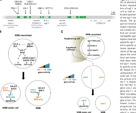
(Figure 2B). First, the activated CED-3protease cleaves the
DCR-1ribonuclease, a double-stranded RNA processing
endo-nuclease, in the middle of thefirst of the 2 RNase III domains; generating a C-terminal cleavage product (tDCR-1) with one and a half RNase III domains (Nakagawaet al.2010). tDCR-1 is capable of binding DNA and making 39hydroxyl DNA nicks (Nakagawaet al.2010; Geet al.2014), which are labeled in the TUNEL assay. Therefore,CED-3cleavage convertsDCR-1
from an RNase to a DNase, which initiates the nuclear DNA degradation process. In the second stage, the mitochondrial endonuclease encoded bycps-6interacting with multiple CRN nucleases, such asCRN-1, and nonnuclease factors, such as the
wormapoptosis-inducing factorhomologWAH-1, to form a multi-nuclease complex (degradeosome) to catalyze stepwise DNA fragmentation; starting from turning the DNA nicks gen-erated by tDCR-1 to single-stranded DNA gaps and double-stranded DNA breaks (Wang et al. 2002; Parrish and Xue 2003; Parrishet al.2003). As a result, inactivation of any of the components in the degradeosome results in accumulation of TUNEL-reactive DNA ends (Wanget al.2002; Parrish and Xue 2003). In the third stage, three DNase II homologs,NUC-1,
CRN-6, and CRN-7, mediate further degradation of
frag-mented nuclear DNA in dying cells, withNUC-1playing the major role in this process (Wuet al.2000; Laiet al.2009; Yu
et al.2015). However, the three DNase II-encoding genes do not appear to affect either the activation or progression of cell death, or the engulfment of cell corpses (Hedgecock et al.
1983; Wuet al.2000; Parrishet al.2001; Parrish and Xue 2003; Laiet al.2009). They are likely involved in the cleanup step of cell death execution. In addition to its cell death function,nuc-1is involved in degradation of DNA derived from ingested bacteria in the intestinal lumen (Sulston 1976; Wuet al.2000).
cps-6andwah-1encode mitochondrial proteins which are similar to human mitochondrial endonuclease G (EndoG) and apoptosis-inducing factor (AIF), respectively (Parrish
et al. 2001; Wang et al.2002). Both EndoG and AIF have been shown to mediate nuclear DNA fragmentation in mam-malian apoptosis (Susinet al.1999; Liet al.2001).WAH-1
can physically associate withCPS-6and enhance the endo-nuclease activity ofCPS-6(Wanget al.2002). Ectopicegl-1 expression induces the release ofWAH-1from mitochondria and its subsequent translocation to nuclei in a CED-3 -dependent manner (Wanget al.2002); suggesting that the role of mitochondria in regulating apoptosis is conserved, at least at the step of cell death execution.
for removal of apoptotic cells (Venegas and Zhou 2007; Wanget al. 2007; Zulliget al. 2007). Interestingly, both
WAH-1and its human homolog AIF, the proapoptotic
fac-tors critical for nuclear DNA degradation, are also involved in promoting PS externalization in apoptotic cells (Susin
et al.1999; Wanget al.2007).WAH-1accomplishes this by binding to theC. elegansphospholipidscramblaseSCRM-1
located on the plasma membrane and activating its bidi-rectional lipid scrambling activity, leading to the exposure of PS on the surface of the dying cell. Consistently, inactiva-tion ofscrm-1results in a mild engulfment defect and RNA interference (RNAi) knockdown ofwah-1enhances the en-gulfment defects of other enen-gulfment mutants (Wanget al.
2007; Hsu and Wu 2010). Sincewah-1acts downstream of ced-3 and the release ofWAH-1from mitochondria during apoptosis is a CED-3-dependent event (Wang et al.2002; Breckenridgeet al.2008), this represents the secondCED-3 -activated cell death execution event mediated byWAH-1
(Figure 2B).
Because loss ofscrm-1only causes a mild engulfment de-fect, other genes may contribute to PS exposure during apo-ptosis. Indeed, another multipass transmembrane protein
CED-8 plays a more important role in externalizing PS on
the surface of the apoptotic cell.ced-8was originally
identi-fied as a gene that affects the kinetics of apoptosis and shares sequence similarity to proteins from the XK transporter fam-ily (Stanfield and Horvitz 2000). Recent studies suggest that
bothCED-8and its human homolog Xkr8 are involved in PS
externalization in apoptotic cells (Y. Z. Chen et al. 2013; Suzukiet al.2013). Importantly,CED-8is cleaved byCED-3
and this cleavage removes a short N-terminal peptide to gen-erate a C-terminal cleavage product (acCED-8) that is both necessary and sufficient to mediate the proapoptotic and PS externalization activities ofCED-8in vivo(Y. Z. Chenet al.
2013). How acCED-8 promotes PS externalization and apo-ptosis is not understood. Inactivation ofced-8enhances the cell corpse engulfment defects in animals that are deficient in either of the two major phagocytosis pathways acting in par-allel inC. elegans(Reddien and Horvitz 2004; Y. Z. Chenet al.
2013; Suzukiet al.2013), indicating thatced-8acts through both pathways to promote phagocytosis.
Surface-exposed PS not only triggers engulfment of apo-ptotic cells by phagocytes, but can also lead to phagocytosis of living cells that ectopically expose PS. This occurs in animals lacking the aminophospholipid translocase transbilayer amphi-path transporter 1 (TAT-1) that maintains PS asymmetry in plasma membrane (Darland-Ransomet al.2008). These ob-servations are confirmed by other studies inC. elegans(Wang
et al.2010; Nawaet al.2012) and by a mammalian study in which inactivation of CDC50A, a cofactor for the human
TAT-1 homolog ATP11C, causes ectopic PS exposure in
living cells and their phagocytosis by macrophages (Segawa
et al.2014). The engulfment of living cells by phagocytes in thetat-1 mutants is blocked by lf mutations inPS receptor family 1(psr-1) andced-1, two phagocyte receptors that rec-ognize surface-exposed PS and act in two major phagocytosis
pathways (Zhou et al. 2001b; Wang et al. 2003; Darland-Ransomet al. 2008; Wanget al.2010; Liet al.2015; Yang
et al.2015), suggesting that externalized PS can serve as an eat me signal for both engulfment pathways.
Mitochondrial elimination: As described above, mitochon-dria play an important role in regulating cell death execution inC. elegans. During apoptosis, mitochondria also undergo dramatic morphological changes, including fragmentation, reorganization of cristae structures, and increased permeabil-ity of the outer mitochondrial membrane (Jagasiaet al.2005; Cereghetti and Scorrano 2006; Parone and Martinou 2006). There are also reports that mitochondria are reduced or lost during apoptosis, which would eliminate cellular energy pro-duction and contribute to the demise of the cell (Skulachev
et al. 2004; Arnoultet al.2005). A comprehensive genetic and cell biological analysis of components of theC. elegans
mitochondrial fission and fusion machinery, the dynamin GTPases DRP-1, FZO-1 (FZO mitochondrial fusion protein related), andeating defective 3 (EAT-3), indicates that de-fects in mitochondrialfission or fusion inC. elegansdo not affect apoptosis activation (Breckenridgeet al.2008). How-ever, loss of DRP-1 or FIS-2 (S. cerevisiae FIS1-related), a homolog of the human Fis1 fission protein, does cause a mild cell death defect that can be detected in sensitized genetic backgrounds, suggesting thatfis-2anddrp-1have minor proapoptotic roles. Genetic epistatic analysis sug-gests that fis-2 and drp-1 act independently of each
an-other and downstream of ced-3 to promote apoptosis.
Analysis by electron microscopy indicates that mitochon-dria normally reduced or eliminated in apoptotic cells per-sist in animals deficient infis-2or drp-1, indicating that
DRP-1 andFIS-2play a role in promoting mitochondrial
elimination during apoptosis (Breckenridge et al.2008). Active CED-3protease can cleaveDRP-1 in vitroand this cleavage is critical forDRP-1’s proapoptotic functionin vivo, but dispensable for its function in mitochondrial fission (Breckenridge et al. 2008). Furthermore, the C-terminal cleavage product ofDRP-1appears to be important for ac-tivating DRP-1’s proapoptotic function, together with the full-lengthDRP-1protein. Therefore,fis-2anddrp-1 rep-resent two novel cell death execution pathways acting
downstream ofced-3to promote mitochondrial
elimina-tion (Figure 2B).
Inactivation of survival signals:In living cells, multiple cell death inhibitors or survival factors work together to maintain the viability and functions of the cell. During apoptosis, these survival factors are inactivated to allow apoptosis to proceed (Danial and Korsmeyer 2004). In C. elegans, the key cell death inhibitor CED-9 is an excellent substrate of CED-3
Another well-known cell survival pathway is the phos-phoinositide 3-kinase (PI3K)/AKT signaling pathway that promotes cell growth, proliferation, and survival in diverse organisms (Luoet al.2003; Cullyet al.2006). How this cru-cial survival pathway is inactivated to promote apoptosis is not well understood. From aCED-3protease suppressor screen,
a CED-3substrate, CNT-1, was identified and found to act
downstream ofCED-3to promote apoptosis (Nakagawaet al.
2014). CNT-1 is cleaved during apoptosis to generate an N-terminal phosphoinositide (PI)-binding cleavage product, tCNT-1. tCNT-1 then translocates from the cytoplasm to the plasma membrane to block AKT binding to phosphatidylino-sitol (3,4,5)-trisphosphate (PIP3), thereby inhibiting AKT
ac-tivation and its prosurvival activity (Nakagawaet al.2014).
CNT-1defines a novel, caspase-activated negative regulator of the AKT survival pathway.
There are probably additionalCED-3substrates that are important for other aspects of cell death execution, such as cytoplasm shrinkage, nuclear membrane breakdown, and cell corpse engulfment. Molecular genetic characterization of additionalCED-3protease suppressors should lead to iden-tification of additionalCED-3substrates andCED-3-activated cell death execution events.
Clearance of Apoptotic Cells
When a cell undergoes apoptosis, eat me signals are rapidly exposed on the surface of the apoptotic cell (Fadok et al.
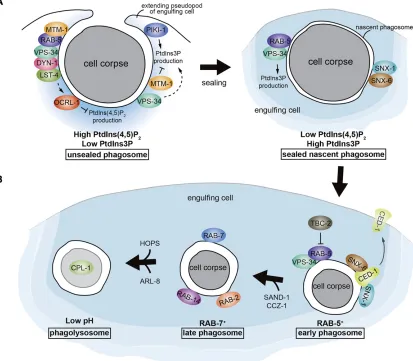
2001). These signals are recognized by receptors on the engulfing cells to trigger the phagocytosis of apoptotic cells (reviewed by Hochreiter-Hufford and Ravichandran 2013). The engulfment process includes membrane extension and cytoskeleton rearrangement of growing pseudopods around an apoptotic cell, and the enclosure of the pseudopods to form a phagosome.
Unlikeflies or humans,C. elegansdoes not have“ profes-sional”phagocytes, such as mobile macrophages; rather, ap-optotic cells are engulfed by their neighboring cells. Cell types such as hypodermal cells (which constitute the external epithelium), muscle cells, and intestinal cells have been shown to function as engulfing cells to remove somatic apo-ptotic cells (Robertson and Thomson 1982; Sulston et al.
1983; Zhouet al.2001b; Hsiehet al.2012). Germ cell corpses are specifically engulfed by gonadal sheath cells, which wrap around the germ line syncytium (Gumiennyet al.2001).
Presentation of eat me signals
Thus far, the best known eat me signal on apoptotic cells is PS, which is externalized from the cytosolic (inner) leaflet to the noncytosolic (outer) leaflet of the plasma membrane during apoptosis (Fadoket al.1992b; Fadeel and Xue 2009). A com-mon feature of all eukaryotic membranes is the asymmetric distribution of different phospholipids in the lipid bilayer. For example, aminophospholipids, phosphatidylethanolamine, and PS are restricted to the inner leaflet of the plasma mem-brane in living cells. Externalization of PS on the cell surface
is a hallmark of apoptosis; exposed PS is a conserved eat me signal that triggers phagocytosis in many organisms, includ-ing C. elegans (Fadeel and Xue 2009). Using a secreted PS-binding protein GFP fusion such as the Annexin V::GFP fusion (sAnxV::GFP), the MFG-E8::GFP, or the secreted GFP::lactadherin fusion (sGFP::LactC1C2) as a PS sensor;
ex-posed PS is detected on the surface of apoptotic cells in
C. elegans (Fadok et al. 2001; Venegas and Zhou 2007; Wang et al. 2007; Zullig et al. 2007; Mapes et al. 2012; Zhanget al.2012).
Two bidirectional phospholipid scramblases,SCRM-1and
SCRM-3, have been implicated in PS exposure on the surface
of apoptotic cells (Venegas and Zhou 2007; Wang et al.
2007). Loss of scrm-1orscrm-3 (also calledplsc-1), which encodes two of the eightC. elegansphospholipid scramblases, results in reduced PS exposure on the surface of apoptotic germ cells and a defect in the removal of apoptotic cells
(Venegas and Zhou 2007; Wang et al. 2007; Hsu and Wu
2010). Loss of scrm-1 or scrm-3 only partially reduces PS exposure in apoptotic cells, suggesting that additional factors or lipid transporters are involved in mediating PS exposure in apoptotic cells. As discussed above, the mitochondrial apoptogenic factor WAH-1also affects PS externalization during apoptosis (Wanget al.2007).WAH-1promotes PS externalization by binding to SCRM-1and activating the phospholipid scrambling activity of SCRM-1(Figure 2B).
TheCED-8protein, a homolog of the XK family transporters,
is critical for mediating PS externalization in somatic apopto-tic cells (Y. Z. Chenet al.2013; Suzukiet al.2013). Cleavage
ofCED-8byCED-3during apoptosis generates a C-terminal
cleavage product, acCED-8, that promotes PS externalization in apoptotic cells and is sufficient to induce ectopic PS exter-nalization in living cells (Y. Z. Chenet al.2013). The ABC transporterCED-7has been proposed to mediate PS exposure in somatic apoptotic cells using the MFG-E8::GFP PS sensor (Venegas and Zhou 2007). However, multiple studies using other PS sensors show thatCED-7does not promote PS ex-ternalization in apoptotic cells (Zulliget al.2007; Mapeset al.
2012; Zhang et al. 2012), and instead, plays a role in the efflux of PS from apoptotic cells (Mapeset al.2012; Zhang
et al.2012).
Surface PS expression on phagocytes
Interestingly, PS exposure was detected not only on the membranes of apoptotic cells but also on those of engulfing cells (Mapeset al.2012; Zhanget al.2012). Externalized PS appears early on the surface of the dying cells and decreases in older or unengulfed apoptotic cells. This decrease in sur-face PS exposure depends on a secreted extracellular protein
transthyretin-related family domain 52 (TTR-52) andCED-7, a homolog of the mammalian ABC1 transporters (Mapeset al.
2012; Zhang et al. 2012). TTR-52 is expressed in and se-creted from the endoderm (Wanget al.2010), whileCED-7
of engulfing cells (Zhou et al.2001b), are required for PS appearance on the surface of the phagocytes (Mapes et al.
2012; Zhanget al.2012). Immunoelectron microscopy anal-ysis of embryos expressing sAnxV::GFP reveals the presence of extracellular PS-containing vesicles between the dying cells and their neighboring cells in aced-7- andttr-52-dependent manner. It has been proposed thatCED-7andTTR-52 pro-mote the efflux of PS from apoptotic cells by generating ex-tracellular PS vesicles, which cause PS appearance on the surface of phagocytes through CED-1(Mapeset al. 2012). Moreover, sGFP::LactC1C2, which labels apoptotic cells but
not phagocytes, prevents sAnxV::GFP from labeling phago-cytes and compromises phagocytosis (Mapes et al. 2012). Therefore, PS expression on the phagocytes is also important for the engulfment of apoptotic cells.
Noseresistant to fluoxetine 5 (NRF-5), a secreted lipid transfer/LPS-binding family protein, is also important for PS appearance on the surface of phagocytes (Zhang et al.
2012). NRF-5 binds TTR-52 and PS, and displays a lipid transfer activity in vitro. NRF-5 may act with TTR-52 and
CED-7to mediate PS transfer from apoptotic cells to engulf-ing cells. How PS expression on phagocytes facilitates apo-ptotic cell clearance is not clear. One possibility is that appearance of PS on the surface of engulfing cells may alter the activity of membrane proteins that are important for the removal of cell corpses, and thus promote the engulfment process. Alternatively, PS may act as a homotypic ligand to tether the apoptotic cell to the engulfing cell through a bi-partite PS-binding bridging molecule and thus facilitates the engulfment process (Mapeset al.2012).
Engulfment receptors and signaling pathways
Forward and reverse genetics have identified20 genes re-quired for the engulfment of apoptotic cells. To assess whether these genes act in the same or separate pathways during the engulfment process, genetic analyses have been performed to position two genes at a time. Double mutants of genes acting in different pathways have a stronger engulf-ment defect (e.g., more persistent cell corpse numbers or longer duration of cell corpses) than those of single mutants alone or double mutants of genes acting in the same pathway (Elliset al.1991). On the basis of such analyses, three par-tially redundant pathways have been established that medi-ate the engulfment process.
The CED-1, CED-6, and CED-7 pathway: This pathway comprises the engulfment receptor CED-1, a homolog of
the mammalian MEGF10 protein (Hamonet al.2006). The
ced-1gene is expressed and functions in engulfing cells, but not in apoptotic cells, during cell corpse engulfment (Zhou
et al.2001b). The CED-1::GFP fusion was found to cluster around apoptotic cells, and this clustering completely de-pends onCED-7(Zhouet al. 2001b) and partially depends on the secreted extracellular protein TTR-52 (Wanget al.
2010). Therefore, the recognition and binding of apoptotic cells by CED-1 requires TTR-52 and CED-7. TTR-52 binds
both PS and the extracellular domain ofCED-1in vitroand likely functions as a bridging molecule that mediates recog-nition of apoptotic cells by cross-linking the exposed PS eat me signal with the engulfment receptorCED-1(Figure 2B). In addition, a recent study suggests that the extracellular re-gion ofCED-1could directly bind PSin vitro, when fused to GST (Liet al.2015).
Upon binding to an apoptotic cell throughTTR-52or di-rectly,CED-1transduces the engulfment signal via the
adap-tor protein CED-6 (homologous to the mammalian GULP
protein) and DYN-1, a member of the large GTPase family that regulate vesicle transport events (Clarket al.1997), to promote the internalization and subsequent degradation of apoptotic cells (Liu and Hengartner 1999; Yuet al.2006; Guo
et al.2010; Wanget al.2010).CED-6contains a phosphotyro-sine binding domain (Liu and Hengartner 1998) and may directly bind to the intracellular domain ofCED-1(Suet al.
2002). DYN-1clusters on pseudopods around an apoptotic cell in aced-1-,ced-6-, andced-7-dependent manner (Yuet al.
2006). Therefore,CED-6may linkCED-1signaling toDYN-1. Using the endosomal marker HGRS-1::GFP to track endo-somes during the engulfment process, Yuet al.(2006) found that endosomes, which displayed a punctate localization pat-tern in the cytoplasm, were gradually recruited to phagocytic cups and phagosomes around apoptotic cells. The incorpora-tion of endosome vesicles to phagocytic membranes requires
CED-1andDYN-1. The role ofdyn-1in internalization of cell corpses is controversial. A parallel study by Kinchen et al.
(2008) showed that clustering of DYN-1 around germ cell corpses was significantly reduced not only inced-1, ced-6, orced-7mutants, but also inced-5,ced-10, orced-12mutants (see below) defective in cell corpse engulfment; suggesting thatDYN-1is recruited at a stage following corpse recogni-tion and internalizarecogni-tion. Consistently, it has been shown that
DYN-1 acts at an early stage in phagosome maturation
(Kinchen et al. 2008; Almendinger et al. 2011; D. Chen
et al. 2013; Cheng et al.2015), particularly in phagosome sealing (see below) (Chenget al.2015). However, Yuet al.
(2006) observedDYN-1clustering around germ cell corpses inced-5,ced-10, orced-12mutants and their ultrastructural studies showed that some germ cell corpses ofdyn-1mutants were either not internalized or internalized but not de-graded, indicating thatdyn-1is important for both internal-ization and degradation of germ cell corpses.
DYN-1also promotes actin assembly at the phagocytic cup
during the engulfment of both embryonic and germ cell corp-ses, probably through different mechanisms (D. Chen et al.