GENETICS OF IMMUNITY
Comparative Genomics RNAi Screen Identi
fi
es
Eftud2 as a Novel Regulator of Innate Immunity
Lesly De Arras,*,†Rebecca Laws,‡Sonia M. Leach,†Kyle Pontis,*,†Jonathan H. Freedman,§ David A. Schwartz,†,** and Scott Alper*,† *Integrated Department of Immunology, and†Integrated Center for Genes, Environment and Health, National Jewish Health and University of Colorado, Denver, Colorado 80206,‡Department of Environmental Health, Boston University School of Public Health, Boston, Massachusetts 02118,§Laboratory of Toxicology and Pharmacology, National Institute of Environmental Health Sciences, National Institutes of Health, Durham, North Carolina 27709, and **Department of Medicine, University of Colorado, Aurora, Colorado 80045
ABSTRACTThe extent of the innate immune response is regulated by many positively and negatively acting signaling proteins. This allows for proper activation of innate immunity to fight infection while ensuring that the response is limited to prevent unwanted complications. Thus mutations in innate immune regulators can lead to immune dysfunction or to inflammatory diseases such as arthritis or atherosclerosis. To identify novel innate immune regulators that could affect infectious or inflammatory disease, we have taken a comparative genomics RNAi screening approach in which we inhibit orthologous genes in the nematode Caenorhabditis elegansand murine macrophages, expecting that genes with evolutionarily conserved function also will regulate innate immunity in humans. Here we report the results of an RNAi screen of approximately half of theC. elegansgenome, which led to the identification of many candidate genes that regulate innate immunity in C. elegans and mouse macrophages. One of these novel conserved regulators of innate immunity is the mRNA splicing regulator Eftud2, which we show controls the alternate splicing of the MyD88 innate immunity signaling adaptor to modulate the extent of the innate immune response.
P
ROPER regulation of the innate immune response is crit-ical to prevent disease. Activation of the innate immune response can involve the recruitment and activation of phagocytic cells that produce antimicrobial compounds as well as cytokines and chemokines that recruit and stimulate other immune cells (Kaufmann 2004). A strong innate immune response also is required for a robust adaptive immune response (Lee and Iwasaki 2007; Iwasaki and Medzhitov 2010). Thus, defects in innate immune signaling can lead to enhanced susceptibility to bacterial or fungal pathogens in human patients (Picard et al. 2011). On the other hand chronic innate immune activation can affect a myriad of diseases with an inflammatory component in-cluding atherosclerosis, asthma, Crohn’s disease, and cancer (Cook et al. 2004; Arcaroli et al. 2005; Karin and Greten2005; Mullick et al. 2005; Goh and Midwood 2012). It is therefore critical that innate immunity be tightly regulated for proper human health, active when needed and inactive when not. Thus, identifying novel regulators of innate im-munity could aid in the development of novel therapeutic and diagnostic options for a range of infectious and infl am-matory diseases (Cook et al. 2004; Connolly and O’Neill 2012).
Several organisms including the nematode Caenorhabdi-tis elegans have been used as models to investigate innate immunity and identify novel innate immunity regulators. While C. elegans lacks an adaptive immune response, it has a robust innate immune response, and has become an established model for innate immunity studies (Engelmann and Pujol 2010; Irazoqui and Ausubel 2010; Pukkila-Worley
and Ausubel 2012). In response to infection,C. elegans pro-duces numerous antimicrobial proteins including lysozymes, saposin-domain-containing proteins, defensin-like molecules, and many others (Ewbank 2006; Ewbank and Zugasti 2011). Production of these antimicrobials occurs in tissues exposed to pathogens and is regulated by conserved innate immune signaling pathways, including a p38 MAPK cascade, Copyright © 2014 by the Genetics Society of America
doi: 10.1534/genetics.113.160499
Manuscript received August 20, 2013; accepted for publication December 15, 2013; published Early Online December 20, 2013.
Supporting information is available online athttp://www.genetics.org/lookup/suppl/ doi:10.1534/genetics.113.160499/-/DC1.
1Corresponding author: National Jewish Health, 1400 Jackson St., Denver, CO 80206.
an insulin signaling pathway, a TGFb cascade, and others (Mochiiet al.1999; Malloet al.2002; Murphyet al.2003; Couillaultet al.2004; Huffmanet al.2004; O’Rourkeet al. 2006; Shapira et al.2006; Troemelet al.2006; Alperet al. 2007). Inhibition or mutation of genes in these conserved signaling pathways diminishes production of antimicrobial genes and enhances susceptibility to infection.
We previously used an RNAi screen to target all of the genes on C. elegans chromosome I to identify genes that regulate expression of a clec-85::gfp fusion in transgenic nematodes (Alper et al. 2008). clec-85 encodes a C-type lectin whose expression is induced by the bacterial pathogen Serratia marcescens(Malloet al.2002).clec-85expression is regulated by several known innate immunity signaling path-ways inC. elegans including the p38 MAPK, insulin signal-ing, and TGFbsignaling pathways in the presence of either pathogenic Pseudomonas aeruginosa or nonpathogenic Escherichia coli (Alper et al. 2007; Alper et al. 2008). We identified several genes on chromosome I that were required for robust clec-85 expression in the presence of the non-pathogenic bacteriumE. coli. Follow-up studies using RNAi in mouse macrophages, and knockouts in C. elegans and mice validated many of these genes as novel regulators of innate immunity (Alper et al. 2008; De Arras et al. 2012, 2013). This demonstrated the utility of this comparative genomics approach for innate immune regulator gene discovery.
Here we extend this approach toC. eleganschromosomes II–IV, representing 10,862 genes and find 32 additional genes that, when inhibited by RNAi, affect expression of clec-85::gfp. In mouse macrophages, we find that 8 of 20 mammalian orthologs of these genes affect lipopolysaccha-ride (LPS)-induced cytokine production in mammalian cells. Overexpression studies confirm that two of these genes, Eftud2 and Atp6v1c1, regulate the response to LPS in mu-rine macrophages. RNAi studies in C. elegans confirm that Eftud2 affects host defense in vivo. We further show that Eftud2, a component of the U5 small nuclear ribonucleopro-tein (snRNP), which works with the rest of the spliceosome to regulate mRNA splicing (Kramer 1996; Fabrizio et al. 1997; Bartels et al. 2002, 2003; Brenner and Guthrie 2006; Small et al. 2006; Sperling et al. 2008; Wahlet al. 2009), regulates the innate immune response in mouse mac-rophages, at least in part, by controlling the alternative mRNA splicing of MyD88, a critical signaling adaptor in multiple Toll-like receptor (TLR) signaling pathways (Kawai and Akira 2010; Takeuchi and Akira 2010).
Materials and Methods
C. elegans chromosome II–IV RNAi screen to identify regulators of clec-85::gfp production
RNAi was performed in liquid culture in 96-well format as described previously (Alper et al. 2007, 2008) using the Ahringer laboratory E. coli RNAi feeding library (Kamath
and Ahringer 2003; Kamathet al.2003). In brief, dsRNA-producing bacteria were inoculated from frozen stocks into LB, grown overnight at 37°, dsRNA synthesis was induced using IPTG, the bacteria were centrifuged and resuspended in nematode growth medium, nematode eggs isolated by bleaching (Wood 1988) were added to the wells, and the nematodes were allowed to grow at 25° in the presence of the dsRNA producing bacteria for 3 days. After this incubation, each nematode was assayed using the COPAS Biosort (Union Biometrica), which assays and reports time offlight (TOF) (nematode length, a mea-sure of nematode age) and greenfluorescence (due to the clec-85::gfp fusion) in each animal. Because nematodes can sometimes get “trapped” in the Biosort and leak be-tween wells, we performed our screen in 48 wells of each 96-well plate with a wash well between screening wells. The data were analyzed in R using slight modifications of our previously published scripts (Alper et al. 2007) (full scripts available upon request). These scriptsfirst remove obvious outliers and noise in the data that are not due to nematodes; then the scripts generate scatterplots and box-plots, which allow us to compare nematode length and fluorescence between RNAi treatments, and report mean fluorescence and the number of worms in each well. For the genomic RNAi screen of chromosomes II–IV, scatter-plots were manually screened for RNAi treatments that diminished nematode fluorescence. RNAi treatments that obviously inhibited nematode development were not con-sidered as potential positives; these were recognized as treatments that hindered bothfluorescence and nematode growth (as reflected in a decrease in time of flight). Be-cause we presumed that any individual RNAi treatment was unlikely to have an effect, each test RNAi on the plate was compared to the other 47 RNAi treatments on that plate to look for differences.
Of the 10,862 RNAi treatments tested in the initial genomic II–IV screen, 430 genes were retested in follow-up assays. These 430 genes were reassayed by RNAi a minimum of two additional times. For the retests, bacteria and nematodes were prepared as described above, although in this case negative control dsRNA-producing bacteria (Dillin et al. 2002) were included on the assay plates. Only those genes that, when inhibited, altered clec-85::gfp fluorescence in both retests were considered positive hits. Table 1 reports the 32 positive genes with mean fluorescence compared to control. Table 1 also displays the mean fluorescence divided by the mean time offlight, which should control for variations in nematode populations. Data for the 430 genes retested, including the 32 positive hits, is presented inSupporting In-formation,Table S1, with the data broken down by each in-dividual test well.
Use of RNAi in mouse macrophages to test candidate genes
Orthologs ofC. elegansgenes were identified using either the Homologene database at the National Center for Biotechnology Information or the Panther database from within WormBase (Miet al.2010; Sayerset al.2012; Yooket al.2012). RNAi was performed largely as described previously (Alperet al.2008). Briefly, either pools of siRNA duplexes or individual siRNA duplexes (Dharmacon) were transfected into the mouse mac-rophage cell line RAW264.7 using the Amaxa 96-well nucleo-fector shuttle. Cells were then plated at either 100,000 cells/ well in 96-well format (for ELISAs) or 200,000 cells/well in 24-well format (for qPCR). Negative control siRNAs included Dharmacon’s nontargeting siRNA pool no. 1 and nontargeting siRNA no. 1. Twenty-four hours after transfection, cells were incubated in the presence of 20 ng/ml LPS (List Biological Labs), and supernatants were collected for ELISA (R&D Sys-tems). In the primary screen, we monitored the production of
IL-6 as we have found this to be a more responsive readout than TNFaor other cytokines in response to most RNAi treat-ments (Alperet al.2008; De Arraset al.2013). The cells were then either subjected to viability analysis usingfluorescien diac-etate as described (Fernandez-Botran and Větvička 2001) or
lysed in RLT buffer (Qiagen) for RNA preparation for qPCR and RT–PCR. RNA was purified using the RNeasy kit (Qiagen), and qPCR was performed using the Quantitect SYBR Green RT– PCR kit (Qiagen) on an ABI 7900 thermocycler. Conditions for qPCR and RT-PCR to monitor MyD88Land MyD88Swere as described previously (De Arras and Alper 2013). Expression levels were normalized usingb-actin as a control; all primer sequences are listed inTable S2.
In one set of experiments, both Eftud2- and MyD88S-specific siRNA were delivered simultaneously. In the controls for this experiment when only a single siRNA was used, an equiva-lent volume of nontargeting control siRNA was included as well.
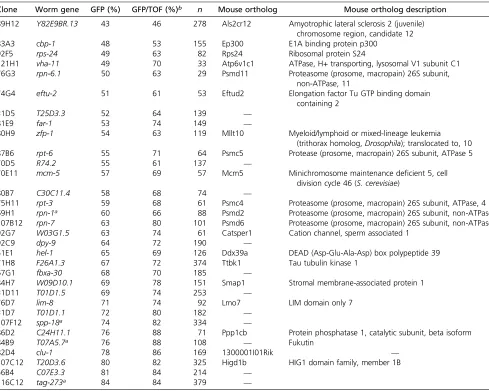
Table 1 Genes onC. eleganschromosomes II–IV that regulateclec-85::gfpexpression
Clone Worm gene GFP (%) GFP/TOF (%)b n Mouse ortholog Mouse ortholog description
89H12 Y82E9BR.13 43 46 278 Als2cr12 Amyotrophic lateral sclerosis 2 (juvenile)
chromosome region, candidate 12
83A3 cbp-1 48 53 155 Ep300 E1A binding protein p300
92F5 rps-24 49 63 82 Rps24 Ribosomal protein S24
121H1 vha-11 49 70 33 Atp6v1c1 ATPase, H+ transporting, lysosomal V1 subunit C1
76G3 rpn-6.1 50 63 29 Psmd11 Proteasome (prosome, macropain) 26S subunit,
non-ATPase, 11
74G4 eftu-2 51 61 53 Eftud2 Elongation factor Tu GTP binding domain
containing 2
31D5 T25D3.3 52 64 139 —
81E9 far-1 53 74 149 —
80H9 zfp-1 54 63 119 Mllt10 Myeloid/lymphoid or mixed-lineage leukemia
(trithorax homolog,Drosophila); translocated to, 10
87B6 rpt-6 55 71 64 Psmc5 Protease (prosome, macropain) 26S subunit, ATPase 5
70D5 R74.2 55 61 137 —
70E11 mcm-5 57 69 57 Mcm5 Minichromosome maintenance deficient 5, cell
division cycle 46 (S. cerevisiae)
80B7 C30C11.4 58 68 74 —
75H11 rpt-3 59 68 61 Psmc4 Proteasome (prosome, macropain) 26S subunit, ATPase, 4
69H1 rpn-1a 60 66 88 Psmd2 Proteasome (prosome, macropain) 26S subunit, non-ATPase, 2
107B12 rpn-7 63 80 101 Psmd6 Proteasome (prosome, macropain) 26S subunit, non-ATPase, 6
92G7 W03G1.5 63 74 61 Catsper1 Cation channel, sperm associated 1
92C9 dpy-9 64 72 190 —
51E1 hel-1 65 69 126 Ddx39a DEAD (Asp-Glu-Ala-Asp) box polypeptide 39
71H8 F26A1.3 67 72 374 Ttbk1 Tau tubulin kinase 1
67G1 fbxa-30 68 70 185 —
84H7 W09D10.1 69 78 151 Smap1 Stromal membrane-associated protein 1
31D11 T01D1.5 69 74 253 —
76D7 lim-8 71 74 92 Lmo7 LIM domain only 7
31D7 T01D1.1 72 80 182 —
107F12 spp-18a 74 82 334 —
86D2 C24H11.1 76 88 71 Ppp1cb Protein phosphatase 1, catalytic subunit, beta isoform
84B9 T07A5.7a 76 88 108 — Fukutin
82D4 clu-1 78 86 169 1300001I01Rik —
107C12 T20D3.6 80 82 325 Higd1b HIG1 domain family, member 1B
56B4 C07E3.3 81 84 214 —
116C12 tag-273a 84 84 379 —
Overexpression studies in mouse macrophages
Plasmids containing full-length cDNAs cloned downstream of the CMV promoter were obtained for Eftud2 (Origene); Rps24, Atp6v1c1, and Mcm5 (Open Biosystems); and negative control chloramphenicol acetyltransferase (CAT, Invitrogen). Transient transfections were performed using 3.75 ml Fugene HD (Roche) with 100,000 cells in 24-well format and contained 300 ng IL-6-luciferase, 100 ng SV40-rluc (Promega), and 600 ng of overexpression plasmid. Twenty-four hours after transfection, cells were stimulated with LPS for 6 hr and then luciferase activity was monitored using the Dual luciferase assay kit (Promega) using SV40-rluc as a normal-ization control for transfection efficiency. Stable Eftud2-overexpressing lines were generated by transfecting the CMV-Eftud2 plasmid (200 ng into 200,000 cells, 12-well format) and selecting with G418.
C. elegans survival assays
Survival assays in the presence of eitherP. aeruginosastrain PA14 (Rahmeet al. 1995; Mahajan-Mikloset al.1999; Tan
et al. 1999) or E. coli strainOP50 (Wood 1988) were per-formed largely as described previously (Alper et al. 2010) with the following modifications. The RNAi hypersensitive strain GR1373 eri-1(mg366) IV (Kennedy et al. 2004) was synchronized by collecting hatchlings at 15°(Wood 1988), the hatchlings were moved to E. coli-expressing dsRNA and incubated at 25°for 24 hr until they were in the late L4 stage, and then the nematodes were exposed to either PA14 or OP50at 25°in the presence of 5mg/ml of the sterilizing agent 5-fluoro-2-deoxyuridine (Mitchell et al. 1979) on modified NGM plates (Powell and Ausubel 2008). In the heat-killed bacteria experiment, a liquidOP50culture was concentrated fivefold, incubated at 80° for 3 hr, and plated on modified NGM supplemented with kanamycin. Inhibition of eftu-2or vha-11in theeri-1mutant background caused the nematodes to develop smaller and thinner than control-treated animals.
Statistical analyses
The analyses of the nematode RNAi data are described above.C. eleganssurvival assays were performed in triplicate and were analyzed using GraphPad Prism 5. All macrophage experiments were performed with a minimum of three in-dependent biological replicates, and in the case of the fol-low-up assays, were performed numerous additional times. Statistical analyses were performed using unpairedt-tests in GraphPad Prism 5; significance was consideredP,0.05.
Results
Genomic RNAi screen of C. elegans chromosomes II–IV
We used anE. colilibrary that expresses dsRNA correspond-ing to most genes in the C. elegans genome (Kamath and Ahringer 2003; Kamathet al.2003) to systematically inhibit each gene on chromosomes II–IV, and then monitored the effect on fluorescence in clec-85::gfp transgenic nematodes
using the COPAS Biosort (essentially a nematodeflow cytom-eter). As a control, we found that clec-85::gfp expression is strongly diminished when either nsy-1ortir-1are inhibited (Figure 1, top). nsy-1 is the MAPKKK that acts upstream of the p38 MAP kinase inC. elegans;tir-1is the soleC. elegans MyD88 family member. Bothnsy-1andtir-1are required for expression of numerous antimicrobial genes and resistance to Gram negative and positive bacteria and fungi (Kim et al. 2002; Couillaultet al.2004; Liberatiet al.2004; Alperet al. 2007; Bolzet al.2010; Muhammedet al.2012).
We screened through10,862 genes by RNAi and mon-itored fluorescence in these nematodes. Of these 10,862 genes tested, we identified 430 genes whose inhibition led to diminishedclec-85::gfpfluorescence without significantly diminishing overall nematode size. RNAi treatments that affect development or overallfitness are predicted to dimin-ish bothfluorescence as well as nematode size (a marker of growth and development) and were not considered further. We retested these 430 gene candidates, performing the assays a minimum of two more times. Of these 430 retested candidates, RNAi-mediated inhibition of 32 genes led to de-creasedclec-85::gfpfluorescence in both retests as well as in the original genomic screen [list of 32 hits in Table 1, box-plots for genes that also affect macrophage immunity (see below) in Figure 1 rows 2 and 3, compilation of all retest data inTable S1]. Thus, we identified 32 genes onC. elegans chromosomes II–IV that affected production of the candi-date antimicrobial gene clec-85 and that are therefore po-tentially novel innate immune regulators. Many of these candidates function in various catabolic pathways (Table S3, analysis performed using the Gather Utility) (Chang and Nevins 2006), suggesting that catabolism could modu-late innate immunity inC. elegans.
RNAi in mouse macrophages to monitor innate immune regulatory function of novel candidate genes
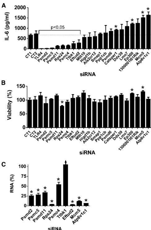
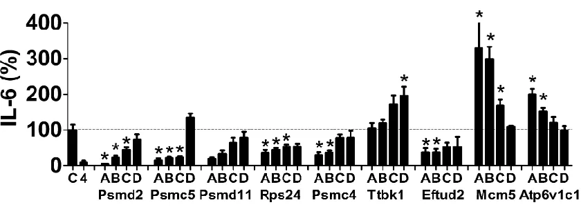
Twenty of these 32 novel candidates identified inC. elegans have an ortholog in mice (Table 1). To examine the poten-tial innate immune regulatory function of these mammalian genes, we inhibited these 20 genes using RNAi in the mouse macrophage cell line RAW264.7, stimulated the cells with LPS, and monitored production of the proinflammatory cy-tokine IL-6. As a positive control, we found that treatment of these cells with siRNA that targeted TLR4, the LPS receptor, strongly inhibited LPS-induced IL-6 production (Figure 2A). Inhibition of 7 of our 20 novel candidates using pools of siRNA duplexes targeting each gene led to a statistically significant decrease in IL-6 production (Figure 2A); in con-trast, inhibition of several other genes significantly increased IL-6 production (Figure 2A). As a control, we found that these siRNA treatments, with the exception of Rps24 siRNA, did not significantly diminish cell viability (Figure 2B).
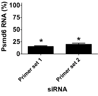
We took several steps to confirm these RNAi data and to confirm that the effects were due to specific inhibition of the corresponding endogenous gene and not an off-target siRNA effect. We focused on the seven genes whose inhibition led to decreased IL-6 production and the two genes whose in-hibition had the strongest effect on increased IL-6 production. First, we used qPCR to demonstrate that the siRNA treat-ments were inhibiting production of the corresponding endogenous gene; we found that gene-specific knockdown was statistically significant for eight of these nine siRNAs, but could not validate knockdown for one gene, Ttbk1 (Figure 2C). To further verify that the effects were due to inhibition of the corresponding endogenous gene, we also demonstrated that at least two individual siRNA duplexes targeting each gene induced a similar phenotype. We were able to validate the data for eight of the nine gene targets, with the exception of Ttbk1, where none of the individual siRNA duplexes di-minished LPS-induced IL-6 production (Figure S1). Multiple siRNA duplexes targeting the same gene induced the same phenotype, which suggested that the altered LPS-induced cy-tokine production was due to inhibition of that gene. Thus, we conclude that with the exception of Ttbk1, eight of these nine novel candidate genes do regulate the extent of LPS-induced IL-6 production. Four of the genes that affected the LPS response were components of the 26S proteasome (Psmd2, Psmc5, Psmd11, Psmc4); however, siRNA-mediated inhibition of another 26S proteasome subunit (Psmd6) did not affect the LPS response (Figure 2A), despite relatively strong Psmd6 knockdown as assayed with either of two sets of qPCR primers (Figure S2).
Overexpression studies in mouse macrophages to monitor innate immune regulatory function of novel candidate genes
Our RNAi studies identified eight potential candidate innate immunity regulators, including six genes whose inhibition diminished LPS-induced IL-6 production (four proteasome subunits Psmd2, Psmc5, Psmd11, and Psmc4; the ribosomal
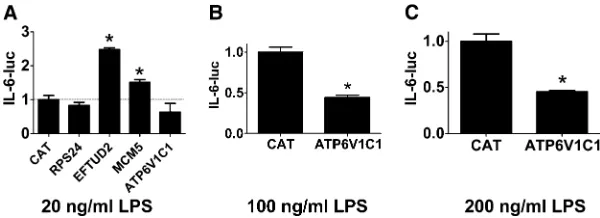
protein Rps24; and the mRNA splicing regulator Eftud2) and two genes whose inhibition enhanced LPS-induced IL-6 production (Mcm5 and Atp6v1c1). To further investigate the potential innate immune regulatory function of these genes, we chose to overexpress some of them and monitor the effect on IL-6 transcription using an IL-6-luciferase reporter construct (Baccam et al. 2003). If overexpression induced a phenotype opposite to that caused by RNAi, that would confirm the importance of that gene in innate immu-nity regulation. In contrast, lack of a phenotype is not nec-essarily informative.
transfected IL-6-luciferase, SV40-rluc for normalization, and plasmids that overexpress each of Rps24, Eftud2, Mcm5, and Atp6v1c1 using the strong CMV promoter into the mouse macrophage cell line RAW264.7 and monitored IL-6-luc production in the presence of 20 ng/ml LPS. As a neg-ative control, we also transfected a plasmid that overex-pressed CAT, which should not affect innate immunity. Rps24 overexpression did not have a significant effect on IL-6-luc production (Figure 3A), and Mcm5 overexpression induced a moderate increase in IL-6-luc production (Figure 3A), which is similar to the effect observed with RNAi, so we cannot draw any further conclusions about the role of Rps24 or Mcm5 in innate immunity based on these overexpression data. In contrast, overexpression of Eftud2 strongly enhanced IL-6-luc production (Figure 3A), a phenotype opposite to that of Eftud2 siRNA. Similarly, Atp6v1c1 overexpression de-creased IL-6-luc expression, a phenotype that also is opposite to the RNAi-induced phenotype, although this decrease was not statistically significant (Figure 3A). To explore the func-tion of Atp6v1c1 further, we overexpressed Atp6v1c1 in the presence of higher doses of LPS and found that Atp6v1c1 did significantly inhibit IL-6-luc expression under those conditions (Figure 3, B and C). Thus, the RNAi and overexpression data indicate that wild-type Eftud2 enhances LPS-induced IL-6 ex-pression while wild-type Atp6v1c1 diminishes LPS-induced IL-6 expression.
eftu-2/Eftud2 regulates host defense in C. elegans
To test the potential innate immune regulatory function of Eftud2 and Atp6v1c1in vivo, we inhibited these two genes by RNAi inC. elegans, exposed these nematodes to the nem-atode and human pathogen P. aeruginosastrain PA14, and then monitored survival of the animals. We inhibited either eftu-2 or vha-11 (the C. elegans orthologs of Eftud2 and Atp6v1c1, respectively) in nematodes harboring a mutation ineri-1, which enhances nematode RNAi sensitivity. Inhibi-tion of either gene diminished survival of nematodes ex-posed to P. aeruginosa compared to animals exposed to control RNAi bacteria (Figure 4A). However, inhibition of vha-11also diminished survival in the presence of the non-pathogenicE. colistrain (Figure 4B). Because liveE. colican be slightly pathogenic to C. elegans (Garsin et al. 2001; Tenor and Aballay 2007), we also monitored survival of vha-11(RNAi)animals in the presence of heat-killedE. coli. Under these conditions, inhibition ofvha-11still diminished
nematode survival (Figure 4C). Thus, we cannot differenti-ate the effects ofvha-11inC. elegansbetween an effect on fitness or an effect on both fitness and host defense. In contrast, nematodes exposed to eftu-2 RNAi lived as long as or slightly longer than control RNAi-treated nematodes in the presence of nonpathogenicE. coli(Figure 4B). Thus, eftu-2/Eftud2 is specifically affecting host defense and not fitness inC. elegans.
Eftud2 is a novel innate immunity regulator in mouse macrophages
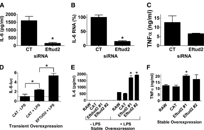
Based on the siRNA and overexpression data in macro-phages and thein vivosurvival data inC. elegans, we chose to investigate the function of Eftud2 further. First, we per-formed additional siRNA experiments in macrophages using a slightly higher dose of Eftud2 siRNA to see if this could enhance the phenotype; we found that this led to a greater inhibition in LPS-induced IL-6 protein production (Figure 5A). Inhibition of Eftud2 also diminished IL-6 mRNA pro-duction (Figure 5B). The effect of Eftud2 was not limited to IL-6 as production of the proinflammatory cytokine TNFa also was diminished when Eftud2 was inhibited by siRNA (Figure 5C). As further confirmation that transient Eftud2 overexpression enhanced IL-6-luciferase expression, we over-expressed Eftud2 in the presence of a second higher LPS dose of 50 ng/ml (Figure 5D) and found that Eftud2 enhanced IL-6 transcription at this higher LPS dose as well. To further test the function of Eftud2, we generated stable RAW264.7 lines overexpressing Eftud2, and as expected, found that stable Eftud2 overexpression enhanced LPS-induced production of IL-6 and TNFa(Figure 5, E and F). All these data indicate that wild-type Eftud2 enhances LPS-induced IL-6 production by enhancing IL-6 expression.
Eftud2 regulates alternative splicing of MyD88 to control the innate immune response in macrophages
Eftud2 is a component of the U5 snRNP, which functions with the rest of the spliceosome to control mRNA splicing (Kramer 1996; Fabrizio et al. 1997; Bartels et al. 2002, 2003; Brenner and Guthrie 2006; Smallet al.2006; Sperling et al. 2008; Wahl et al. 2009). We previously found that SF3a and SF3b, which function with the U2 snRNP to con-trol mRNA splicing (Krameret al.2005; Sperlinget al.2008; Collins et al. 2009; Rino and Carmo-Fonseca 2009; Wahl
response (De Arras and Alper 2013). SF3a and SF3b affect innate immunity in mouse macrophages, at least in part, by regulating alternative splicing of MyD88 (De Arras and Alper 2013). MyD88 is a signaling adaptor that functions down-stream of most TLRs (Kawai and Akira 2010; Takeuchi and Akira 2010). Full-length MyD88 is encoded by a five-exon mRNA (long form or MyD88L). However, a shorter MyD88 mRNA (MyD88S) that lacks exon 2 encodes a negative reg-ulator of TLR signaling (Janssens et al. 2002; Burns et al. 2003; Janssenset al.2003; Adib-Conquyet al.2006; Mendoza
-Barberaet al.2009). Inhibition of SF3a or SF3b using siRNA or a pharmacological agent diminishes LPS-induced IL-6 and TNFa production, at least in part, by enhancing production of the negatively acting MyD88S splice form (De Arras and Alper 2013). We therefore chose to monitor if the Eftud2 mRNA splicing regulator likewise affects alternative splicing of MyD88.
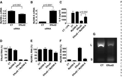
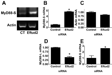
To monitor MyD88Land MyD88Slevels when Eftud2 was inhibited, we used a qPCR assay and primer sets that can distinguish between the two mRNA splice forms (De Arras and Alper 2013). We inhibited Eftud2 using siRNA, stimu-lated the cells with 20 ng/ml LPS for 6 hr, and then moni-tored the two MyD88 mRNA splice forms by qPCR. Eftud2 inhibition led to a moderate decrease in MyD88Llevels and a substantial increase in MyD88Slevels (Figure 6, A and B). In contrast, Eftud2 inhibition did not affect all mRNA splic-ing events as intron 3 of actin was excised normally (data not shown).
We also used semiquantitative RT–PCR and subsequent agarose gel electrophoresis to monitor the two MyD88 mRNA splice forms. RT–PCR using primers specific to the MyD88SmRNA splice form demonstrated that inhibition of Eftud2 by RNAi led to increased MyD88Sproduction (Figure S3, A and B), confirming the qPCR data. In contrast, pro-duction ofb-actin was not significantly affected (Figure S3, A and C). Using primers that bracketed exon 2, we also were able to amplify products corresponding to both MyD88Land MyD88S simultaneously. As observed previously (De Arras and Alper 2013), there is substantially more MyD88L than MyD88S in unstimulated cells (Figure 6G). Inhibition of Eftud2 by RNAi led to a significant decrease in MyD88L and a concomitant increase in the smaller MyD88S form (Figure 6G andFigure S3, D and E).
The increased amount of the MyD88S inhibitory splice form could explain in whole or in part the weakened LPS response when Eftud2 is inhibited. To directly test if Eftud2’s effect on innate immunity is mediated by changes
in MyD88Slevels, we chose to inhibit the MyD88S mRNA splice form when Eftud2 was inhibited in an attempt to re-store LPS-induced IL-6 production. To decrease MyD88S mRNA levels, we used a siRNA that targets the unique exon 1–exon 3 splice junction that is present in MyD88Sbut ab-sent in MyD88L (De Arras and Alper 2013). Treatment of cells with this MyD88SsiRNA leads to a moderate increase in LPS-induced IL-6 production (Figure 6C; in other experi-ments not shown, this increase was more significant). We found that treatment of cells with Eftud2 siRNA alone strongly diminished LPS-induced IL-6 production while treatment of cells with both Eftud2 siRNA and MyD88S siRNA simultaneously strongly rescued LPS-induced IL-6 production Figure 6C). As a control, we verified that the siRNAs were inducing similar knockdown under the various conditions (Figure 6, D–F). This suggests that much of the effect of Eftud2 on the LPS response is mediated by altered MyD88 mRNA splicing.
Discussion
Identification of novel conserved regulators of innate immunity using comparative genomics
Our hypothesis is that genes with evolutionarily conserved innate immune regulatory function inC. elegansand murine macrophages will likely also affect humans and are there-fore candidate regulators of human infectious and infl am-matory disease. Our comparative genomics RNAi screening approach in C. elegansand mouse macrophages has identi-fied many such candidate innate immune regulators (this study and Alper et al. 2008; De Arras et al. 2013). One advantage of this approach is that it may overcome the high false-positive rate present in mammalian RNAi screens due to off-target effects (Editorial 2003). Thus far, we have val-idated many of these dual RNAi hits (nematodes and macro-phages) using knockout mutations in C. elegans and have validated two dual RNAi hits using mouse knockouts (Alper et al.2008; De Arraset al.2012, 2013).
levels in the Gene Expression Omnibus database, data not shown). One unanswered question is: Are these genes reg-ulating innate immunity in a similar fashion in both species? This is particularly relevant as many of the candidates iden-tified in the current study are components of the 26S pro-teasome, which regulates innate immunity inDrosophilaand mammals by degrading IkB, thereby allowing NFkB activa-tion (Couxet al.1996; Foley and O’Farrell 2004). However, whileC. eleganshas an IkB homolog, as yet no direct NFkB homolog has been identified (Pujolet al.2001). It could be that another gene serves a similar function inC. elegans, or it could be that the 26S proteasome regulates innate immunity in C. elegans by a different mechanism. Nevertheless, this comparative genomics approach seems to be an efficient way tofind novel genes that matter.
There is precedent in C. elegans for innate immunity genes inC. elegansacting in both conserved and novel fash-ion. The p38 MAPK pathway acts similarly in bothC. elegans and mammals (Kimet al.2002). In contrast, while the Xbp1 transcription factor enhances host defense in both mammals andC. elegans, it does so by different mechanisms: by acting as a TLR-signaling dependent transcriptional activator that functions with NFkB in mice (Martinonet al.2010) and by playing a detoxification role in the immune response in C. elegans(Richardsonet al.2010). Thus, while we see a high degree of“conservation of function”of innate immunity reg-ulators, it is as yet unclear if there is“conservation of mech-anism,”although given the reported differences inC. elegans innate immunity, we speculate that many of these genes may use different mechanisms in the different species to effect host defense.
Many of our candidate genes encoded components of the 26S proteasome, which is already the target of potential therapeutic pharmacological options (Elliott et al. 2003; Wang and Maldonado 2006; Calzadoet al.2007). We note that while siRNA-mediated inhibition of several 26S protea-some subunits strongly diminished LPS-induced cytokine
production, the inhibition of Psmd6 did not. This raises the possibility of either differential function or differential sensitivity of different 26S proteasome subunits to inhibi-tion, suggesting that targeting individual subunits of the 26S proteasome using siRNA could be another approach to treat inflammatory diseases.
The role of Atp6v1c1 in innate immunity regulation
Our RNAi and overexpression data indicate that Atp6v1c1 limits the innate immune response by diminishing IL-6 transcription. Atp6v1c1 is the VMA5 subunit of the vacuolar H+-ATPase (V-ATPase) (Smithet al.2003). The V-ATPase is a large multiprotein complex that mediates acidification of intracellular organelles and can therefore affect numerous biological processes (Stevens and Forgac 1997; Forgac 1999; Nishi and Forgac 2002). Atp6v1c1 helps mediate the assembly of the catalytic and membrane portions of the V-ATPase (Hoet al.1993; Droryet al.2004). Inhibition of the V-ATPase in macrophages using the pharmacological agent bafilomycin A1 enhances LPS-induced NFkB activa-tion and cytokine producactiva-tion (Bidani and Heming 1995; Conboy et al. 1999), which is consistent with our RNAi and overexpression data. This suggests that activating indi-vidual V-ATPase subunits could also be a potential therapeu-tic option for inflammatory diseases.
The Eftud2 mRNA splicing regulator controls innate immunity by altering MyD88 mRNA splicing
P granule development (Meissner et al.2009; Updike and Strome 2009). In humans, mutations in Eftud2 cause cra-niofacial conditions including mandibulofacial dysostosis and esophageal atresia (Gordon et al. 2012; Lines et al. 2012; Luquettiet al.2013; Voigtet al.2013).
In the present study, we found that inhibition of Eftud2 diminished LPS-induced IL-6 and TNFa production while overexpression of Eftud2 enhanced cytokine production. At least part of this effect occurs at the IL-6 transcriptional level. Importantly, inhibition of Eftud2 did not affect overall cell viability, suggesting that at this level of gene inhibition, the innate immune response is far more sensitive to Eftud2 levels than are other general cell functions.
These data are reminiscent of the effects of the SF3a and SF3b complexes that interact with the U2 snRNP to facilitate mRNA splicing (Kramer et al. 2005; Sperling et al. 2008; Collins et al. 2009; Rino and Carmo-Fonseca 2009; Wahl
et al. 2009). We previously reported that inhibition of SF3a or SF3b by RNAi or a pharmacological agent diminished LPS-induced cytokine production in mouse macrophages (De Arras and Alper 2013). SF3a and SF3b regulate innate immunity in part by modulating alternative splicing of MyD88 (De Arras and Alper 2013). Thefive exon full-length long form MyD88Lencodes a critical signaling adaptor in multiple TLR response pathways. In contrast, the shorter splice form MyD88Sthat lacks the 135-bp exon 2 encodes an in-frame protein that is a negative regulator of TLR signaling, which prevents activation of the downstream signaling IRAK proteins (Janssens et al. 2002; Burns et al. 2003; Mendoza-Barbera
et al. 2009). Because of the similarity in the effects of
SF3a/b and Eftud2 inhibition, we monitored MyD88 mRNA splicing when Eftud2 was inhibited and found that the in-hibitory splice form MyD88S was increased dramatically, which is consistent with the resulting diminished cytokine response. Moreover, we were able to partially rescue the defect caused by Eftud2 inhibition by normalizing MyD88S levels with a second MyD88S-specific siRNA. Thus, SF3a, SF3b, and Eftud2 all are required for retention of exon 2 in MyD88, as this exon is lost and MyD88Sis produced when any of these spliceosome components are inhibited. This re-sult is consistent with other reports that show that inhibition of specific spliceosome subunits leads to alternative splicing and exon skipping (Corrioneroet al.2011; Fanet al.2011; An and Henion 2012; Visconte et al.2012). Moreover, the cur-rent data show that regulators of the U2 snRNP (SF3a and SF3b), which affect 39splice site choice, and the U5 snRNP (Eftud2), which joins the spliceosome at a later stage of as-sembly (Newman 1997; Turneret al.2004; Wahlet al.2009), both have similar effects on MyD88 mRNA splicing.
The C. elegans and mouse orthologs of Eftud2, SF3a1, and SF3b1 exhibit 70, 45, and 67% identity, respectively. While Eftud2 regulates innate immunity in both species, it is unclear how much mechanistic conservation there is. While the sole C. elegansMyD88 family member tir-1 also exhibits alternate splicing, no alternate functions have been reported for these splice forms in innate immunity, and the relevant intron–exon junctions in MyD88 do not appear to be conserved intir-1(data not shown).
MyD88Slevels are increased10-fold in monocytes from septic patients (Adib-Conquy et al. 2006), suggesting that
Figure 6Eftud2 regulates innate immunity by controlling the alter-native mRNA splicing of MyD88. (A and B) Cells were treated with either Eftud2 siRNA or control (CT) nontargeting siRNA, were stimulated with 20 ng/ml LPS for 6 hr, and MyD88L and MyD88S
mRNA levels were monitored by qPCR. (C–F) Cells were treated with the indicated siRNAs, were stimulated with 20 ng/ml LPS for 6 hr, and either IL-6 was moni-tored by ELISA (C) or Eftud2, MyD88L, and MyD88S mRNA
levels were monitored by qPCR (D–F). mRNA levels were normal-ized such that 100% indicates mRNA in the presence of control RNAi in the absence of LPS. (G) Cells were exposed to either Eftud2 siRNA or control nontar-geting siRNA (CT), were then treated with 20 ng/ml LPS for 6 hr, RNA was extracted, RT–PCR was performed to amplify the in-dicated mRNAs, and the amplified products were analyzed by agarose gel electrophoresis. Representative image is shown of PCR products using primers that bracket MyD88 exon 2 and that therefore simultaneously generate a 280-bp product corresponding to MyD88Land a 145-bp product
a better understanding of the regulation of MyD88 mRNA splicing could contribute to our understanding and ability to treat inflammatory disease. The identification of Eftud2 as a novel regulator of MyD88 mRNA splicing and innate im-munity should further this approach.
Acknowledgments
We thank Gail Bishop for the IL-6-luciferase reporter construct. This work was supported by R21ES019256 from the National Institute of Environmental Health Sciences, the Intramural Research Programs of the National Heart Lung and Blood Institute, the National Institute of Environmental Health Sciences (Z01ES102045), and a Butcher Seed grant.
Literature Cited
Adib-Conquy, M., C. Adrie, C. Fitting, O. Gattolliat, R. Beyaertet al., 2006 Up-regulation of MyD88s and SIGIRR, molecules inhib-iting Toll-like receptor signaling, in monocytes from septic pa-tients. Crit. Care Med. 34: 2377–2385.
Alper, S., S. J. McBride, B. Lackford, J. H. Freedman, and D. A. Schwartz, 2007 Specificity and complexity of theC. elegans innate immune response. Mol. Cell. Biol. 27: 5544–5553. Alper, S., R. Laws, B. Lackford, W. A. Boyd, P. Dunlap et al.,
2008 Identification of innate immunity genes and pathways using a comparative genomics approach. Proc. Natl. Acad. Sci. USA 105: 7016–7021.
Alper, S., M. McElwee, J. Apfeld, B. Lackford, J. Freedmanet al., 2010 Germline proliferation regulates distinct signaling path-ways in C. elegansto control lifespan and innate immunity. J. Biol. Chem. 285: 1822–1828.
An, M., and P. D. Henion, 2012 The zebrafish sf3b1b460 mutant reveals differential requirements for the sf3b1 pre-mRNA pro-cessing gene during neural crest development. Int. J. Dev. Biol. 56: 223–237.
Arcaroli, J., M. B. Fessler, and E. Abraham, 2005 Genetic poly-morphisms and sepsis. Shock 24: 300–312.
Baccam, M., S. Y. Woo, C. Vinson, and G. A. Bishop, 2003 CD40-mediated transcriptional regulation of the IL-6 gene in B lympho-cytes: involvement of NF-kappa B, AP-1, and C/EBP. J. Immunol. 170: 3099–3108.
Bartels, C., C. Klatt, R. Luhrmann, and P. Fabrizio, 2002 The ribosomal translocase homologue Snu114p is involved in un-winding U4/U6 RNA during activation of the spliceosome. EMBO Rep. 3: 875–880.
Bartels, C., H. Urlaub, R. Luhrmann, and P. Fabrizio, 2003 Mutagenesis suggests several roles of Snu114p in pre-mRNA splicing. J. Biol. Chem. 278: 28324–28334.
Bidani, A., and T. A. Heming, 1995 Effects of bafilomycin A1 on functional capabilities of LPS-activated alveolar macrophages. J. Leukoc. Biol. 57: 275–281.
Bolz, D. D., J. L. Tenor, and A. Aballay, 2010 A conserved PMK-1/ p38 MAPK is required in C. elegans tissue-specific immune re-sponse to Y. pestis infection. J. Biol. Chem. 285: 10832–10840. Brenner, T. J., and C. Guthrie, 2006 Assembly of Snu114 into U5 snRNP requires Prp8 and a functional GTPase domain. RNA 12: 862–871.
Burns, K., S. Janssens, B. Brissoni, N. Olivos, R. Beyaert et al., 2003 Inhibition of interleukin 1 receptor/Toll-like receptor signaling through the alternatively spliced, short form of MyD88 is due to its failure to recruit IRAK-4. J. Exp. Med. 197: 263–268.
Calzado, M. A., S. Bacher, and M. L. Schmitz, 2007 NF-kappaB inhibitors for the treatment of inflammatory diseases and can-cer. Curr. Med. Chem. 14: 367–376.
Chang, J. T., and J. R. Nevins, 2006 GATHER: a systems approach to interpreting genomic signatures. Bioinformatics 22: 2926– 2933.
Collins, L. J., C. G. Kurland, P. Biggs, and D. Penny, 2009 The modern RNP world of eukaryotes. J. Hered. 100: 597–604. Conboy, I. M., D. Manoli, V. Mhaiskar, and P. P. Jones,
1999 Calcineurin and vacuolar-type H+-ATPase modulate macrophage effector functions. Proc. Natl. Acad. Sci. USA 96: 6324–6329.
Connolly, D. J., and L. A. O’Neill, 2012 New developments in Toll-like receptor targeted therapeutics. Curr. Opin. Pharmacol. 12: 510–518.
Cook, D. N., D. S. Pisetsky, and D. A. Schwartz, 2004 Toll-like receptors in the pathogenesis of human disease. Nat. Immunol. 5: 975–979.
Corrionero, A., B. Minana, and J. Valcarcel, 2011 Reduced fi -delity of branch point recognition and alternative splicing in-duced by the anti-tumor drug spliceostatin A. Genes Dev. 25: 445–459.
Couillault, C., N. Pujol, J. Reboul, L. Sabatier, J. F. Guichouet al., 2004 TLR-independent control of innate immunity in Caeno-rhabditis elegans by the TIR domain adaptor protein TIR-1, an ortholog of human SARM. Nat. Immunol. 5: 488–494. Coux, O., K. Tanaka, and A. L. Goldberg, 1996 Structure and
functions of the 20S and 26S proteasomes. Annu. Rev. Biochem. 65: 801–847.
De Arras, L., and S. Alper, 2013 The Sf3a mRNA splicing complex mediates a MyD88-dependent negative feedback loop that lim-its the innate immune response. PLoS Genet. 9: e1003855. De Arras, L., I. V. Yang, B. Lackford, D. W. Riches, R. Prekeriset al.,
2012 Spatiotemporal Inhibition of Innate Immunity Signaling by the Tbc1d23 RAB-GAP. J. Immunol. 188: 2905–2913. De Arras, L., A. Seng, B. Lackford, M. Keikhaee, B. Bowermanet al.,
2013 An evolutionarily conserved innate immunity protein in-teraction network. J. Biol. Chem. 288: 1967–1978.
Dillin, A., D. K. Crawford, and C. Kenyon, 2002 Timing requirements for insulin/IGF-1 signaling in C. elegans. Science 298: 830–834. Drory, O., F. Frolow, and N. Nelson, 2004 Crystal structure of
yeast V-ATPase subunit C reveals its stator function. EMBO Rep. 5: 1148–1152.
Elliott, P. J., T. M. Zollner, and W. H. Boehncke, 2003 Proteasome inhibition: a new anti-inflammatory strategy. J. Mol. Med. 81: 235–245.
Engelmann, I., and N. Pujol, 2010 Innate immunity in C. elegans. Adv. Exp. Med. Biol. 708: 105–121.
Ewbank, J. J., 2006 Signaling in the immune response (January 23, 2006),WormBook, ed. TheC. elegansResearch Community, WormBook, doi/10.1895/wormbook.1.83.1, http://www. wormbook.org.
Ewbank, J. J., and O. Zugasti, 2011 C. elegans: model host and tool for antimicrobial drug discovery. Dis. Model. Mech. 4: 300–304. Fabrizio, P., B. Laggerbauer, J. Lauber, W. S. Lane, and R. Luhrmann, 1997 An evolutionarily conserved U5 snRNP-specific protein is a GTP-binding factor closely related to the ribosomal translocase EF-2. EMBO J. 16: 4092–4106.
Fan, L., C. Lagisetti, C. C. Edwards, T. R. Webb, and P. M. Potter, 2011 Sudemycins, novel small molecule analogues of FR901464, induce alternative gene splicing. ACS Chem. Biol. 6: 582–589.
Fernandez-Botran, R., and V. Větvička, 2001 Methods in Cellular Immunology, CRC Press, Boca Raton.
Forgac, M., 1999 Structure and properties of the vacuolar (H+)-ATPases. J. Biol. Chem. 274: 12951–12954.
Garsin, D. A., C. D. Sifri, E. Mylonakis, X. Qin, K. V. Singhet al., 2001 A simple model host for identifying Gram-positive viru-lence factors. Proc. Natl. Acad. Sci. USA 98: 10892–10897. Goh, F. G., and K. S. Midwood, 2012 Intrinsic danger: activation
of Toll-like receptors in rheumatoid arthritis. Rheumatology (Oxford) 51: 7–23.
Gordon, C. T., F. Petit, M. Oufadem, C. Decaestecker, A. S. Jourdain et al., 2012 EFTUD2 haploinsufficiency leads to syndromic oe-sophageal atresia. J. Med. Genet. 49: 737–746.
Ho, M. N., K. J. Hill, M. A. Lindorfer, and T. H. Stevens, 1993 Isolation of vacuolar membrane H(+)-ATPase-deficient yeast mutants; the VMA5 and VMA4 genes are essential for assembly and activity of the vacuolar H(+)-ATPase. J. Biol. Chem. 268: 221–227.
Huffman, D. L., L. Abrami, R. Sasik, J. Corbeil, F. G. van der Goot et al., 2004 Mitogen-activated protein kinase pathways defend against bacterial pore-forming toxins. Proc. Natl. Acad. Sci. USA 101: 10995–11000.
Irazoqui, J., and F. Ausubel, 2010 99th Dahlem Conference on infection, inflammation, and chronic inflammatory disorders: Caenorhabditis elegansas a model to study tissues involved in host immunity and microbial pathogenesis. Clin. Exp. Immunol. 160: 48–57.
Iwasaki, A., and R. Medzhitov, 2010 Regulation of adaptive im-munity by the innate immune system. Science 327: 291–295. Janssens, S., K. Burns, J. Tschopp, and R. Beyaert,
2002 Regulation of interleukin-1- and lipopolysaccharide-in-duced NF-kappaB activation by alternative splicing of MyD88. Curr. Biol. 12: 467–471.
Janssens, S., K. Burns, E. Vercammen, J. Tschopp, and R. Beyaert, 2003 MyD88S, a splice variant of MyD88, differentially mod-ulates NF-kappaB- and AP-1-dependent gene expression. FEBS Lett. 548: 103–107.
Kamath, R. S., and J. Ahringer, 2003 Genome-wide RNAi screen-ing in Caenorhabditis elegans. Methods 30: 313–321.
Kamath, R. S., A. G. Fraser, Y. Dong, G. Poulin, R. Durbin et al., 2003 Systematic functional analysis of the Caenorhabditis el-egans genome using RNAi. Nature 421: 231–237.
Karin, M., and F. R. Greten, 2005 NF-kappaB: linking infl amma-tion and immunity to cancer development and progression. Nat. Rev. Immunol. 5: 749–759.
Kaufmann, S. H. E., R. Medzhitov, S. Gordon, 2004 The Innate Immune Response to Infection. ASM Press, Washington D.C. Kawai, T., and S. Akira, 2010 The role of pattern-recognition
receptors in innate immunity: update on Toll-like receptors. Nat. Immunol. 11: 373–384.
Kennedy, S., D. Wang, and G. Ruvkun, 2004 A conserved siRNA-degrading RNase negatively regulates RNA interference in C. elegans. Nature 427: 645–649.
Kim, D. H., R. Feinbaum, G. Alloing, F. E. Emerson, D. A. Garsin et al., 2002 A conserved p38 MAP kinase pathway in Caeno-rhabditis elegans innate immunity. Science 297: 623–626. Kramer, A., 1996 The structure and function of proteins involved
in mammalian pre-mRNA splicing. Annu. Rev. Biochem. 65: 367–409.
Kramer, A., F. Ferfoglia, C. J. Huang, F. Mulhaupt, D. Nesicet al., 2005 Structure-function analysis of the U2 snRNP-associated splicing factor SF3a. Biochem. Soc. Trans. 33: 439–442. Lee, H. K., and A. Iwasaki, 2007 Innate control of adaptive
immu-nity: dendritic cells and beyond. Semin. Immunol. 19: 48–55. Liberati, N. T., K. A. Fitzgerald, D. H. Kim, R. Feinbaum, D. T.
Golenbocket al., 2004 Requirement for a conserved Toll/in-terleukin-1 resistance domain protein in the Caenorhabditis el-egans immune response. Proc. Natl. Acad. Sci. USA 101: 6593– 6598.
Lines, M. A., L. Huang, J. Schwartzentruber, S. L. Douglas, D. C. Lynchet al., 2012 Haploinsufficiency of a spliceosomal GTPase encoded by EFTUD2 causes mandibulofacial dysostosis with mi-crocephaly. Am. J. Hum. Genet. 90: 369–377.
Luquetti, D. V., A. V. Hing, M. J. Rieder, D. A. Nickerson, E. H. Turner et al., 2013 “Mandibulofacial dysostosis with micro-cephaly”caused by EFTUD2 mutations: expanding the pheno-type. Am. J. Med. Genet. A. 161A: 108–113.
Mahajan-Miklos, S., M. W. Tan, L. G. Rahme, and F. M. Ausubel, 1999 Molecular mechanisms of bacterial virulence elucidated using a Pseudomonas aeruginosa-Caenorhabditis elegans path-ogenesis model. Cell 96: 47–56.
Mallo, G. V., C. L. Kurz, C. Couillault, N. Pujol, S. Granjeaudet al., 2002 Inducible antibacterial defense system in C. elegans. Curr. Biol. 12: 1209–1214.
Martinon, F., X. Chen, A. H. Lee, and L. H. Glimcher, 2010 TLR activation of the transcription factor XBP1 regulates innate im-mune responses in macrophages. Nat. Immunol. 11: 411–418. Meissner, B., A. Warner, K. Wong, N. Dube, A. Lorch et al.,
2009 An integrated strategy to study muscle development and myofilament structure in Caenorhabditis elegans. PLoS Genet. 5: e1000537.
Mendoza-Barbera, E., M. A. Corral-Rodriguez, A. Soares-Schanoski, M. Velarde, S. Macieiraet al., 2009 Contribution of globular death domains and unstructured linkers to MyD88.IRAK-4 het-erodimer formation: an explanation for the antagonistic activity of MyD88s. Biochem. Biophys. Res. Commun. 380: 183–187. Mi, H., Q. Dong, A. Muruganujan, P. Gaudet, S. Lewis et al.,
2010 PANTHER version 7: improved phylogenetic trees, or-thologs and collaboration with the Gene Ontology Consortium. Nucleic Acids Res. 38: D204–D210.
Mitchell, D. H., J. W. Stiles, J. Santelli, and D. R. Sanadi, 1979 Synchronous growth and aging of Caenorhabditis elegans in the presence offluorodeoxyuridine. J. Gerontol. 34: 28–36. Mochii, M., S. Yoshida, K. Morita, Y. Kohara, and N. Ueno,
1999 Identification of transforming growth factor-beta- regu-lated genes in Caenorhabditis elegans by differential hybridization of arrayed cDNAs. Proc. Natl. Acad. Sci. USA 96: 15020–15025. Muhammed, M., B. B. Fuchs, M. P. Wu, J. Breger, J. J. Coleman et al., 2012 The role of mycelium production and a MAPK-mediated immune response in the C. elegans-Fusarium model system. Med. Mycol. 50: 488–496.
Mullick, A. E., P. S. Tobias, and L. K. Curtiss, 2005 Modulation of atherosclerosis in mice by Toll-like receptor 2. J. Clin. Invest. 115: 3149–3156.
Murphy, C. T., S. A. McCarroll, C. I. Bargmann, A. Fraser, R. S. Kamath et al., 2003 Genes that act downstream of DAF-16 to influence the lifespan of Caenorhabditis elegans. Nature 424: 277–283.
Newman, A. J., 1997 The role of U5 snRNP in pre-mRNA splicing. EMBO J. 16: 5797–5800.
Nishi, T., and M. Forgac, 2002 The vacuolar (H+)-ATPases– nature’s most versatile proton pumps. Nat. Rev. Mol. Cell Biol. 3: 94–103.
O’Rourke, D., D. Baban, M. Demidova, R. Mott, and J. Hodgkin, 2006 Genomic clusters, putative pathogen recognition mole-cules, and antimicrobial genes are induced by infection of C. elegans with M. nematophilum. Genome Res. 16: 1005–1016. Picard, C., J. L. Casanova, and A. Puel, 2011 Infectious diseases in
patients with IRAK-4, MyD88, NEMO, or IkappaBalpha defi -ciency. Clin. Microbiol. Rev. 24: 490–497.
Powell, J. R., and F. M. Ausubel, 2008 Models of Caenorhabditis elegans infection by bacterial and fungal pathogens. Methods Mol. Biol. 415: 403–427.
Pukkila-Worley, R., and F. M. Ausubel, 2012 Immune defense mechanisms in the Caenorhabditis elegans intestinal epithe-lium. Curr. Opin. Immunol. 24: 3–9.
Pulverer, B., 2003 Whither RNAi? Nat Cell Biol 5: 489–90. Rahme, L. G., E. J. Stevens, S. F. Wolfort, J. Shao, R. G. Tompkins
et al., 1995 Common virulence factors for bacterial pathoge-nicity in plants and animals. Science 268: 1899–1902. Richardson, C. E., T. Kooistra, and D. H. Kim, 2010 An essential
role for XBP-1 in host protection against immune activation in C. elegans. Nature 463: 1092–1095.
Rino, J., and M. Carmo-Fonseca, 2009 The spliceosome: A self-organized macromolecular machine in the nucleus? Trends Cell Biol. 19: 375–384.
Sayers, E. W., T. Barrett, D. A. Benson, E. Bolton, S. H. Bryantet al., 2012 Database resources of the National Center for Biotech-nology Information. Nucleic Acids Res. 40: D13–D25.
Shapira, M., B. J. Hamlin, J. Rong, K. Chen, M. Ronen et al., 2006 A conserved role for a GATA transcription factor in reg-ulating epithelial innate immune responses. Proc. Natl. Acad. Sci. USA 103: 14086–14091.
Shen, J., J. Reis, D. C. Morrison, C. Papasian, S. Raghavakaimal et al., 2006 Key inflammatory signaling pathways are regu-lated by the proteasome. Shock 25: 472–484.
Small, E. C., S. R. Leggett, A. A. Winans, and J. P. Staley, 2006 The EF-G-like GTPase Snu114p regulates spliceosome dynamics mediated by Brr2p, a DExD/H box ATPase. Mol. Cell 23: 389–399.
Smith, A. N., R. C. Lovering, M. Futai, J. Takeda, D. Brownet al., 2003 Revised nomenclature for mammalian vacuolar-type H+ -ATPase subunit genes. Mol. Cell 12: 801–803.
Sperling, J., M. Azubel, and R. Sperling, 2008 Structure and func-tion of the Pre-mRNA splicing machine. Structure 16: 1605– 1615.
Stevens, T. H., and M. Forgac, 1997 Structure, function and reg-ulation of the vacuolar (H+)-ATPase. Annu. Rev. Cell Dev. Biol. 13: 779–808.
Takeuchi, O., and S. Akira, 2010 Pattern recognition receptors and inflammation. Cell 140: 805–820.
Tan, M. W., S. Mahajan-Miklos, and F. M. Ausubel, 1999 Killing of Caenorhabditis elegans by Pseudomonas aeruginosa used to model mammalian bacterial pathogenesis. Proc. Natl. Acad. Sci. USA 96: 715–720.
Tenor, J. L., and A. Aballay, 2007 A conserved Toll-like receptor is required for Caenorhabditis elegans innate immunity. EMBO Rep. 9: 103–109.
Troemel, E. R., S. W. Chu, V. Reinke, S. S. Lee, F. M. Ausubelet al., 2006 p38 MAPK regulates expression of immune response genes and contributes to longevity in C. elegans. PLoS Genet. 2: e183.
Turner, I. A., C. M. Norman, M. J. Churcher, and A. J. Newman, 2004 Roles of the U5 snRNP in spliceosome dynamics and catalysis. Biochem. Soc. Trans. 32: 928–931.
Updike, D. L., and S. Strome, 2009 A genomewide RNAi screen for genes that affect the stability, distribution and function of P granules in Caenorhabditis elegans. Genetics 183: 1397–1419. Visconte, V., H. J. Rogers, J. Singh, J. Barnard, M. Bupathiet al.,
2012 SF3B1 haploinsufficiency leads to formation of ring side-roblasts in myelodysplastic syndromes. Blood 120: 3173–3186. Voigt, C., A. Megarbane, K. Neveling, J. C. Czeschik, B. Albrecht et al., 2013 Oto-facial syndrome and esophageal atresia, in-tellectual disability and zygomatic anomalies: expanding the phenotypes associated with EFTUD2 mutations. Orphanet J. Rare Dis. 8: 110.
Wahl, M. C., C. L. Will, and R. Luhrmann, 2009 The spliceosome: design principles of a dynamic RNP machine. Cell 136: 701–718. Wang, J., and M. A. Maldonado, 2006 The ubiquitin-proteasome system and its role in inflammatory and autoimmune diseases. Cell. Mol. Immunol. 3: 255–261.
Wood, W. B., 1988 The Nematode Caenorhabditis elegans, Cold Spring Harbor Laboratory Press, Cold Spring Harbor, NY. Yook, K., T. W. Harris, T. Bieri, A. Cabunoc, J. Chan et al.,
2012 WormBase 2012: more genomes, more data, new web-site. Nucleic Acids Res. 40: D735–D741.
GENETICS
Supporting Information
http://www.genetics.org/lookup/suppl/doi:10.1534/genetics.113.160499/-/DC1
Comparative Genomics RNAi Screen Identi
fi
es
Eftud2 as a Novel Regulator of Innate Immunity
Lesly De Arras, Rebecca Laws, Sonia M. Leach, Kyle Pontis, Jonathan H. Freedman, David A. Schwartz, and Scott Alper
Figure S3 RT-PCR demonstrates that Eftud2 regulates the alternative mRNA splicing of MyD88. Cells were exposed to either Eftud2 siRNA or control non-targetting siRNA (CT), were then treated with 20 ng/ml LPS for six hours, RNA was extracted, RT-PCR was performed to amplify the indicated mRNAs, and the amplified products were analyzed by agarose gel electrophoresis. (A) Representative images of PCR products specific for either MyD88S or actin. (B,C) Quantitation of MyD88S and actin from
three independent experiments. (E,F) RT-PCR using primers that bracket MyD88 exon 2 was performed to simultaneously amplify both MyD88L and MyD88S (Figure 6G). Depicted is quantitation of MyD88L and MyD88S from three independent
Table S1 Complete C. elegans RNAi retest data. Compilation of all data from each individual well for all retests and original genomic screen (indicated by *). GFP % calculated compared to control RNAi wells on retest plates and compared to average of whole plate for genomic screen. ** indicates gene that differs from library annotation based on our sequencing. Data from TOF bin 150-350. Wells with n<5 in the retests for this bin are not displayed.
Table S2 Oligonucleotides used for qPCR and RT-PCR
Gene Left primer Right primer
actin CTGAACCCTAAGGCCAACCGTG CCGTCTCCGGAGTCCATCACAATG IL-6 CTTGGGACTGATGCTGGTGAC GCCTCCGACTTGTGAAGTGGTATAGACAGG Psmd2 GCT ACC TGT AAT GGG AGA TTC CAA GTC CAG C GAT GGT CTG AAG GAT GGT GGA AGT GAC
ATC TCC
Psmc5 CTG TCC AAG ATT GAA GAA CTC CAG TTG ATT G CAG CTG CAG CTC CTC CCG CAA CAG GCG AAC TTT TG
Psmd11 GGC TCA AGC TAG CAA GAA CAG ATC ACT GGC AG
CGG ATC AGA TTC TGT TCC AGT AAG TTA TCG TAC AG
Rps24 CCT CTT TTC CTC CTC TCC AGC TCC GGC GCC GTA G
CTC TGA AGC AGA CGG TTG GTC ATG AAC TTC CTG G
Psmc4 CGA CAG CAG CAT CAT GAT GCT CAC CTC AGA CCA G
CAC CTC CTG CTT CTG GAT GTC CAT GCC TCC G
Ttbk1 CTG GAC CAC ATT GCC AGC CTC GAC TAC TTC ACC
CTC CCA GTC AAA GGC CTC GTT CTC GGC AAT GCC
Eftud2 CAT AGT CCA GGA GGA AGA CAC CCA GCC CCT CAC AG
CTC AGA GTT GTC CAT CAG ATC TGC TAA GAA ATC CA
Mcm5 TGC TCA AGT CAG ATG CCA GCC CGT CGA GCA TTC GG
GCT GAA ATG ATG ATG CCA GGG ATC TTC ACC AGG TG
Atp6v1c1 CGG ATG AAC TGG CTA AAC TGG ATG CAT TTG TAG
GCT GTC CTC CAG CAC ATC AGC CAT GTA CTG AG
Psmd6 #1 CAG TAT GAT CGC CTT AGA AAG GCC GGA CCT CAG G
AAC AGA TAC TGC CGA ACT GCT GGA AGA CTG TG
Psmd6 #2 GGT GTG GGT GTG GAC TTC ATT GAC CAG GAG CTG
GCC AGT TCT TGC TAT CAG GTC TGT TGG TCT CCA CG
MyD88-S GGAGCTGAAGTCGCGCATCGGACAAAC GTCTGTTCTAGTTGCCGGATCATCTCCTGCAC MyD88-L GTCTGTTCTAGTTGCCGGATCATCTCCTGCAC CCACCCTTGATGACCCCTAGGACAAAC MyD88-S PCR TTGTTGGATGCCTGGCAGGGGCGCTCTGGC GTTCCGGCGTTTGTCCGATGC MyD88-L/S
PCR
CTGTCGGCAGGCTGCTAGAGCTGCTGGCC CATCTCCTGCACAAACTCGATATCGTTGGGGC
oligo-dT from Promega
Table S3 Pathways that affect clec-85 expression in C. elegans
# Annotation p value
1 GO:0019941 [9]: modification-dependent protein catabolism 0.005 2 GO:0006511 [10]: ubiquitin-dependent protein catabolism 0.005
3 GO:0044257 [7]: cellular protein catabolism 0.009
4 GO:0006508 [8]: proteolysis and peptidolysis 0.009
5 GO:0030163 [6]: protein catabolism 0.010
6 GO:0043285 [5]: biopolymer catabolism 0.010
7 GO:0044265 [6]: cellular macromolecule catabolism 0.011
8 GO:0009057 [5]: macromolecule catabolism 0.011
9 GO:0044248 [5]: cellular catabolism 0.012
10 GO:0009056 [4]: catabolism 0.012
11 GO:0019538 [5]: protein metabolism 0.013
12 GO:0044267 [6]: cellular protein metabolism 0.013
13 GO:0044260 [5]: cellular macromolecule metabolism 0.014
14 GO:0043283 [4]: biopolymer metabolism 0.014
15 GO:0043170 [4]: macromolecule metabolism 0.015
16 GO:0044238 [4]: primary metabolism 0.034
17 GO:0044237 [4]: cellular metabolism 0.038
Table S4 Survival data from Fig. 4
Panel A RNAi Median survival (hrs) n P value P. aeruginosa
Control 74 134
eftu-2 48.5 106 <0.0001
vha-11 15 131 <0.0001
Panel B RNAi Median survival (days) n P value E. coli
Control 12 88
eftu-2 13 83 0.0001
vha-11 4 91 <0.0001
Panel C RNAi Median survival (days) n P value
Live E. coli Control 14 124
Live E. coli vha-11 6 126 < 0.0001
Heat killed E. coli Control 15 92 < 0.0001