CHROMOSOME STRUCTURE X. AN X-RAY E X P E R I M E N T t
B. R. NEBEL
New York State Agricdtural Experiment Station Geneva, New York
Received April 30, 1936 INTRODUCTION
T WAS recently found (NEBEL and RUTTLE, in press) that Tradescantia
I
reflexa Raf. shows four threads1 per chromosome during all somatic as well as during all premeiotic stages.Multiplication of threads occurs a t metaphase during somatic division (fig. I). I n meiosis of T . reflexa the four threads multiply a t early inter- kinesis and do not do so again until the metaphase of the first pollen divi- sion (fig. 2 ) . The leptotene thread is four-partite. This corresponds to the four-partite structure of chromosomes in mitotic prophases. The chromo- some a t interkinesis contains eight threads per univalent while a t the quartet stage each chromosome again contains four threads. The orienta- tion of these threads disregarding the coils is equidistant for only a short time during telophase. During the remainder of the cycle the chromosome consists essentially of two well-defined chromatids which can only with special precautions be resolved into longitudinal half-chromatids. This orientation is represented by figures I and 2.
To demonstrate the quadripartite nature of the presynaptic or leptotene chromosome X-rays were used in order to determine whether such treat- ment might bring about lesions affecting only one of the constituent threads of a chromosome. If it is possible to influence the discrete threads individually, the cytological study of their subsequent behavior may well complete the demonstration of the quadripartite nature of the presynaptic chromosome. These lesions, while related to the peculiar spatial and func- tional order of the four threads in a chromosome, seemed to vary in type with variation in dosage, as will be explained below.
LITERATURE AND PRELIMINARY DISCUSSION
X-rays have been used to determine the number of chromonemata in chromosomes by several investigators. According to the majority the chromosome is bipartite during most stages of the cycle. The present find-
t
Approved by the Director of the N. Y. State Agricultural Experiment Station for publication as Journal paper No. 140. April 29, 1936.606 B. R. NEBEL
ing that the chromosome is four-partite may perhaps be harmonized with that of other investigators under the following premises:
(a) Tradescantia is favorable material with large chromosomes; other material such as Drosophila may have chromosomes which cannot be divided into four parts by either optical or genetical means.
(b) Most workers raying liliaceous plants have not recognized four-par- tite chromosomes as a normal phenomenon and have therefore not been looking for results in their experiments which might indicate their pres- ence.
(c) Half-chromatids are broken only under certain conditions of dosage and at certain stages. Whether they can be broken in mitosis as well as in meiosis is not certain.
LEWITZKY and ARARATIAN (1931), radiating somatic cells of Secale, found after 2 days a large number of fragments some of which were very
small. These small fragments they believe may have come from parts of the chromosome which are naturally attenuated (the kinetochore and its neighborhood). According to the present view they may come from frag- mentation of one of the four constituent threads of the chromosome. STONE (1933) and MATHER and STONE (1933), raying somatic tissue of liliaceous plants, observed only breaks of entire chromosomes. MATHER
(1934) rayed cymes of Tradescantia and observed the resulting chromo- some changes from 3 to 23 days after the treatment. Although MATHER did not use methods which would show the threads plainly, his figure 18, showing first pollen grain divisions nine days after radiation, indicates that the chromosome is four-partite before meiosis. SAX and EDMONDS
(1933) give a time schedule for the development of the pollen grains in Tradescantia, according to which the material which MATHER shows in his figure 18 was apparently rayed before synapsis. Small fragments of about half the diameter of a chromatid are also present in figure I I of HUSKINS and HUNTER (1935), who radiated microspores of Trillium after the second reduction division and observed the first division in the pollen grain. These results indicate that breaks in half-chromatids may be ob- served in radiation of nuclei of the somatic type. MARSHAK (1935) con- cludes from his work on Gasteria that the chromosome previous to synap-
sis is “ a t least two-partite.”
According to earlier findings (NEBEL and RUTTLE in press), the chromo- some in Tradescantia is visibly four-partite from the last premeiotic meta- phase until early interkinesis (fig. 2 ) . Each constituent thread then
CHROMOSOME STRUCTURE IN TRADESCANTIA 607
direct descendant of one chromatid of pre-meiosis. Any surviving frag- ments of half-chromatids produced a t pre-meiosis would, if they divided normally, appear as longitudinal half-chromosome (chromatid) fragments in the early metaphase of the first division of the pollen grain. When the original fragments survive but are too small to be capable of division, they will show up in the pollen grain division as minute bodies of about one- fourth the diameter of a normal chromosome.
The observations of LEVITZKY and ARARATIAN (1931) on somatic cells of rye can be interpreted according to the present view point. The small fragments shown in their figure 2 plate I from divisions fixed two days after radiation may be fragments of half-chromatids. The au- thors vainly seek an explanation of these very small fragments. MATHER
(1934) also shows small fragments in his figure 18. These, as well as the large chromosome pieces which consist of only one chromatid of prometa- phase of the pollen grain division, are interpreted to be the direct descend- ants of breaks of half-chromatids, which occurred during presynapsis.
HUSKINS
and HUNTER (1935) likewise probably obtain breaks in half- chromatids of Trillium, indicating that the chromosomes of this plant are four-partite a t all stages. MARSHAK (1935) considers the chromosome a t least two-partite before synapsis. MARSHAK was working with relatively low dosages of X-rays which according to the present results give onlya very low frequency of half-chromatid breaks or none a t all. For this reason MARSHAK’S findings are considered not to contradict the present work. Thus it may be said that the data of the literature while not directly
so interpreted by the respective investigators may be in actual agreement with the fact that the chromosomes of many monocots are four-partite a t all stages.
608 B. R. NEBEL
MATERIALS AND METHODS
Cymes (flowering heads) of plants of T . re$exa were radiated with 50, 200, 500 and 1000 r units respectively. A Coolidge tube running a t 180
k.v.p. and 3 milliamperes furnished the radiation. All buds containing stages later than diakinesis were removed previous to exposure.
It
is as- sumed that the majority of the sporocytes subjected to radiation contained presynaptic nuclei for the following reason. In T . rejexa reduction divi- sion roughly follows a daily cycle, and successively younger buds in a vigor- ous half-cyme show stages separated by a definite interval. From leptotene to first metaphase requires approximately 24 hours. If all buds later than diakinesis are removed in a given cyme, it is safe to say that all remaining buds are in presynapsis a t that time. In less vigorous cymes such as were used in the present experiment, successively younger buds show even greater time intervals.Smear preparations were made in the following way: pretreated in
4
normal Ringer 30 seconds a t p H 7.5; fixed in 3:1 alcohol acetic acid; stained with carmine alum lake in 45 percent acetic acid; dehydrated for two minutes each in alcohol acetic acid 2 : I , 4: I , IO: I , absolute alcohol, clove oil; mounted in diaphane. This method yields slides which will keep for a few months. The use of clove oil was suggested by M. L. RUTTLE (unpublished). Had i t not been for this method the half-chromatid breaks described in this paper would undoubtedly have escaped observation.OBSEpVATIONS
The microscopic evidence of half-chromatid and chromatid lesions of presynapsis is shown in figure 3, A-D. These chromosomes are from cells radiated during presynapsis; A, B and
c
are from second anaphase, D isfrom prometaphase of the first division in the pollen grain. The chromo- some of second anaphase contains four threads, two in each chromatid. In figure 3 , A and B there are visible asymmetries between sister chroma- tids. Since each chromatid of the second anaphase is the equivalent of a half-chromatid of the cycle preceding first metaphase, lesions as shown in figure 3, A and B are termed half-chromatid lesions of presynapsis. Figure 3 c shows an entire chromosome of second anaphase, which forms a slight angle a t the insertion region. On the vertical arm there are two achromatic spots on sister chromatids which are due to a lesion of a presynaptic chrom- atid. Figure 3~ is from prometaphase of the first division in the pollen grain. The chromosome is obviously split a t this stage into the two prospec- tive chromosomes of anaphase and each half contains two threads which are about to become four. The asymmetric pieces towards IO and 2 o’clock
CHROMOSOME STRUCTURE I N TRADESCANTIA 609 asymmetry of prometaphase chromosomes of the first division in the pollen grain corresponds to lesions which affected half-chromatids of presynapsis. With reference to figure 3 , A and E it may be said that they are presented
as examples of a phenomenon which appeared only under the specific conditions of radiation of 2 0 0 r units and above. The origin of each single case might be questioned but the occurrence of this type of lesion exclu- sively under the condition of the experiment renders the individual case significant.
A B C D A B C D E
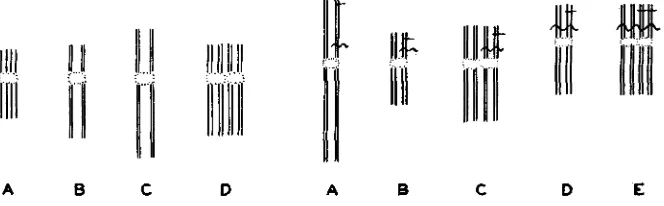
FIGURE I.-Diagram of Mitosis. A, telo- phase with threads equidistant. B, interphase. C, prophase. D, metaphase. Multiplication of threads becomes visible during metaphase as soon as the prospective chromosomes of ana- phase are ready to separate. Coiling has been disregarded; also the threads are not actually arranged in a single plane.
FIGURE 2.-Diagram of number of threads in a single chromosome in meiosis and meta- phase of first division in the pollen grain. A. leptotene. B, first metaphase. C, interkine- sis. At this stage multiplication of threads be- comes visible; each chromosome contains eight threads which separate at the second division.
D, quartet stage (only one of the two chromo- somes which separate in the second division is shown). E, metaphase in first division of the pollen grain. Another multiplication occurs here. “Lesions” have been entered by cross bars, the straight horizontal bar illustrates the history of a lesion affecting a half chromatid of presynapsis. The wavy cross bar illustrates a lesion affecting one chromatid of presynap- sis. A half-chromatid lesion of presynapsis be- comes a chromatid lesion a t second anaphase and a chromosome lesion after the metaphase of the first division in the pollen grain. A chromatid lesion of presynapsis becomes a chromosome lesion at second division etc.
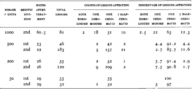
Table I is a summary of the observations. The effect of radiation was
observed primarily during first and second reduction division. Turning to figure 2, it can be seen that a lesion affecting one chromatid a t leptotene
(fig. 2A) would be observed as such during first division, (fig. 2 ~ ) but would during late metaphase and anaphase of the second division appear as a lesion affecting a chromosome (fig. 2 ~ ) . At the division in the pollen
610 B. R. NEBEL
TABLE I
Effect of X-rays on presynaptic chromosomes observed during first and second anaphase
AOURS COUNTS OF LESIONS AFFECTINQ PERCENTAQE OF LESIONS AFFECTING DOSAQE MEIOTIC AFTER TOTAL
T UNITS DIVI- TREAT- LESIONS BOTA O N E O N E I A A L F - BOTA O N E O N E I AALF- SION MENT AOMO- CARO- CARO- CHRO- AOMO- CARO- CARO- CARO-
L O G W S MOSOME MATID MATID LOGUES MOSOME MATID MATID
1000 2nd 60.5 SI 2 18 51 I O 2 . 5 2 2 63 12.3
2 42 2 5 157 2 1
4 . 4 91.2 4 . 4 2 . 7 85.7 11.6
2 0 0 1st 26 35 2 32 I 5.7 91.4 2.9
2nd 26 I 2 0 9 109 2 7.5 90.8 1.7
50 1st 29 55 55
2nd 29 32 1 32
100
3 97
A lesion affecting one half-chromatid at leptotene becomes a lesion of half a chromosome in second division and of a whole chromosome in the anaphase of the division in the pollen grain. I n table I observations made
FIGURE 3.-Material radiated during presynapsis. A, B and C are from second anaphase,
A, showing one chromatid asymmetrically attached to the end of another chromosome, B, show- ing one chromatid partly deleted. Half-chromosome (chromatid) lesions of second anaphase are the result of half-chromatid lesions of presynapsis. C illustrates two achromatic spots a t the same level of a chromosome a t second anaphase. These are the results of a lesion affecting one chromatid
of presynapsis. D is from prometaphase of the first division in the pollen grain. The asymmetric pieces towards IO and 2 o’clock are prospective anaphasic elements. Their sister halves have be- come detached. This corresponds to half-chromatid lesions of presynapsis. Camera lucida X 2400.
CHROMOSOME STRUCTURE I N TRADESCANTIA 611
on first and second divisions are reduced to the condition of the chromo- some a t first metaphase, which in Tradescantia is identical with the condi- tion in leptotene or any other presynaptic stage after the last archesporial division. Thus if a t second anaphase a lesion is observed which a t that stage involves a single chromatid, this is entered in table I as a lesion hav- ing affected one half-chromatid.
The term lesion is used to include any type of cytological abnormality, such as achromatic spots, breaks or translocations, here again using the last term to describe the transfer of chromosomes or parts of chromosomes to previously unrelated parts of other chromosomes.
A lesion which affected a half-chromatid observed in second division could not be distinguished from a lesion affecting three half-chromatids. Breaks affecting three half-chromatids a t first division were, however, not observed. Achromatic spots affecting a half-chromatid only were not ob- served. Achromatic spots seemed always to affect both halves of a chroma- tid (fig. 3c) and appeared in second division side by side as a clearly defined pair of spots or as narrow achromatic band across the width of a chromo- some of second anaphase.
The data in table I seem to indicate that the effect of radiation changes with increasing dosage. At low dosages the effect is confined almost com- pletely to lesions of chromatids. At higher dosages there is an increasing number of other lesions. Single half-chromatid lesions increase and lesions involving more than one chromatid a t a given level become more numer-
Since higher dosages of radiation merely represent longer exposures to the same type of radiation it is hard to understand why a t the low dosage the types of lesions were narrowed to nearly a single class.
To explain the results it is suggested that under longer exposures the reaction of the chromatic elements is no longer a simple one. Under pro- longed exposure heat may be generated in the cell, sensitive processes of metabolism may be invalidated sufficiently to change the reaction of chromatin toward radiation. In a general sense prolonged radiation causes a change in the environment of the chromosome threads, which in turn changes the type of reaction of the chromosomes toward radiation. Under low dosage, with no change in the environment, the unit of reaction within the chromosome is the chromatid. This is in keeping with the fact that crossing over is a reaction between chromatids, not between half- chromatids. It is also in keeping with the observation that half-chromatids are very closely united, perhaps by a common matrix.2 It is assumed that
I have suggested earlier (NEBEL 1932) that each chromonema is surrounded by its own in- dividual matrix. In a general sense this is still maintained. If the diameter of the gene string
6 1 2 B. R. NEBEL
sister half-chromatids form a physiological unit, which under moderate radiation reacts as a unit. Under increased radiation, due to a change in the environment the equilibrium between half-chromatids may become up- set, and the normal physiological and spatial distance between related chromatids and chromosomes may also become disturbed. The effect of heavy radiation is similar to that of heat which causes clumping and stick- ing of chromatic elements which normally form no contacts.
The present evidence is purely cytological but chromatic changes may entail genetic changes. The finding of STUBBE (1935) that the genom of
Epilobium hirsutum is more sensitive to radiation when carried in E . luteum plasma than when carried in its own plasma may be quoted as an analogy for the instability of the genom under changes of environment.
The existence of half-chromatid breaks is not taken as proof but merely as a confirmation of the morphological observation that meiotic chroma- tids are formed and split in the last premeiotic metaphase in Tradescantia.
It would be possible to explain the data differently but any alternative explanation-leads into additional difficulties.
MARSHAK- (1935) found a linear progression for lesions with dosage. The present material confirms MARSHAK’S finding. A special study of
this phase was not possible but when a graph was drawn from the number of lesions observed under various dosages using only those cells in which all chromosomes could be seen, this graph showed 2 to 3 lesions per cell with
50
r units, 4 to 5 with 2 0 0 , 8 to I O with 500 and I O to 18 with 1000 r units respectively. The graph is not given here because the numbers of perfect cells available for counting was low. Lesions involving a chromatid were counted as one, lesions involving a half-chromatid likewise. Lesions in- volving more than one chromatid were counted as one only where sister chromatids clearly appeared involved a t the same level.With higher dosages counts of lesions will appear low, for the reason that if a fragment becomes a micronucleus i t will hide additional lesions which i t may contain. Counts of lesions will appear high for the following reason.
It is assumed that a single hit may a t higher dosage spread “horizontally” -at right angles to the long axis of the chromosome. The first division separates homologous chromatids and the second division separates sister
CHROMOSOME STRUCTURE I N TRADESCANTIA 613 chromatids. This separation makes it hard to detect lesions in correspond- ing regions of sister and homologous chromatids. The effect of what may have been a single hit will thus be considered multiple.
These two phenomena may cancel one another. With this in mind the obtained linearity of the curve of number of lesions per cell plotted against dosage suggests that under higher dosages the effect of a single hit may occasionally spread a t right angles to the longitudinal axis of a chromo- some, and also may, in contrast to the effect of low dosages, remain con- fined to a single half-chromatid. A few observations were made on the first somatic division in the pollen grain from cymes rayed with 500 r units. The divisions were observed 14 days after radiation. This time interval indicates that a t the time of radiation the material was in presynapsis. Several divisions showed an abundance of fragments involving only one chromatid of prometaphase. From diagrams A to E of figure 2 it may be seen that such breaks would be expected from the lesion of half-chromatids of presynapsis.
The occurrence of half-chromatid breaks as observed a t first and second division was followed thru successive days and could be traced for about a week after treatment. After this time half-chromatid breaks were no longer observed. This one would expect if the maximum time limit be- tween reduction division and the last preceding archesporial division for any one cell were around 170 hours. This corresponds to the time schedule given by
SAX
and EDMONDS (1933).SUMMARY
Radiation of cymes of Tradescantia rejexa Raf. was applied to pre- synaptic stages of microsporocytes. The effect of the radiation was ob- served during first and second reduction divisions. With low dosage the experiment indicates that the chromosome is split in two previous to synapsis. With higher dosage the experiment suggests that occasionally the chromosome is split into four previous to synapsis.
This is interpreted as follows: Each chromosome is composed of two split chromatids (or four half-chromatids) a t all stages of presynapsis. In normal material the two half chromatids that constitute a given chromatid lie very close to one another and do not react individually. (The process of crossing over in Tradescantia is a reaction between chromatids in which every two half-chromatids behave as a physiological and mechanical unit.) Under low dosage of X-rays half-chromatids are physiologically unable to show separate reactions.
With higher dosage of radiation ( 2 0 0 r units and more) the spatial and
614 B. R. NEBEL
threads differentially. It is also indicated that with higher dosage chro- matic reactions a t a certain level of the chromosome may spread, involving sister and even homologous loci.
The differential effect of higher dosage is thus attributed to action upon the immediate environment of the chromatin, perhaps to heat or to a general interference with the normal metabolism of the nucleus which in turn changes the type of reaction of the chromatin.
ACKNOWLEDGMENT
The present study was carried out under a grant from the Rockefeller Foundation. I am indebted to Dr. F. SCHRADER, Dr. M. DEMEREC, Dr. C. P. HASKINS and Dr. K. SAX for suggestions and criticism.
Dr. WM. E. ACHILLES, M.D., Geneva, N. Y . , kindly furnished the facili- ties for radiation.
LITERATURE CITED
HASKINS, C. P., 1935 A determination of the magnitude of the cell “sensitive volume” associated with the white-eye mutation in X-rayed Drosophila. Proc. Nat. Acad. Sci. 21 : 561-566. HUSKINS, C. L. and HUNTER, A. W. S., 1935 The effects of X-radiation on chromosomes in the
microscopores of TriUium erectum L. Proc. Roy. SOC London Ser. B. 117: 22-33.
KUWADA, Y. and NAKAKURA, T., 1934 Behavior of chromonemata in mitosis 11. Artificial un- ravelling of coiled chromonemata. Cytologia 5: 244-247.
LEWITZKY, G. A. and ARARATIAN, A. G., 1931 Transformation of chromosomes under the influ- ence of X-rays. Bull. Appl. Bot. 27: 289-303.
MARSHAK, A., 1935 The effect of X-rays on chromosomes in different stages of meiosis. J. Gen. Physiol. 19: 179-198.
MATHER, K. and STONE, L. H. A., 1933 The effect of X-radiation on somatic chromosomes. J. Genet. 28: 1-24.
MATHER, K., 1934The behavior of meiotic chromosomes after X-radiation. Hereditas 19: 303-322. MOORE, W. G., 1934 A comparison of the frequencies of visible mutations produced by X-ray
NEBEL, B R. and RUTTLE, M. L., 1936 Chromosome structure XI. in press.
PATTERSON, J. T., 1933 The mechanism of mosaic formation in Drosophila. Genetics 18: 32-52. 1935 The question of delayed breakage in the chromosomes of Drosophila. J. Exp. Zool. 70: 233-243.
SAX K. and EDMONDS, H. W., 1933 Development of the male gametophyte in Tradescantia. Bot. Gaz. 95: 156-163.
STONE, L. H. A., 1933 The effect of X-radiation on the mitotic divisions of certain plants. Annals of Bot. 47: 815-816.
STUBBE, H., 1935 Uber den Einfluss artfremden Plasmas auf die Konstanz der Gene. Z.i.A.V. 70: 161-169.
WHITE, M. J. D., 1935 The effects of X-rays on mitosis in the spermatogonial division of Locusla
migratoria L. Proc. Roy. Soc. London Ser. B. rrg: 61-83.