DOI: 10.1534/genetics.110.120782
Lambda Red Recombineering in
Escherichia coli
Occurs Through a Fully
Single-Stranded Intermediate
J. A. Mosberg,*
,†,1,2M. J. Lajoie*
,†,1,2and G. M. Church*
*Department of Genetics, Harvard Medical School, Boston, Massachusetts 02115 and†Program in Chemical Biology, Harvard University, Cambridge, Massachusetts 02138
Manuscript received July 12, 2010 Accepted for publication August 30, 2010
ABSTRACT
The phage lambda-derived Red recombination system is a powerful tool for making targeted genetic changes inEscherichia coli, providing a simple and versatile method for generating insertion, deletion, and point mutations on chromosomal, plasmid, or BAC targets. However, despite the common use of this system, the detailed mechanism by which lambda Red mediates double-stranded DNA recombination remains uncertain. Current mechanisms posit a recombination intermediate in which both 59 ends of double-stranded DNA are recessed bylexonuclease, leaving behind 39overhangs. Here, we propose an alternative in which lambda exonuclease entirely degrades one strand, while leaving the other strand intact as single-stranded DNA. This single-stranded intermediate then recombines via beta recombinase-catalyzed annealing at the replication fork. We support this by showing that single-stranded gene insertion cassettes are recombinogenic and that these cassettes preferentially target the lagging strand during DNA replication. Furthermore, a double-stranded DNA cassette containing multiple internal mismatches shows strand-specific mutations cosegregating roughly 80% of the time. These observations are more consistent with our model than with previously proposed models. Finally, by using phosphorothioate linkages to protect the lagging-targeting strand of a double-stranded DNA cassette, we illustrate how our new mechanistic knowledge can be used to enhance lambda Red recombination frequency. The mechanistic insights revealed by this work may facilitate further improvements to the versatility of lambda Red recombination.
O
VER the past decade, lambda Red recombination (‘‘recombineering’’) has been used as a power-ful technique for making precisely defined insertions, deletions, and point mutations in Escherichia coli, requiring as few as 35 bp of homology on each side of the desired alteration (Thomason et al. 2007a;Sharanet al. 2009). With this system, single-stranded
DNA (ssDNA) oligonucleotides have been used to efficiently modify E. coli chromosomal targets (Ellis
et al. 2001; Costantino and Court 2003), BACs
(Swaminathanet al.2001), and plasmids (Thomason
et al.2007b), as well as to rapidly optimize a metabolic pathway coding for the production of lycopene (Wang
et al.2009). Furthermore, linear double-stranded DNA (dsDNA) recombineering has been used to replace chromosomal genes (Murphy1998; Murphyet al.2000),
to disrupt gene function (Datsenko and Wanner
2000), and to develop novel cloning methods (Lee
et al. 2001; Liand Elledge2005). Large-scale dsDNA
recombineering projects include creating a library of single-gene knockout E. coli strains (Babaet al.2006)
and removing 15% of the genomic material from a singleE. coli strain (Posfaiet al.2006). Linear dsDNA
recombineering has also been used to insert heterol-ogous genes and entire pathways into the E. coli chromosome (Zhang et al. 1998; Wang and Pfeifer
2008) and BACs (Leeet al.2001; Warminget al.2005),
including those used for downstream applications in eukaryotes (Chaveroche et al. 2000; Bouvier and
Cheng2009). However, despite the broad use of this
method, the mechanism of lambda Red recombination has not achieved scientific consensus, particularly in the case of dsDNA recombination. A clearer under-standing of the mechanism underlying this process could suggest ways to improve the functionality, ease, and versatility of lambda Red recombination.
Three phage-derived lambda Red proteins are neces-sary for carrying out dsDNA recombination: Gam, Exo, and Beta. Gam prevents the degradation of linear dsDNA by the E. coli RecBCD and SbcCD nucleases; lambda exonuclease (Exo) degrades dsDNA in a 59to 39manner, leaving single-stranded DNA in the recessed regions; and Beta binds to the single-stranded regions
Supporting information is available online athttp://www.genetics.org/ cgi/content/full/genetics.110.120782/DC1.
Available freely online through the author-supported open access option.
1These authors contributed equally to this work.
2Corresponding authors: Department of Genetics, Harvard Medical School, 77 Avenue Louis Pasteur, New Research Bldg., Room 238, Boston, MA 02115. E-mail: [email protected] and mlajoie@ fas.harvard.edu
produced by Exo and facilitates recombination by promoting annealing to the homologous genomic target site (Sawitzkeet al.2007). Current mechanisms
claim that Exo binds to both 59ends of the dsDNA and degrades in both directions simultaneously to produce a double-stranded region flanked on both sides by 39
overhangs (Sharan et al. 2009; Szczepanska 2009).
However, a comprehensive explanation of how this construct ultimately recombines with the chromosome has not yet been advanced.
Initially, it was proposed that this recombination occurs via strand invasion (Thaler et al.1987).
How-ever, it has more recently been shown that strand invasion is unlikely to be the dominant mechanism in the absence of long regions of homology, as recombi-nation remains highly proficient in arecA-background
(Yu et al. 2000). Furthermore, a detailed analysis of
lambda Red recombination products showed character-istics consistent with strand annealing rather than a strand invasion model (Stahl et al. 1997). Finally,
lambda Red dsDNA recombination has been shown to preferentially target the lagging strand during DNA replication, which suggests strand annealing rather than strand invasion (Limet al.2008; Poteete2008).
To explain these results, Courtet al.(2002) proposed
a strand-annealing model for insertional dsDNA re-combination (Figure 1A), in which one single-stranded 39end anneals to its homologous target at the replica-tion fork. The replicareplica-tion fork then stalls, due to the presence of a large dsDNA nonhomology (i.e., the insertion cassette). The stalled replication fork is ulti-mately rescued by the other replication fork traveling in the opposite direction around the circular bacterial chromosome. The other 39end of the recombinogenic DNA anneals to the homology region exposed by the second replication fork, forming a crossover structure, which is then resolved by unspecified E. coli enzymes (Courtet al.2002).
The Court mechanism was challenged by Poteete
(2008), who showed that the dsDNA recombination of a linear lambda phage chromosome occurs readily onto a unidirectionally replicating plasmid, which does not have the second replication fork required by the Court mechanism (Court et al. 2002). Thus, Poteete
pro-posed an alternate mechanism (Poteete2008), termed
‘‘replisome invasion’’ (Figure 1B), in which a 39overhang of the Exo-processed dsDNA first anneals to its comple-mentary sequence on the lagging strand of the recom-bination target. Subsequently, this overhang displaces the leading strand, thereby serving as the new template for leading-strand synthesis. The resulting structure is resolved by an unspecified endonuclease, after which the recombinogenic DNA becomes the template for the synthesis of both new strands. In the context of recom-bineering using a linear dsDNA cassette, the author indicates that a second strand-switching event must occur at the other end of the incoming dsDNA.
While Poteete’s mechanism addresses some of the weaknesses of the Court mechanism, it remains largely speculative. This mechanism does not identify the endonuclease responsible for resolving the structure after the first template switching event, nor does it explain how the recombinogenic DNA and replication machinery form a new replication fork. Additionally, this template-switching mechanism would have to oper-ate two times in a well-controlled manner, which may not be consistent with the high-recombination frequen-cies often observed (Murphy et al. 2000) for lambda
Red-mediated dsDNA insertion. Finally, little experi-mental evidence has been advanced to directly support this hypothesis.
Figure 1.—Previously proposed lambda Red-mediated dsDNA recombination mechanisms. Heterologous dsDNA is shown in green; Exo is an orange oval, and Beta is a yellow oval. In both mechanisms the recombination intermediate is pro-posed to be a dsDNA core flanked on either side by 39ssDNA overhangs. (A) The Court mechanism posits that (1) Beta fa-cilitates annealing of one 39 overhang to the lagging strand of the replication fork. (2) This replication fork then stalls and backtracks so that the leading strand can template switch onto the synthetic dsDNA. The heterologous dsDNA blocks further replication from this fork. (3) Once the second repli-cation fork reaches the stalled fork, the other 39end of the in-tegration cassette is annealed to the lagging strand in the same manner as prior. Finally, the crossover junctions must be re-solved by unspecified E. coli enzymes (Court et al. 2002). (B) The Poteete mechanism suggests that (1) Beta facilitates 39overhang annealing to the lagging strand of the replication fork and (2) positions the invading strand to serve as the new template for leading-strand synthesis. This structure is resolved by an unspecified host endonuclease (red triangle), and (3) the synthetic dsDNA becomes template for both lagging and leading-strand synthesis. A second template switch must then occur at the other end of the synthetic dsDNA (Poteete 2008). The figure was adapted from the references cited.
To address the deficiencies in these mechanisms, we propose that lambda Red dsDNA recombination pro-ceeds via a ssDNA intermediate rather than a dsDNA core flanked by 39overhangs (Figure 2). In this mech-anism, Exo binds to one of the two dsDNA strands and degrades that strand completely, leaving behind full-length ssDNA. This ssDNA then anneals to its homology target at the lagging strand of the replication fork and is incorporated as part of the newly synthesized strand as if it were an Okazaki fragment. This process is analogous to the accepted mechanism for the lambda Red-mediated recombination of ssDNA oligonucleoti-des (Court et al. 2002) and, therefore, unifies the
mechanisms for ssDNA and dsDNA recombination. Notably, our mechanism uses one replication fork for the incorporation of a full-length heterologous cassette, thereby addressing Poteete’s criticism of the Court mechanism.
The degradation of an entire strand by lambda Exo is feasible, given the highly processive nature of the enzyme (Subramanianet al.2003). Whereas previously
proposed mechanisms assume that both dsDNA ends are degraded approximately simultaneously, our hy-pothesis implies that some dsDNA molecules will be entirely degraded to ssDNA before a second Exo can bind to the other end. In this article, we demonstrate that single-stranded DNA is a viable recombinogenic intermediate with lagging-strand bias. Furthermore, we show that genetic information from one strand of a recombinogenic dsDNA cassette cosegregates during lambda Red-mediated recombination. These results provide strong support of our proposed mechanism.
MATERIALS AND METHODS
The preparation of the various DNA constructs used in this study is detailed in the supplementalmaterials and methods section (supporting information, File S1). These DNA con-structs were recombined into EcNR2 cells (E. coli MG1655 DmutSTcatD(ybhB-bioAB)T[lcI857D(cro-ea59)TtetR-bla]) in a
similar manner as previously described (Wang et al.2009). Briefly, cells were grown in a rotator drum at 32°in LB-min media (10 g tryptone, 5 g yeast extract, 5 g sodium chloride/ liter water) until they reached an OD600of 0.4 – 0.6. At this time, the expression of the lambda Red proteins was induced by vigorously shaking the cells in a 42°water bath for 15 min. Cells were then chilled on ice, washed twice with deionized water, and resuspended in 50ml of deionized water containing the desired DNA construct. For the experiment investigating strand bias, 20 ng of DNA was recombined. For all other experiments, 50 ng was used. The DNA was then introduced into the cells via electroporation (Bio-Rad Gene Pulser; 0.1 cm cuvette, 1.78 kV, 25mF, 200 ohm). After electroporation, cells were recovered in 3 ml LB-min media for 3 hr in a rotator drum at 32°.
Recombinants were identified by plating 50 ml or 1 ml (concentrated to 50 ml) of undiluted recovery culture on selective media (LB-min with 30 mg/ml kanamycin sulfate, 95 mg/ml spectinomycin, or 10 mg/ml Zeocin). The total viable cell count was determined by plating 50ml of a 104 dilution of the recovery culture (in LB-min) onto LB-min1
20mg/ml chloramphenicol plates. For experiments involving lacZgene disruption, the plates also contained Fisher Chromo-Max X-Gal/IPTG solution at the manufacturer’s recommended concentration. Recombination frequencies were determined by dividing the extrapolated number of recombinants by the total viable cell count. All experiments assessing recombination frequency were performed in triplicate, except the series of recombinations in which phosphorothioate placement was altered—these were performed in duplicate. The recombina-tion frequencies determined for each replicate were averaged and the error of the mean was taken to be sx¼sx=
ffiffiffiffiffi
N
p
default parameters by MATLAB. Following the lacZTkanR
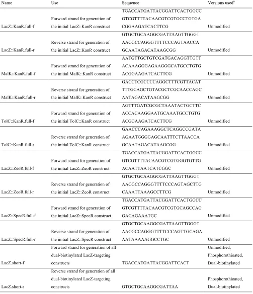
mismatched dsDNA recombinations, mismatch amplification mutation assay (MAMA) PCR (see File S1for detailed de-scription) was used for genotypic analysis. A complete list of primers used in the study can be found in the supporting information (Table S1).
RESULTS
Testing the predicted ssDNA recombination inter-mediate:We designed alacZTkanRcassette (1.2 kb),
consisting of a kanamycin resistance gene (kanR) flanked by 45-bp regions homologous to thelacZgene on theE. coli chromosome. Successful kanR insertion disrupted LacZ function, so proper targeting of the lacZTkanR cassette could be verified by selecting on
kanamycin and assaying for the inability to cleave X-Gal in order to release a blue chromophore. This dsDNA construct was generated by PCR and converted into ssDNA using a biotin capture and DNA melting protocol (Poundet al.2009), as detailed inFile S1. PAGE analysis
confirmed the purity of the lacZTkanR ssDNA
con-struct, as no dsDNA band was readily detected. This construct was then recombined into EcNR2 (Wang
et al.2009). ThelacZTkanRssDNA construct was found
to yield 1.3310564.53106recombinants per viable
cell, in comparison with 1.9310467.53105for the
corresponding dsDNA construct. Both ssDNA and dsDNA gave over 99% white colonies, indicating correct targeting of the recombinogenic cassette.
This result confirms that ssDNA—the predicted in-termediate for our mechanism—is recombinogenic. It is, however, 14.8-fold less recombinogenic than the corre-sponding dsDNA. We hypothesize that this disparity is caused by ssDNA secondary structure and/or the lack of Exo–Beta synergy. Previous work has demonstrated that ssDNA oligonucleotides longer than 90 bases and/or having secondary structure with DG , 12 kcal/mol are likely to have substantially reduced recombination frequency (Wanget al.2009). Thus, we expect
second-ary structure to significantly diminish the recombina-tion frequency of this1.2-kb cassette. Additionally, it has previously been suggested (Dattaet al.2008) that
Exo and Beta act synergistically, with Exo facilitating the binding of Beta to recessed regions of ssDNA. Since Exo does not readily bind to ssDNA, this synergistic action cannot occur; therefore, recombination frequency may decrease. However, even in light of these considera-tions, our predicted ssDNA intermediate is highly capable of recombination.
To confirm that the observed recombinants arose from the ssDNA rather than from dsDNA contamination, this recombination experiment was repeated in SIMD90 (Dattaet al.2008), a strain of E. colicontaining Beta,
but lacking Exo and Gam. In the absence of Exo and Gam, dsDNA recombination frequency should decline significantly due to increased dsDNA degradation and
inefficient processing into ssDNA. In this strain,lacZ
T-kanRssDNA demonstrated a recombination frequency of 1.8 3 104, in comparison with a recombination
fre-quency of only 8.7 3 107 for dsDNA (a 209-fold
difference). This result indicates that the observed recombinants in EcNR2 also arose from ssDNA.
Investigating the strand bias of the recombination intermediate: We propose that the long ssDNA inter-mediate recombines by annealing at the replication fork in the same manner as ssDNA oligonucleotides (Court
et al. 2002). It has been demonstrated that lagging-targeting oligonucleotides recombine with substantially greater frequency than the corresponding leading-targeting oligonucleotides, due to the greater accessi-bility of the lagging strand for annealing (Liet al.2003).
To test whether long ssDNA recombines in the same manner, we investigated whether several pairs of lagging-targeting and leading-lagging-targeting ssDNA insertion cas-settes demonstrated a similar strand bias. We controlled for the effect of differential secondary structure be-tween the two strands by recombining three differ-ent antibiotic resistance markers intolacZ—kanamycin (lacZTkanR), zeocin (lacZTzeoR), and spectinomycin
(lacZTspecR). Additionally, to demonstrate that strand
bias was not caused by replichore-specific context or transcriptional direction, we constructed two additional kanR cassettes. To this end, tolCTkanR targets a gene
located on the opposite replichore from lacZ, and malKTkanRtargets a gene transcribed from the
oppo-site strand of the chromosome as lacZ. As shown in Figure 3, the lagging-targeting strand was substantially more recombinogenic than the leading-targeting strand for all of the tested constructs. As previously observed for oligonucleotides (Elliset al.2001), there appears to
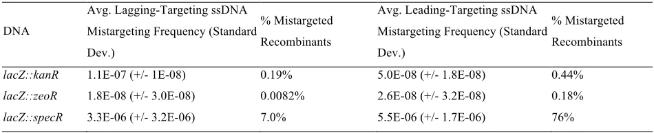
be a significant amount of locus-specific and sequence-specific variability in recombination frequency. Interest-ingly, a significant number of mistargeted recombinants (antibiotic-resistant colonies that retained LacZ func-tion) were observed for bothlacZTspecRstrands (Table
S2; discussion inFile S2). Mistargeted (LacZ1
) colonies were not scored as recombinants and do not affect the broader interpretation of our results. The overall results of this set of experiments clearly indicate a robust lagging-strand bias, likely arising from the greater accessibility of the lagging strand during DNA replication. This supports our claim that long ssDNA insertion constructs recombine by annealing at the replication fork in a manner similar to ssDNA oligonucleotides.
Testing mechanistic predictions by tracking designed mutations: The prior experiments provide strong indirect evidence supporting our proposed ssDNA annealing mechanism. To more directly test the pre-dictions of this mechanism, we designed a lacZTkanR
dsDNA cassette with internal mismatches (Figure 4), which enables us to empirically determine which strand provided genetic information during recombination.
This construct was generated by annealing two strands of ssDNA and purifying the resulting dsDNA by agarose gel extraction. In each of the flankinglacZ homology regions, this construct contains two sets of adjacent dinucleotide mismatches that differentiate the two strands. At these loci, neither strand’s sequence matches the targeted chromosomal copy oflacZ. Thus, one can infer which strand has recombined by observing which strand-specific alleles are present.
Our proposed ssDNA annealing mechanism can be distinguished from the prevailing dsDNA recombina-tion mechanisms on the basis of the results of this experiment. Our mechanism predicts that the muta-tions contained on a single strand will be inherited together and that the mutations arising from the lagging-targeting strand will be observed more fre-quently than those from the opposite strand. Con-versely, as detailed in Figure 4, the previously proposed mechanisms predict that the alleles on the 39 ends of both strands would be incorporated.
This mismatchedlacZTkanRcassette was transformed
into EcNR2, which is deficient for mismatch repair. Recombinants were identified by plating on kanamycin, and colonies were screened using MAMA PCR (Qiang
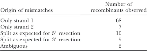
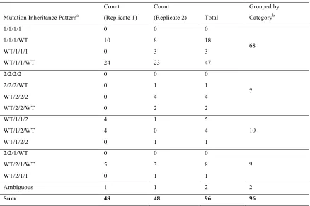
et al.2002) to identify which strand-specific mutations were inherited in each colony. Two replicates were performed, and 48 colonies were screened for each recombination (Table 1; detailed results in Table S3). The accuracy of the MAMA PCR assay was confirmed by sequencing the relevant regions of several colonies and by performing a complementary MAMA PCR assay to detect unaltered wild-type alleles at the targeted loci. In line with our predictions, we found that roughly 80% of the colonies inherited mismatch alleles from only one strand. Furthermore, of these colonies, 91% in-herited mismatch alleles specifically from the lagging-targeting strand, strongly supporting our ssDNA annealing mechanism.
Half of the remaining 20% of the colonies showed an inheritance pattern consistent with resection from both 59 ends, and the other half was consistent with resection from both 39ends. Resection from the 59ends is predicted by the previously proposed mechanisms and indicates that one of these mechanisms may also operate as a disfavored process. However, Exo has not been
Figure3.—Strand bias in lambda Red ssDNA insertion re-combination. Recombination frequencies were assessed for several leading-targeting and lagging-targeting complemen-tary ssDNA pairs. Lagging-targeting strands were found to be more recombinogenic than leading-targeting strands. An asterisk indicatesP,0.05.
Figure 4.—Strand-specific mismatch alleles were used to identify the strand of origin for each recombined mutation. The mismatched lacZTkanR cassette contained two consecutive
shown to degrade dsDNA in a 39 / 59 manner, even though our results imply that this occurs nearly as often as 59 / 39 resection. A plausible explanation for this discrepancy is that the colonies possessing alleles from both strands have instead undergone two sequential recombination events according to our proposed mech-anism. The first recombination would proceed normally, and the second recombination would involve a partially degraded complementary strand. This second recombina-tion event would be expected to occur quite frequently—-after the first recombination event, the kanR gene is present in the genome, providing a large region of homology to which remaining fragments ofkanRssDNA can anneal in subsequent rounds of replication.
Interestingly, mutations arising from loci 1 and 4 (Figure 4) are observed only rarely in the studied recombinants. This result suggests that a significant portion of the DNA may be undergoing slight exo-nuclease degradation from both the 59and 39ends, or that annealed strands are processed at the replication fork in a manner that degrades or excludes the distal ends of the recombined DNA. This is consistent with a previous observation that mutations placed on the ends of a 90-bp oligonucleotide are inherited at a substan-tially lower frequency than mutations placed nearer to the center of the same strand. Elucidating the basis of this phenomenon may shed more light on the detailed mechanism of lambda Red recombination. Nevertheless, the results from this experiment provide direct evidence that our proposed mechanism is the dominant process by which lambda Red dsDNA recombination occurs.
Phosphorothioate placement alters recombination frequency:Leveraging our increased understanding of lambda Red dsDNA recombination, we enhanced re-combination frequency by over an order of magni-tude. Since the lagging-targeting strand is the most important recombinogenic species, we reasoned that protecting this strand would improve recombination frequency. It is known that phosphorothioate bonds diminish the ability of many exonucleases to degrade DNA (Liu and Liu 2010). To test whether altering
phosphorothioate placement changes the resulting re-combination frequencies, we made four variants of the mismatchedlacZTkanRdsDNA cassette, as denoted
in Figure 5. These cassettes were recombined into EcNR2, and recombination frequencies were deter-mined (Figure 5).
These results show that protecting the lagging-targeting strand with phosphorothioate bonds increases the frequency of dsDNA recombination, whereas protecting the leading-targeting strand has no effect. This further supports our proposed mechanism, since alternating which of the two strands is protected by phosphoro-thioates would not be expected to have differential effects if resection occurred from both 59ends. Addi-tionally, our results unexpectedly show that lagging-targeting strand protection and dual protection yield
approximately equivalent recombination frequencies. This suggests that phosphorothioation does not signif-icantly inhibit in vivo Exo degradation, as dual pro-tection would prohibit processing by Exo if this were the case. Instead, it is likely that placing phosphorothioates on the lagging-targeting strand protects it from host exonuclease degradation after Exo processing. This result demonstrates how our improved mechanistic knowledge of lambda Red recombination can facilitate rational improvements of the process.
DISCUSSION
This work provides strong empirical support for the proposed mechanism that lambda Red dsDNA recom-bination operates through a full-length ssDNA inter-mediate. This mechanism appears to be the dominant means of lambda Red dsDNA recombination, although other mechanisms may still occur as minor processes. Notably, a replisome invasion mechanism (Poteete
2008) involving a fully single-stranded intermediate is not directly refuted by our work, although a strand-annealing model is favorable due to its well-precedented (Stahlet al.1997; Courtet al.2002) and parsimonious
nature.
While our mechanism has not previously been postu-lated as the manner by which the lambda Red system recombines large dsDNA segments, it is consistent with numerous results observed by other groups. By anneal-ing two staggered oligonucleotides, Yu et al. (2003)
previously generated a 106-bp construct consisting of a dsDNA core flanked by 39overhangs—the recombina-tion intermediate predicted by the canonical model of lambda Red dsDNA recombination. As expected, re-combination of this construct did not depend on the presence of Exo; however, even in the presence of Exo, the recombination frequency was roughly 4000-fold lower than that of its corresponding dsDNA. Given that the construct with 39 overhangs is postulated to be a downstream intermediate of this dsDNA, this result casts doubt upon the claim that the tested construct is indeed the predominant recombination intermediate. However, this result is explained by our proposed mechanism—only the intact dsDNA can generate the full-length ssDNA needed to undergo recombination, as
TABLE 1
Tracking cosegregation in mismatched dsDNA recombination
Origin of mismatches
Number of recombinants observed
Only strand 1 68
Only strand 2 7
Split as expected for 59resection 10 Split as expected for 39resection 9
Ambiguous 2
neither individual strand of the construct containing 39
overhangs is sufficient for recombination (Yu et al.
2003). We suggest that this 39 overhang construct recombines by a separate and disfavored process. This is supported by the fact that this proposed recombina-tion intermediate had no greater recombinarecombina-tion fre-quency than the corresponding structure with 59(rather than 39) overhangs. It is unlikely that either of these structures represents the predominant intermediate of dsDNA recombination.
Muyrers et al. (2000) have also provided evidence
contrary to a dsDNA recombination intermediate con-taining 39 overhangs. The authors created a dsDNA construct in which phosphorothioate linkages placed between an antibiotic resistance gene and its flanking genome homology regions were used to prevent exo-nuclease degradation beyond these homology regions. Two 59-to-39 exonucleases other than Exo were then used in vitro to resect the 59ends of this construct to generate the putative intermediate for dsDNA recom-bination. However, it was found that none of the tested resection conditions could produce a construct that would recombine in the absence of Exo. In contrast, the
predicted intermediate in our proposed mechanism is highly recombinogenic—even when preparedin vitro.
Additionally, other prior work supports our proposed mechanism by reinforcing the processive nature of Exo. Hillet al. (1997) showed that nonreplicating lambda
phage inE. coliis capable of converting linear dsDNA into ssDNA, creating single-stranded regions that span more than 1.4 kb. They also demonstrated thatexois sufficient for generating these regions of ssDNA, which are similar in length to the 1.2 kb constructs used in this experiment. An additional implication of this result is that a single-stranded intermediate is also present during crosses involving an intact lambda chromosome. These results suggest that our proposed mechanism may apply for natural lambda Red recombination between phage and bacterial chromosomes. By extension, this model may also describe crosses between the phage chromo-some and a plasmid (Poteete2008), as plasmids present
an accessible lagging strand at the replication fork in the same manner as the bacterial chromosome.
The results of Limet al.(2008) further reinforce that
Exo generates long strands of ssDNA. These researchers created a dsDNA construct in which two antibiotic resistance genes were attached via a genome homol-ogy region and flanked with two additional regions of genome homology. Using this cassette, only about 10% of recombinants incorporated both resistance genes, while a majority of recombinants incorporated only one of the two. This implies that a majority of recombina-tion events involved the central homology region, which is roughly 1 kb away from either end of the dsDNA construct. Given that strand annealing requires exposed ssDNA, this result further suggests that Exo can be substantially processive in vivo, degrading large stretches of DNA rather than short flanking segments. Indeed, limits to the processivity of Exo could explain why recombination frequency declines substantially for increasing dsDNA insertion sizes, but not for increasing chromosomal deletion sizes (Marescaet al.2010).
Finally, while this article was in revision, Maresca
et al.(2010) published complementary results, in which strand-specific 59phosphorylation and phosphorothioa-tion were used to bias Exo degradaphosphorothioa-tion to each strand of a selectable cassette. For recombination events follow-ing bothin vitroandin vivo digestion, the authors ob-served a lagging-targeting strand bias. Building upon these observations, the authors identified ssDNA as a recombinogenic species and proposed a mechanism consistent with the one advanced in this manuscript. These results provide substantial validation of our model. Our experiment involving a mismatched dsDNA cassette extends this work by showing that information from a single strand cosegregates during lambda Red-mediated dsDNA recombination. More importantly than simply showing a lagging-targeting strand bias, this ex-periment provides direct evidence of a single-stranded intermediate in lambda Red dsDNA recombination.
Figure5.—Testing the effect of strand protection on re-combination frequency. FourlacZTkanRcassettes were tested
Our proposed mechanism may also describe other recombineering processes mediated by lambda Red. One example is gap repair, in which linearized plasmid DNA is used to capture chromosomal DNA (Leeet al.
2001; Court et al. 2002). Notably, while a detailed
mechanism has not yet been advanced for lambda Red-facilitated gap repair, our model involving a single-stranded intermediate provides a plausible explanation. Given a full-length ssDNA intermediate, the linearized plasmid would anneal to the chromosomal target with its homology regions facing one another. The 39-end homology would then be elongated in the direction of the 59-end homology, thereby introducing the chromo-somal DNA of interest into the plasmid. The linear single-stranded plasmid would be circularized by ligase in the same manner as Okazaki fragment joining. The circular ssDNA would then be liberated from the chromosome, possibly during chromosomal replication. Residual ssDNA from the other strand of the linearized plasmid may be necessary to prime replication in order to convert the circular single-stranded plasmid into double-stranded DNA. Notably, this mechanism ac-counts for the gap repair of large (.80 kb) genomic sequences (Zhanget al.2000), since the two homology
regions could anneal with multiple Okazaki fragments between them. These fragments would then be joined by the natural lagging-strand replication mechanism.
In conclusion, a large body of evidence from our current work and from previously published studies supports our claim that the predominant mechanism for lambda Red dsDNA recombination involves the annealing of a full-length ssDNA intermediate to the lagging strand of the replication fork. However, it is possible that previously suggested mechanisms involv-ing the resection of both 59ends still operate as minor processes. The mismatched dsDNA approach described in this work may be a powerful platform from which to further explore the extent to which any such minor recombination mechanisms may operate.
The mechanism of lambda Red recombination has long been a matter of debate (Szczepanska2009). This
work posits and supports a novel mechanism, which may reveal improved recombination parameters that increase the frequency and robustness of recombineering. Just as the mechanistic understanding of Red-mediated oligo-nucleotide recombination facilitated its optimization and use in novel and powerful applications (Wanget al.2009),
similar innovations may provide for transformative appli-cations of lambda Red dsDNA recombination.
The authors thank Farren Isaacs and Harris Wang for helpful discussions regarding the lambda Red recombination system and MAMA protocols. Francois Vigneault provided expert advice on gen-erating single-stranded DNA. John Aach and Tara Gianoulis helped with statistical analysis. Tara Gianoulis, Srivatsan Raman, Farren Isaacs, and Uri Laserson provided valuable feedback regarding this manuscript, and Jaron Mercer assisted in conducting experiments. TheE. colistrain SIMD90 was a gracious gift from Donald Court, National Cancer Institute—Frederick. This work was funded by the U.S. Department of
Energy. M.J.L. is supported by a U.S. Department of Defense National Defense Science and Engineering Graduate Fellowship.
LITERATURE CITED
Baba, T., T. Ara, M. Hasegawa, Y. Takai, Y. Okumura et al.,
2006 Construction of Escherichia coli K-12 in-frame, single-gene knockout mutants: the Keio collection. Mol. Syst. Biol.2:
2006.0008.
Bouvier, J., and J. G. Cheng, 2009 Recombineering-based
proce-dure for creating Cre/loxP conditional knockouts in the mouse. Curr. Protoc. Mol. Biol.85:23.13.1–23.13.27.
Chaveroche, M. K., J. M. Ghigoand C.d’Enfert, 2000 A rapid
method for efficient gene replacement in the filamentous fungus Aspergillus nidulans. Nucleic Acids Res.28:E97.
Costantino, N., and D. L. Court, 2003 Enhanced levels of lambda
Red-mediated recombinants in mismatch repair mutants. Proc. Natl. Acad. Sci. USA100:15748–15753.
Court, D. L., J. A. Sawitzkeand L. C. Thomason, 2002 Genetic
engineering using homologous recombination. Annu. Rev. Genet.36:361–388.
Datsenko, K. A., and B. L. Wanner, 2000 One-step inactivation of
chromosomal genes in Escherichia coli K-12 using PCR products. Proc. Natl. Acad. Sci. USA97:6640–6645.
Datta, S., N. Costantino, X. Zhou and D. L. Court,
2008 Identification and analysis of recombineering functions from Gram-negative and Gram-positive bacteria and their phages. Proc. Natl. Acad. Sci. USA105:1626–1631.
Ellis, H. M., D. Yu, T. DiTizioand D. L. Court, 2001 High
effi-ciency mutagenesis, repair, and engineering of chromosomal DNA using single-stranded oligonucleotides. Proc. Natl. Acad. Sci. USA98:6742–6746.
Hill, S. A., M. M. Stahland F. W. Stahl, 1997 Single-strand DNA
intermediates in phage lambda’s Red recombination pathway. Proc. Natl. Acad. Sci. USA94:2951–2956.
Lee, E. C., D. Yu, J. Martinez de Velasco, L. Tessarollo, D. A.
Swing et al., 2001 A highly efficient Escherichia coli-based
chromosome engineering system adapted for recombinogenic targeting and subcloning of BAC DNA. Genomics73:56–65. Li, M. Z., and S. J. Elledge, 2005 MAGIC, an in vivo genetic method
for the rapid construction of recombinant DNA molecules. Nat. Genet.37:311–319.
Li, X. T., N. Costantino, L. Y. Lu, D. P. Liu, R. M. Wattet al.,
2003 Identification of factors influencing strand bias in oligonucleotide-mediated recombination in Escherichia coli. Nucleic Acids Res.31:6674–6687.
Lim, S. I., B. E. Minand G. Y. Jung, 2008 Lagging strand-biased
ini-tiation of red recombination by linear double-stranded DNAs. J. Mol. Biol.384:1098–1105.
Liu, X. P., and J. H. Liu, 2010 The terminal 59phosphate and
prox-imate phosphorothioate promote ligation-independent cloning. Protein Sci.19:967–973.
Maresca, M., A. Erler, J. Fu, A. Friedrich, Y. Zhang et al.,
2010 Single-stranded heteroduplex intermediates in lambda Red homologous recombination. BMC Mol. Biol.11:54.
Murphy, K. C., 1998 Use of bacteriophage lambda recombination
functions to promote gene replacement in Escherichia coli. J. Bacteriol.180:2063–2071.
Murphy, K. C., K. G. Campelloneand A. R. Poteete, 2000
PCR-mediated gene replacement in Escherichia coli. Gene 246:
321–330.
Muyrers, J. P., Y. Zhang, F. Buchholz and A. F. Stewart,
2000 RecE/RecT and Redalpha/Redbeta initiate double-stranded break repair by specifically interacting with their respec-tive partners. Genes Dev.14:1971–1982.
Posfai, G., G. Plunkett, 3rd, T. Feher, D. Frisch, G. M. Keilet al.,
2006 Emergent properties of reduced-genome Escherichia coli. Science312:1044–1046.
Poteete, A. R., 2008 Involvement of DNA replication in phage
lambda Red-mediated homologous recombination. Mol. Micro-biol.68:66–74.
Pound, E., J. R. Ashton, H. A. Becerril and A. T. Woolley,
2009 Polymerase chain reaction based scaffold preparation for the production of thin, branched DNA origami nanostruc-tures of arbitrary sizes. Nano Lett.9:4302–4305.
Qiang, Y. Z., T. Qin, W. Fu, W. P. Cheng, Y. S. Liet al., 2002 Use of a
rapid mismatch PCR method to detect gyrA and parC mutations in ciprofloxacin-resistant clinical isolates of Escherichia coli. J. Antimicrob. Chemother.49:549–552.
Sawitzke, J. A., L. C. Thomason, N. Costantino, M. Bubunenko,
S. Dattaet al., 2007 Recombineering: in vivo genetic
engineer-ing inE. coli,S. enterica, and beyond. Methods Enzymol.421:171– 199.
Sharan, S. K., L. C. Thomason, S. G. Kuznetsovand D. L. Court,
2009 Recombineering: a homologous recombination-based method of genetic engineering. Nat. Protoc.4:206–223. Stahl, M. M., L. Thomason, A. R. Poteete, T. Tarkowski,
A. Kuzminovet al., 1997 Annealing vs.invasion in phage
lambda recombination. Genetics147:961–977.
Subramanian, K., W. Rutvisuttinunt, W. Scottand R. S. Myers,
2003 The enzymatic basis of processivity in lambda exonucle-ase. Nucleic Acids Res.31:1585–1596.
Swaminathan, S., H. M. Ellis, L. S. Waters, D. Yu, E. C. Leeet al.,
2001 Rapid engineering of bacterial artificial chromosomes us-ing oligonucleotides. Genesis29:14–21.
Szczepanska, A. K., 2009 Bacteriophage-encoded functions
en-gaged in initiation of homologous recombination events. Crit. Rev. Microbiol.35:197–220.
Thaler, D. S., M. M. Stahland F. W. Stahl, 1987 Double-chain-cut
sites are recombination hotspots in the Red pathway of phage lambda. J. Mol. Biol.195:75–87.
Thomason, L., D. L. Court, M. Bubunenko, N. Costantino,
H. Wilsonet al., 2007a Recombineering: genetic engineering
in bacteria using homologous recombination. Curr. Protoc. Mol. Biol.78:1.16.1–1.16.24.
Thomason, L. C., N. Costantino, D. V. Shawand D. L. Court,
2007b Multicopy plasmid modification with phage lambda Red recombineering. Plasmid58:148–158.
Wang, H. H., F. J. Isaacs, P. A. Carr, Z. Z. Sun, G. Xu et al.,
2009 Programming cells by multiplex genome engineering and accelerated evolution. Nature460:894–898.
Wang, Y., and B. A. Pfeifer, 2008 6-Deoxyerythronolide B
produc-tion through chromosomal localizaproduc-tion of the deoxyerythrono-lide B synthase genes inE. coli.Metab. Eng.10:33–38.
Warming, S., N. Costantino, D. L. Court, N. A. Jenkinsand N. G.
Copeland, 2005 Simple and highly efficient BAC
recombineer-ing usrecombineer-ing galK selection. Nucleic Acids Res.33:e36.
Yu, D., H. M. Ellis, E. C. Lee, N. A. Jenkins, N. G. Copelandet al.,
2000 An efficient recombination system for chromosome engi-neering inEscherichia coli.Proc. Natl. Acad. Sci. USA97:5978–5983.
Yu, D., J. A. Sawitzke, H. Ellis and D. L. Court,
2003 Recombineering with overlapping single-stranded DNA oligonucleotides: testing a recombination intermediate. Proc. Natl. Acad. Sci. USA100:7207–7212.
Zhang, Y., F. Buchholz, J. P. Muyrersand A. F. Stewart, 1998 A
new logic for DNA engineering using recombination in Escheri-chia coli.Nat. Genet.20:123–128.
Zhang, Y., J. P. Muyrers, G. Testaand A. F. Stewart, 2000 DNA
cloning by homologous recombination inEscherichia coli.Nat. Biotechnol.18:1314–1317.
GENETICS
Supporting Information
http://www.genetics.org/cgi/content/full/genetics.110.120782/DC1
Lambda Red Recombineering in
Escherichia coli
Occurs Through a Fully
Single-Stranded Intermediate
J. A. Mosberg, M. J. Lajoie and G. M. Church
J. A. Mosberg et al.
2 SI
FILE S1
Supporting Materials and Methods
Preparation of DNA constructs: PCR primers were ordered from Integrated DNA Technologies and are listed and described in Table S1. All primers were ordered with standard desalting, except dual-biotinylated primers, which were HPLC-purified. Antibiotic resistance insertion cassettes were generated using long PCR primers containing 45 bp genome homology regions on the 5′ end, followed by roughly 20 bp of homology to the antibiotic resistance gene to be amplified. The insertion cassettes were designed such that the resistance gene was inserted 46 base pairs into the coding DNA sequence in the case of lacZ, and directly after the start codon in the cases of malK and tolC. This set of PCRs was performed using Qiagen HotStarTaq Plus Master Mix. Final primer concentrations were 0.4 µM, and templates were resuspended bacterial colonies bearing the desired resistance gene (the tn903aphA1 gene for kanamycin resistance, the Sh ble gene for zeocin resistance, and the tn21aadA1 gene for
spectinomycin resistance - each cassette contained promoter and terminator sequences flanking the resistance gene). These PCRs were heat activated at 95 °C for 6:00, and then cycled 30 times using a denaturation step of 94 °C for 0:30, an annealing step of 56 °C for 0:30, and an extension step of 72 °C for 2:30. After a final 5:00 extension step at 72 °C, PCRs were held at 4 °C, then purified via 1% agarose gel extraction using the Qiagen gel extraction kit. DNA samples were quantitated using a NanoDrop™ ND1000 spectrophotometer.
These constructs were used as template for subsequent PCRs to generate dual-biotinylated dsDNA constructs. In each reaction, one primer contained a 5′ dual-biotin tag. The other primer was unmodified, or contained four 5′ phosphorothioate bonds. Phosphorothioate bonds were used in the experiment comparing leading-targeting and lagging-targeting ssDNA, with the rationale that this would increase recombination frequency by mitigating exonuclease degradation. PCR conditions were as above, but with 1 µM primers, a 1:30 extension step, and 0.1 ng of the relevant insertion construct used as template. PCR products were purified using the Qiagen PCR purification kit.
J. A. Mosberg et al. 3 SI
pH 5.0 NaOAc, and adding an additional rinse with Buffer PE. The purity of the resulting ssDNA was confirmed by PAGE. To this end, 10 ng of purified ssDNA was loaded onto a 6% TBE non-denaturing PAGE gel (Invitrogen) and post-stained with SYBR® Gold (Invitrogen).
A similar strategy was employed for creating the internally mismatched lacZ::kanR dsDNA cassette. Two dual-biotinylated dsDNA constructs were generated, each intended to give rise to one of the two strands of the final mismatched construct. These constructs were generated in an analogous manner to those described above, with mutations arising from the PCR primers as described in Table S1. These dual-biotinylated dsDNA constructs were used to produce ssDNA in the same manner as above. The dual-biotin tags were arranged such that complementary strands were purified from the two constructs, so that they could be annealed together in order to form the dsDNA construct diagrammed in Figure 4. Purified strands were annealed in equimolar amounts (25 nM) in 5 mM Tris, 0.25 M NaCl, pH 8.0. Samples were annealed by heating to 95 °C, and then cooling the samples in a thermocycler. The temperature was decreased by 1 °C every two minutes to a final temperature of 25 °C. The resulting annealed dsDNA was purified from a 1% agarose gel using the Qiagen gel extraction kit.
In order to generate phosphorothioated variants of the lacZ::kanR mismatched dsDNA construct, the two lacZ::kanR dsDNA constructs containing the designed mutations were each amplified as above, this time using phosphorothioated primers opposite the dual-biotinylated primers. The resulting dsDNA constructs were used to purify phosphorothioated strands of ssDNA as above. The phosphorothioated ssDNA and the previously produced unmodified ssDNA strands were then each used in annealing reactions, set up so as to generate all four combinations of dsDNA (as given in Figure 5). Annealing and purification was carried out as described above.
Analysis of mismatched dsDNA recombinants by MAMA PCR: The mismatch amplification mutation assay (MAMA) PCR method was used to analyze the genotypes of mismatched lacZ::kanR dsDNA recombinants (Qiang et al. 2002). We used 2-bp mismatches in our mismatched lacZ::kanR cassette in order to increase the specificity of our MAMA primers and to decrease the chances of spontaneous point mutations confounding our results. We designed four primers for each mismatch locus (Figure 4): a forward primer corresponding to the strand 1 allele, a forward primer corresponding to the strand 2 allele, a forward primer corresponding to the wild type allele, and a universal reverse primer. Primers were designed with the 2-bp mismatches at the 3' ends so that amplification would only occur when these two nucleotides matched the recombinant colony's genotype. The L1 amplicon was 799 bp, L2 was 591 bp, L3 was 372 bp, and L4 was 205 bp. Primers were designed with a target Tm of 62 °C, and a gradient PCR (annealing temperature between 62 °C and 68 °C) determined that the optimal annealing
J. A. Mosberg et al.
4 SI
monoclonal kanR colony to stationary phase and performing a 1/100 dilution of this culture into PCR-grade water. Our 20 µL MAMA PCR reactions consisted of 10 µL Qiagen multiplex PCR master mix, 5 µL PCR grade water, 4 µL primer mix (1 µM each), and 1 µL template. PCRs were heat activated at 95 °C for 15:00, and then cycled 27 times using a denaturation step of 94 °C for 0:30, an annealing step of 64 °C for 0:30, and an extension step of 72 °C for 1:20. After a final 5:00 extension step at 72 °C, PCRs were held at 4 °C until they were analyzed on a 1.5% agarose gel stained using ethidium bromide. Strand 1 reactions for a given recombinant were loaded adjacent to corresponding strand 2 reactions for easy visual comparison. All 48
J. A. Mosberg et al. 5 SI
FILE S2
Mistargeted Recombinants in ssDNA Recombination
A significant number of mistargeted recombinants (antibiotic-resistant colonies that retained LacZ function) were observed for both single-stranded lacZ::specR cassettes in the experiments tracking strand bias in ssDNA recombination (Table S2). Such mistargeting also occurred with lacZ::specR dsDNA, butonly rarely with the other lacZ-targeting cassettes. These recombinants likely arise when microhomology sequences within the specR gene anneal to regions of the E. coli chromosome other than lacZ. The observed strand bias for mistargeting may be due to differing secondary structure between the two strands. Alternatively, a leading strand bias may be observed for mistargeting, since mistargeted annealing to regions on the lagging strand could outcompete correctly targeted annealing to the less accessible leading strand. Mistargeted (LacZ+) colonies were not
J. A. Mosberg et al.
6 SI
TABLE S1
Primer Sequences Used in this Study
Name Use Sequence Versions useda
LacZ::KanR.full-f
Forward strand for generation of the initial LacZ::KanR construct
TGACCATGATTACGGATTCACTGGCC GTCGTTTTACAACGTCGTGCCTGTGA
CGGAAGATCACTTCG Unmodified
LacZ::KanR.full-r
Reverse strand for generation of the initial LacZ::KanR construct
GTGCTGCAAGGCGATTAAGTTGGGT AACGCCAGGGTTTTCCCAGTAACCA
GCAATAGACATAAGCGG Unmodified
MalK::KanR.full-f
Forward strand for generation of the initial MalK::KanR construct
AATGTTGCTGTCGATGACAGGTTGTT ACAAAGGGAGAAGGGCATGCCTGTG
ACGGAAGATCACTTCG Unmodified
MalK::KanR.full-r
Reverse strand for generation of the initial MalK::KanR construct
GACCTCGCCCCAGGCTTTCGTTACAT TTTGCAGCTGTACGCTCGCAACCAGC
AATAGACATAAGCGG Unmodified
TolC::KanR.full-f
Forward strand for generation of the initial TolC::KanR construct
AGTTTGATCGCGCTAAATACTGCTTC ACCACAAGGAATGCAAATGCCTGTG
ACGGAAGATCACTTCG Unmodified
TolC::KanR.full-r
Reverse strand for generation of the initial TolC::KanR construct
GAACCCAGAAAGGCTCAGGCCGATA AGAATGGGGAGCAATTTCTTAACCA
GCAATAGACATAAGCGG Unmodified
LacZ::ZeoR.full-f
Forward strand for generation of the initial LacZ::ZeoR construct
TGACCATGATTACGGATTCACTGGCC GTCGTTTTACAACGTCGTGGGTGTTG
ACAATTAATCATCGGC Unmodified
LacZ::ZeoR.full-r
Reverse strand for generation of the initial LacZ::ZeoR construct
GTGCTGCAAGGCGATTAAGTTGGGT AACGCCAGGGTTTTCCCAGTAGCTTG
CAAATTAAAGCCTTCG Unmodified
LacZ::SpecR.full-f
Forward strand for generation of the initial LacZ::SpecR construct
TGACCATGATTACGGATTCACTGGCC GTCGTTTTACAACGTCGTGCAGCCAG
GACAGAAATGC Unmodified
LacZ::SpecR.full-r
Reverse strand for generation of the initial LacZ::SpecR construct
GTGCTGCAAGGCGATTAAGTTGGGT AACGCCAGGGTTTTCCCAGTTGCAGA
AATAAAAAGGCCTGC Unmodified
LacZ.short-f
Forward strand for generation of all dual-biotinylated LacZ-targeting
constructs TGACCATGATTACGGATTCACT
Unmodified, Phosphorothioated, Dual-biotinylated
LacZ.short-r
Reverse strand for generation of all dual-biotinylated LacZ-targeting
constructs GTGCTGCAAGGCGATTAA
J. A. Mosberg et al. 7 SI
MalK::KanR.short-f
Forward strand for generation of dual-biotinylated MalK::KanR
constructs AATGTTGCTGTCGATGACAGG
Phosphorothioated, Dual-biotinylated
MalK::KanR.short-r
Reverse strand for generation of dual-biotinylated MalK::KanR
constructs GACCTCGCCCCAGGC
Phosphorothioated, Dual-biotinylated
TolC::KanR.short-f
Forward strand for generation of dual-biotinylated TolC::KanR
constructs AGTTTGATCGCGCTAAATACTG
Phosphorothioated, Dual-biotinylated
TolC::KanR.short-r
Reverse strand for generation of dual-biotinylated TolC::KanR
constructs GAACCCAGAAAGGCTCAGG
Phosphorothioated, Dual-biotinylated
MM.LacZ::KanR.AA-f
Forward strand for generation of Construct AA (For the creation of mismatched LacZ::KanR)
TGACCATGAAAACGGATTCACTGGC CGTCGTTAAACAACGTCGTGCCTGTG
ACGGAAGATCACTTCG Unmodified
MM.LacZ::KanR.AA-r
Reverse strand for generation of Construct AA (For the creation of mismatched LacZ::KanR)
GTGCTGCAAAACGATTAAGTTGGGT AACGCCAAAGTTTTCCCAGTAACCA
GCAATAGACATAAGCGG Unmodified
MM.LacZ::KanR.CC-f
Forward strand for generation of Construct CC (For the creation of mismatched LacZ::KanR)
TGACCATGACCACGGATTCACTGGC CGTCGTTCCACAACGTCGTGCCTGTG
ACGGAAGATCACTTCG Unmodified
MM.LacZ::KanR.CC-r
Reverse strand for generation of Construct CC (For the creation of mismatched LacZ::KanR)
GTGCTGCAACCCGATTAAGTTGGGTA ACGCCACCGTTTTCCCAGTAACCAGC
AATAGACATAAGCGG Unmodified MM.AA.short-f
Forward strand for generation of
dual-biotinylated Construct AA TGACCATGAAAACGGATTCAC
Unmodified, Phosphorothioated MM.AA.short.DB-r
Reverse strand for generation of
dual-biotinylated Construct AA GTGCTGCAAAACGATTAAGTTG Dual-biotinylated MM.CC.short.DB-f
Forward strand for generation of
dual-biotinylated Construct CC TGACCATGACCACGGATTC Dual-biotinylated MM.CC.short-r
Reverse strand for generation of
dual-biotinylated Construct CC GTGCTGCAACCCGATTAAG
Unmodified, Phosphorothioated
Kan.L1.AA.set1
Forward MAMA PCR primer corresponding to the forward strand specific mismatch at
position 1 in MM.LacZ::KanR CAGGAAACAGCTATGACCATGAAA Unmodified
Kan.L2.AA.set1
Forward MAMA PCR primer corresponding to the forward strand specific mismatch at
J. A. Mosberg et al.
8 SI
Kan.L3.TT.set1
Forward MAMA PCR primer corresponding to the forward strand specific mismatch at
position 3 in MM.LacZ::KanR ATTGCTGGTTACTGGGAAAACTT Unmodified
Kan.L4.TT.set1
Forward MAMA PCR primer corresponding to the forward strand specific mismatch at
position 4 in MM.LacZ::KanR GGCGTTACCCAACTTAATCGTT Unmodified
Kan.L1.AA.set2
Forward MAMA PCR primer corresponding to the reverse strand specific mismatch at position 1 in
MM.LacZ::KanR GGAAACAGCTATGACCATGACC Unmodified
Kan.L2.AA.set2
Forward MAMA PCR primer corresponding to the reverse strand specific mismatch at position 2 in
MM.LacZ::KanR TCACTGGCCGTCGTTCC Unmodified
Kan.L3.TT.set2
Forward MAMA PCR primer corresponding to the reverse strand specific mismatch at position 3 in
MM.LacZ::KanR GCTGGTTACTGGGAAAACGG Unmodified
Kan.L4.TT.set2
Forward MAMA PCR primer corresponding to the reverse strand specific mismatch at position 4 in
MM.LacZ::KanR GCGTTACCCAACTTAATCGGG Unmodified
Kan.L1.TT.setWT
Forward MAMA PCR primer corresponding to the wild type allele at position 1 in
MM.LacZ::KanR CAGGAAACAGCTATGACCATGATT Unmodified
Kan.L2.TT.setWT
Forward MAMA PCR primer corresponding to the wild type allele at position 2 in
MM.LacZ::KanR GATTCACTGGCCGTCGTTTT Unmodified
Kan.L3.CC.setWT
Forward MAMA PCR primer corresponding to the wild type allele at position 3 in
MM.LacZ::KanR GCTGGTTACTGGGAAAACCC Unmodified
Kan.L4.CC.setWT
Forward MAMA PCR primer corresponding to the wild type allele at position 4 in
J. A. Mosberg et al. 9 SI
Kan.L1.rev
Reverse MAMA PCR primer that is complementary to
Kan.L1.AA.set1, Kan.L1.AA.set2,
and Kan.L1.TT.setWT ATGCATTTCTTTCCAGACTTGTTCA Unmodified
Kan.L2.rev
Reverse MAMA PCR primer that is complementary to
Kan.L2.AA.set1, Kan.L2.AA.set2, and Kan.L2.TT.setWT
GCATCAACAATATTTTCACCTGAATC
A Unmodified
Kan.L3.rev
Reverse MAMA PCR primer that is complementary to
Kan.L3.TT.set1, Kan.L3.TT.set2,
and Kan.L3.CC.setWT CTGTAGCCAGCTTTCATCAACA Unmodified
Kan.L4.rev
Reverse MAMA PCR primer that is complementary to
Kan.L4.TT.set1, Kan.L4.TT.set2, and Kan.L1.CC.setWT;
Sequencing primer for MAMA PCR validation
AGGGGACGACGACAGTATC
Unmodified Kan.L1.L2.seq
Sequencing primer for MAMA
PCR validation TAGCTCACTCATTAGGCACC Unmodified
a Phosphorothioated primers contain four phosphorothioate linkages on the 5′ end. Dual-biotinylated primers contain a dual-biotin
J. A. Mosberg et al.
10 SI
TABLE S2
Mistargeting Frequencies from ssDNA Strand Bias Recombination Experiment
DNA
Avg. Lagging-Targeting ssDNA Mistargeting Frequency (Standard Dev.)
% Mistargeted Recombinants
Avg. Leading-Targeting ssDNA Mistargeting Frequency (Standard Dev.)
% Mistargeted Recombinants
lacZ::kanR 1.1E-07 (+/- 1E-08) 0.19% 5.0E-08 (+/- 1.8E-08) 0.44%
lacZ::zeoR 1.8E-08 (+/- 3.0E-08) 0.0082% 2.6E-08 (+/- 3.2E-08) 0.18%
J. A. Mosberg et al. 11 SI
TABLE S3
Full MAMA PCR Results from Mismatched lacZ::kanR Recombination
Mutation Inheritance Patterna
Count (Replicate 1)
Count
(Replicate 2) Total
Grouped by Categoryb 1/1/1/1 0 0 0
1/1/1/WT 10 8 18 WT/1/1/1 0 3 3 WT/1/1/WT 24 23 47
68
2/2/2/2 0 0 0 2/2/2/WT 0 1 1 WT/2/2/2 0 4 4 WT/2/2/WT 0 2 2
7
WT/1/1/2 4 1 5 WT/1/2/WT 4 0 4 WT/1/2/2 0 1 1
10 2/2/1/WT 0 0 0
WT/2/1/WT 5 3 8 WT/2/1/1 0 1 1
9 Ambiguous 1 1 2 2
Sum 48 48 96 96
a Loci 1-4 are listed in order. “1” indicates inheritance from strand 1, “2” indicates inheritance from strand 2, and
“WT” indicates no mutation (i.e. a wild type allele)