University of Warwick institutional repository: http://go.warwick.ac.uk/wrap
This paper is made available online in accordance with
publisher policies. Please scroll down to view the document
itself. Please refer to the repository record for this item and our
policy information available from the repository home page for
further information.
To see the final version of this paper please visit the publisher’s website.
Access to the published version may require a subscription.
Author(s): John P. CLARKSON, John STAVELEY, Kath PHELPS,
Caroline S. YOUNG and John M. WHIPPS
Article Title: Ascospore release and survival in
Sclerotinia sclerotiorum
Year of publication: 2003
Link to published
Ascospore release and survival in
Sclerotinia sclerotiorum
John P. CLARKSON1*, John STAVELEY2, Kath PHELPS1, Caroline S. YOUNG3and John M. WHIPPS1 1Horticulture Research International,Wellesbourne,Warwick CV35 9EF,UK.
2Department of Applied Sciences,Geography and Archaeology,University College Worcester,Henwick Grove,
Worcester WR2 6AJ,UK.
3ADAS Consulting Ltd,ADAS Wolverhampton, ‘Woodthorne’,Wergs Road,Wolverhampton WV6 8TQ,UK.
E-mail:john.clarkson@hri.ac.uk
Received 8 October 2002 ; accepted 16 December 2002.
The release and survival of ascospores of a UKSclerotinia sclerotiorumisolate were studied. Apothecia placed in a spore clock apparatus with different lighting regimes at 15xC released ascospores continuously with an increasing rate for the duration of experiments (72–84 h). Spore release was not confined to light or dark periods in alternating regimes and occurred in continuous dark or light. Ascospores were released in both saturated air (90–95 % rh) and at 65–75 % rh. High temperature and rh were detrimental to ascospore survival but spore viability was maintained for longer periods than previously reported. The significance of these results in relation to disease control is discussed.
I N T R O D U C T I O N
Sclerotinia sclerotiorum is a nectrotrophic pathogen
with worldwide distribution known to infect over 400 species of plants (Boland & Hall 1994). Important crops affected include oilseed rape, sunflower, tobacco and a range of vegetables such as lettuce, bean, cabbage, cauliflower, carrot and potato as well as a
number of flower crops. The epidemiology ofS.
sclero-tiorum has been investigated for a number of crops,
particularly bean and oilseed rape (canola) (Abawi & Grogan 1979, Boland & Hall 1987) where ascospores are the primary infection source. Ascospores are re-leased by apothecia which develop following carpo-genic germination of sclerotia at or near the soil surface. Studies have shown that environmental factors governing sclerotial germination and development of apothecia include soil temperature, soil moisture and light (Phillips 1987, Bardin & Huang 2001). Ascospores require wetness for germination and infection, with senescent tissues generally serving as a nutrient source for subsequent disease development (Bardin & Huang 2001).
In contrast with the information available on carpogenic germination of sclerotia, there are few studies on the effect of environmental factors on
S. sclerotiorumascospore release and survival, despite
these being potentially important factors in the life cycle of the pathogen and the development of disease. The structure and development of the apothecium of
S. sclerotiorum, however, has been well documented
(Saito 1973, Jones 1974, Kosasih & Willetts 1975,
Jayachandran et al. 1987) while the induced forcible
ejection or ‘ puffing ’ of ascospores was observed and first photographed by Dickson & Fisher (1923) fol-lowed by Harthill & Underhill (1976). This puffing phenomenon, also observed in a variety of other
asco-mycetes, was studied by Ingold using Ascobolus
crenulatus as a model. He developed a ‘ spore clock ’
apparatus and found that puffing could be induced by sudden changes in environmental conditions such as illumination after a dark period or a decrease in relative humidity or temperature (Ingold 1971). In addition, a
continuous low background discharge ofA. crenulatus
ascospores was observed in saturated air. Similarly, Raynal (1990), in one of the few studies on spore
dis-charge inSclerotiniaspp., also reported that there was
continuous regular release of ascospores of S.
trifo-liorum in saturated air. Puffing of spore clouds by
apothecia of S. sclerotiorum is easily induced by
removing the lid of a sealed container containing apothecia kept in a saturated environment. However, if this puffing is due to a sudden decrease in humidity or pressure, then it may be questionable whether this * Corresponding author.
Mycol. Res.107(2) : 213–222 (February 2003). fThe British Mycological Society 213
phenomenon happens regularly in the field where sud-den changes are unlikely to occur, although it has been observed on occasion (Newton & Seqeuira 1972).
Asco-spore release in S. sclerotiorumis hence poorly
under-stood with forcible ejection by puffing assumed to be the main type of spore discharge. This is perhaps be-cause this is generally observed in the laboratory when contained apothecia are suddenly exposed to the air.
The information available on the effect of
environ-mental factors on the survival of S. sclerotiorum
ascospores is similarly scant, and only one study has tested a range of temperatures and humidities (Caesar & Pearson 1983). Here, increasing temperature and humidity were shown to increase mortality such that
most spores were killed within a few daysin vitro. In
the field, increased mortality of ascospores on bean leaves in the top of the canopy was positively correlated
with hours above 21xC.
Ascospore release and survival are therefore key
stages in the lifecycle of S. sclerotiorumand hence
in-formation on environmental factors which affect these stages not only increases our understanding of the pathogen, but can also contribute to future disease control strategies.
The aim of the work reported here was to determine the effect of environmental conditions on both
asco-spore release and survival for a UK isolate ofS.
sclero-tiorum. Firstly, the potential for S. sclerotiorum to
release ascospores in a continuous manner rather than by puffing was investigated using a spore clock appar-atus similar to that used by Ingold & Marshall (1963).
Secondly, the survival of ascospores was assessedin vitro
using similar conditions to those tested by Caesar & Pearson (1983) to determine whether a UK isolate of
S. sclerotiorumresponded in the same way.
M A T E R I A L S A N D M E T H O D S
Sclerotinia sclerotiorumisolate and production of sclerotia
The isolate ofSclerotinia sclerotiorumused in this study
(isolate 13 ; IMI 390053) was derived from sclerotia from diseased lettuce plants grown on a Cheshire peat soil (Turbary Moor Series). Original isolations were made by surface sterilising the collected sclerotia in 50 % v/v sodium hypochlorite and 70 % ethanol for 4 min with agitation followed by two washes in sterile distilled water (SDW) for 1 min. Sclerotia were then bisected, placed on potato dextrose agar (PDA ; Oxoid)
and incubated for 4 wk at 20x. Sclerotia formed in
cul-ture were then removed, stored at 10xand used as a
stock supply for all further cultures. Isolate 13 was sel-ected from 20 original sclerotial isolations for its ability to produce uniform sclerotia in culture from which further sclerotia produced would germinate carpo-genically to form apothecia under the appropriate conditions. To produce large numbers of sclerotia for
apothecial production, two agar plugs (approx. 2 mm2)
from the edge of four-day-oldS. sclerotiorumcolonies
derived from stock sclerotia were used to inoculate sterile wheat grain (25 g, wheat grain, 50 g water
auto-claved at 121x for 15 min) in 500 ml conical flasks.
Flasks were shaken gently by hand twice a week to encourage formation of uniform sclerotia and prevent clumping of wheat grain and mycelium while
incubat-ing at 18x(Sansford & Coley-Smith 1992). After 4 wk,
mature sclerotia were formed which were then
incu-bated in the flasks at 4xfor a further 4 wk, as a cold
con-ditioning treatment, to ensure carpogenic germination and production of apothecia (Mylchreest & Wheeler 1987, Sansford & Coley-Smith 1992). After this period, sclerotia were wet sieved to recover those of 2–5 mm, and the wheat grain floated off. Finally the sclerotia were dried in an air-flow cabinet overnight after which they were ready for use.
Production of apothecia
Pre-conditioned sclerotia (30) of isolate 13 were evenly placed on 100 g John Innes No 1 compost (GEM
gar-dening, Accrington ; pasteurised at 110xfor 30 min) in
clear plastic boxes (600 ml ; Malsar Kest, London). They were then covered with a further 30 g pasteurised compost (0.5 cm depth), the boxes sealed and placed
in a cooled glasshouse at 15–22xor in a controlled
en-vironment cabinet at 15x(12 h light/dark). The
com-post moisture content was maintained at 30 % (w/w) by adding an appropriate amount of water initially and maintaining the weight of each box by further additions each week. Apothecia appeared after approx. 3–5 wk.
Apparatus and methodology used to study ascospore release
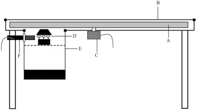
dismantled and the disc removed so that any further spore release through puffing would not affect the spore count in the other sectors. The chamber (E) which housed the apothecia had a slatted floor, below which was a reservoir which contained either water to
main-tain almost saturated air around the apothecia (>90 %
rh), or a saturated salt solution to maintain a different rh. The rh in the chamber was monitored using a humidity probe (Vaisala, Newmarket ; F). The entire apparatus was placed in a controlled environment
cabinet at 15x where lighting duration could be
con-trolled.
Preparation of apothecia for ascospore release experiments
Apothecia for use in the spore clock apparatus were produced in compost in a controlled environment
cabinet at 15xwith a 12 h light and 12 h dark regime as
described before. Initially, single sclerotia with three apothecia were selected for Experiments 1–3 (see be-low). These apothecia were 5–7 mm diam having just reached maturity. The cap was fully formed but not crenullated. In order to increase potential ascospore release and overcome the problem that three apothe-cia from a single sclerotium were often of different maturity, subsequent experiments used larger single apothecia (8–10 mm diam) from individual sclerotia. These apothecia were more mature and the caps were crenellated. Selected sclerotia with apothecia were embedded in damp sand in a clear plastic round ‘ polypot ’ (4 cm diam, 3.5 cm deep ; Kartell Plastics UK, Cambridge) and covered with another inverted polypot. Apothecia were then kept for 24 h in this condition under the same (synchronous) 12 h light regime as used for the spore clock apparatus. Just before use in experiments, spore release by puffing was induced in the apothecia at the end of a light period by removing the inverted polypot lid. Apothecia were
then ready for use and transferred to the spore clock apparatus in the polypots. Those apothecia not releas-ing a visible spore cloud were rejected.
Experiments on ascospore release
Effect of light regime
The first set of four experiments investigated the effect of light regime on ascospore release from apothecia of
Sclerotinia sclerotiorum at 15xand 90–95 % rh. Each
experiment was repeated three times (Table 1). Experi-ment 1 was set up to determine if ascospores were re-leased only during an 8 h light period. Three apothecia attached to a single sclerotium were exposed to a 12 h dark 12 h light regime but with time sectors rotated
every 16 and 8 h (Fig. 2a). Initially, the apothecia were
placed in the apparatus in the dark (acetate time sector 1) and the disc rotated to the next sector after 16 h. Thereafter the apothecia was exposed to acetate time sector 2 for 8 h (entirely in the light) and then to sector
3 for 16 h (4 h light+12 h dark) and so on. Hence, time
sectors 2, 4 and 6 were exposed entirely in the light (8 h) and sectors 3 and 5 to 4 h light and 12 h dark. The experiment was stopped at the end of the light period after 72 h (time sector 6). The same set-up was used
for experiments 2 and 3 (Fig. 2a, Table 1) except that
the light regimes were continuous light and continu-ous dark repectively. The disc was rotated at the same times so that time sectors were exposed for 8 or 16 h for comparison with Experiment 1. At the end of each experiment, ascospores were removed from each acet-ate time sector and counted using a haemocytometer as described before. Experiment 4 again tested the ef-fects of a 12 h light and 12 h dark but with synchron-ous movement of the acetate time sectors on the
rotating disc (Fig. 2b). Hence, each sector was exposed
to apothecia for either 12 h entirely in the dark or 12 h entirely in the light. For this experiment, three apothecia attached to three separate sclerotia of
C D
E F
B
[image:4.595.130.447.60.236.2]A
Fig. 1.Spore clock apparatus. A, perspex rotating disc ; B, removeable perspex lid ; C, motor ; D, apothecia ; E, detachable perspex chamber ; F, humidity probe.
S. sclerotiorum were placed in the spore clock appar-atus chamber in single poly pots. The experiment was stopped at the end of the dark period after 84 h (time sector 7).
Effect of relative humidity
Experiment 5 tested whether ascospores were released at rh levels below almost saturated air (90–95 % rh) at
15x. A 12 h light and 12 h dark regime was used with
movement of the time sectors on the rotating disc every
24 h (Fig. 2c). The spore clock apparatus reservoir
contained a saturated salt solution which maintained the apothecia at 65–70 % rh as measured by the hu-midity probe. Five apothecia from individual sclerotia were placed in the chamber (three in one polypot and two in another) and the experiment terminated at the end of the dark period after 168 h (7 d, time sector 7). Instead of removing spores from the acetate at the end of the experiment and counting, spore release in each acetate time sector was assessed visually for each of the five apothecia. This was a quicker method of de-termining whether there were large differences in spore discharge between treatments. The following score system was used : 0, no visible release ; 1, very light spore release (just visible) ; 2, light spore release ; 3,
average spore release ; 4, heavy spore release. The ex-periment was repeated 3 times. To compare ascospore release from Experiment 5 with release at 90–95 % rh, Experiment 6 was set up in the same way but with water in the apparatus reservoir to maintain saturated air (90–95 % rh) over the apothecia. Ascospore release was then assessed using the same visual scale.
Collection of ascospores and spore survival studies
For spore survival studies, ascospores were first ob-tained by trapping them on acetate sheets. The acetate was rubbed with a cloth to create a static charge and then held over a plastic box containing mature apo-thecia. When the box lid was removed, the resulting spore cloud impacted onto the acetate resulting in a monolayer of ascospores. In spore survival experi-ments, these acetate sheets were taped to the lids of clear plastic boxes (600 ml ; Malsar Kest) containing 150 ml of various saturated salt solutions in order to create rh levels within each box. Six rh levels were tested in the range 33–96 % at temperatures of 3, 5, 15,
20, 25 and 30xin a first experiment and 5, 10, 15, 20, 25
and 30xin a repeat experiment. The rh in each box was
[image:5.595.70.546.69.340.2]measured using a probe (Rotronic Instruments, Crawley) as the target rh varied with temperature Table 1.Summary of treatments in experiments carried out using the spore clock apparatus.
Experiment
no. Light regime
rh ( %)
Exposure time in each acetate time sector (h)
No. apothecia/ no. sclerotia used
Expt duration (h)
Spore release assessment method
1 12 h light/dark 90–95 8 or 16 3/1 72 Count
2 continuous dark 90–95 8 or 16 3/1 72 Count
3 continuous light 90–95 8 or 16 3/1 72 Count
4 12 h light/dark 90–95 12 3/3 84 Count
5 12 h light/dark 65–75 24 5/5 168 Visual
6 12 h light/dark 90–95 24 5/5 168 Visual
(a) (b) (c)
Fig. 2.Periods of apothecial exposure to acetate sectors attached to the perspex rotating disc from the spore clock apparatus for different experiments : (a) Experiments 1–3, (b) Experiment 4, and (c) Experiments 5–6. In each experiment, acetate was attached to the underside of the disc and divided into 8 sectors (1–8) as shown. The chamber containing apothecia (see Fig. 1, E) was completely covered by acetate sector 1 at the start of each experiment and subsequently rotated to give the following exposure times :a(Experiments 1–3) ; acetate sectors 1, 3 and 7 exposed for 16 h and sectors 2, 4 and 6 for 8 h.
(Winston & Bates 1960) and between experiments. To test spore viability at intervals, a small piece of acetate
(approx. 1 cm2
) was cut out from each temperature/ humidity treatment sheet and placed face down with 0.5 ml SDW on a PDA plate. The acetate piece was then gently spread around the plate with the water to remove spores. Germination of ascospores was then
assessed under the microscope (r100) after incubating
plates at 20xfor 16 h. A spore was considered to have
germinated if one or more germ tubes was longer than half the spore length. For each humidity/temperature combination, 100 spores were assessed from three random areas on each PDA plate. Experiment 1 was terminated after 16 wk and Experiment 2 after 21 wk.
Statistical Analyses
In the ascospore release experiments, spore count per apothecium for each acetate time sector was calculated
and converted to a rate of release per apothecium hx1
. Rates of spore release were then transformed to log-arithms. Analyses of variance (ANOVA) were then performed on these data or visual scores treating the same time sectors from each of the three repeat ex-periments as replicates. Standard error of the difference between the means (SEM) derived from ANOVA was calculated. For the ascospore survival experiments mean percentage germination was calculated at each assessment time for each of the temperature/humidity
3·5
3·0
2·5
2·0
1·5
1·0
0·5
0·0
0 12 24 36 48 60 72 84
Time sector
Light/dark periods SEM
Lo
g10
spores per apothecium h
–
1
h (a)
3·5
3·0
2·5
2·0
1·5
1·0
0·5
0·0
0 12 24 36 48 60 72 84
Time sector SEM
Lo
g10
spores per apothecium h
–
1
h (b)
1 2 3 4 5 6
3·5
3·0
2·5
2·0
1·5
1·0
0·5
0·0
0 12 24 36 48 60 72 84
Time sector SEM
Lo
g10
spores per apothecium h
–
1
h (c)
[image:6.595.122.451.58.522.2]1 2 3 4 5 6
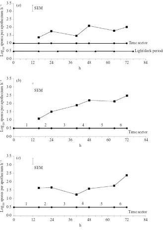
Fig. 3.Effect of different light regimes on rate of spore release from three apothecia from a single sclerotia ofSclerotinia sclerotiorumat 90–95 % rh and 15xC.a, 12 h light/dark ;b, continuous light ;c, continuous dark. Data are means of three experiments and represent the rate of spore release (log10spores per apothecium hx
1
) for each of the 8 h or 16 h time periods shown.SEM, standard error of the difference between the means derived from ANOVA.
treatments. Time to 50 % mortality was calculated for each treatment by interpolation between appropriate time periods.
R E S U L T S
Effect of light regime on ascospore release
In saturated air (90–95 % rh) there was continuous
ascospore release over 72 h at 15xwhether apothecia
from a single sclerotium were exposed to fluctuating
light and dark (Experiment 1 ; Fig. 3a), continuous
light (Experiment 2 ; Fig. 3b) or continuous dark
(Experiment 3 ; Fig. 3c). For all the light regimes there
was a significant increase in the rate of spore release
(Pf0.05) between the first time sector (16 h) and the
last (72 h) with a range of 10–316 ascospores released
per apothecium hx1. When apothecia from individual
sclerotia were exposed to acetate time sectors for 12 h synchronised in the 12 light/dark regime over 84 h at
15x and 90–95 % rh, there was again release in both
the light and dark periods (Experiment 4 ; Fig. 4). The rate of ascospore release also increased significantly
(Pf0.05) between the first time sector (12 h) and the
last (84 h) with a range of 100–1600 ascospores released
per apothecium hx1
. This confirmed that ascospore discharge was not confined to light or dark periods.
Effect of relative humidity on ascospore release
In both saturated air (90–95 % rh) and at 65–75 % rh,
apothecia released ascospores continuously at 15x in
the 12 h light and dark regime (Experiments 5 and 6 ; Fig. 5). For both humidity levels, there was an initial
significant increase (Pf0.05) in spore number score
after 36 h followed by regular release. However, during the 168 h (7 d) duration of the experiment however there was a significant decline in the spore number score between 36 h and 168 h for apothecia at 65–75 %
rh (Pf0.05), but this was not observed for apothecia
at 90–95 % rh.
Effect of temperature and humidity on ascospore survival
Ascospore survival at temperatures below 15x was
consistently high irrespective of rh (data not shown).
3·5
3·0
2·5
2·0
1·5
1·0
0·5
0·0
0 12 24 36 48 60 72 84
Time sector SEM
Lo
g10
spores per apothecium h
–
1
h
1 2 3 4 5 6
[image:7.595.132.486.67.206.2]Light/dark periods 7
Fig. 4.Effect of 12 h light/dark regime on rate of spore release from three apothecia from three sclerotia ofSclerotinia sclerotiorumat 90–95 % rh and 15xC. Data are means of three experiments and represent the rate of spore release (log10spores per apothecium hx1
) for each of the 12 h periods shown.SEM, standard error of the difference between the means derived from ANOVA.
4·0
3·5
3·0
2·5
2·0
1·5
1·0
0·5
0·0
0 12 24 36 84 108 120 168
Time sector SEM
Spore release score
h
1 2 3 4 5 6
Light/dark periods 7
SEM
[image:7.595.136.488.280.418.2]48 60 72 96 132 144 156
Briefly, at temperatures of 3 and 5x, ascospore germi-nation was 80 % or above over the 16 wk of Exper-iment 1, except at the highest rh tested for each temperature, which was 94 and 96 % rh respectively. For these rh values, the time to 50 % mortality was
9.5 wk at 3xand 6.6 wk at 5x. However, in Experiment
2, ascospore germination was 80 % or above for all rhs
tested at 5 and 10x, but the highest final rh values
achieved were 91 % and 93 % respectively. For
tem-peratures of 15xand above, high humidity combined
with increasing temperature reduced ascospore survival (Fig. 6). For rh values above 70 %, at least 50 %
mor-tality was observed at 15, 20, 25 and 30xby the end of
the experiments. At 30x, 50 % of spores were killed
within 2–3 wk for rh>80 % while at 40–70 % rh, 50 %
mortality occurred after 4–6 wk (Fig. 7). At 25x, 50 %
mortality occurred at 2–7 wk at 80–93 % rh and
8–15 wk at 40–70 % rh. At 20 and 15x, few spores were
killed below 60 % rh. 50 % mortality occurred at 15–18 wk at 75 % rh and after 3 wk at 92 % rh. Although Figs 6–7 clearly show that both final asco-spore viability and rate of loss of viability are related
both to temperature and humidity, we were unable to derive a model to describe these relationships satisfac-torily. This was in part due to 50 % mortality being not reached for many of the temperature and humidity treatments for the duration of the experiments.
D I S C U S S I O N
This is the first time that continual discharge of
Sclero-tinia sclerotiorum ascospores has been quantified in
the laboratory, and the results showed that spore re-lease occurred both in light and dark. The greater rates of ascospore release per apothecium were observed for more mature apothecia with crenullated cups and for experiments with a single apothecium on an indi-vidual sclerotium. Raynal (1990) also observed greater
spore release in S. trifoliorumfor larger apothecia. In
addition, Raynal calculated that an apothecium of
S. trifoliorumcould release up to 5r106spores over its
lifetime of 20 d at 15xwhile Abawi & Grogan (1979)
report thatS. sclerotiorumapothecia may release up to
100 90 80 60 50 40 30 20 10 0 70
30 °C
2 4 6 8 10 1214 16 18 20 22 100 80 70 60 90 50 40 30
Humidity (% RH)
Time (weeks) Ascospore ger mination 100 90 80 60 50 40 30 20 10 0 70
25 °C
2 4 6 8 10 1214 16 18 20 22 100 80 70 60 90 50 40 30
Humidity (% RH)
Time (weeks) Ascospore ger mination 100 90 80 60 50 40 30 20 10 0 70
20 °C
2 4 6 8 10 1214 16 18 20 22 100 80 70 60 90 50 40 30
Humidity (% RH)
Time (weeks) Ascospore ger mination 100 90 80 60 50 40 30 20 10 0 70
15 °C
2 4 6 8 10 1214 16 18 20 22 100 80 70 60 90 50 40 30
Humidity (% RH)
Time (weeks) Ascospore ger
[image:8.595.87.485.59.438.2]mination
Fig. 6.Effect of humidity onSclerotinia sclerotiorumascospore viability (as measured by % germination) at 15–30xC. Data plotted are from two experiments (&, Experiment 1 ;m, Experiment 2). There was little decrease in viability at temperatures below 15x(see Results).
3r107
ascospores. In comparison, at the maximum
rate of 1600 ascospores hx1
observed for S.
sclero-tiorumapothecia in this study, corresponds to a release
of 7.6r105
spores over 20 d.
Ascospore release did not decrease during dark periods in our experiments and this is in contrast to
some other ascomycetes such as Ascobolus crenulatus
where although there is a steady ‘ background ’ dis-charge in the dark, levels of spore release are much greater in the light (Ingold 1971). Raynal (1990) also
reported that light was more conducive toS. trifoliorum
ascospore release than dark at 15xalthough no data
were presented. Ascospore release byS. sclerotiorumor
S. trifoliorum in the field also shows a diurnal
distri-bution pattern with peak levels occurring in the middle of the day (Harthill 1980, Raynal 1990, McCartney & Lacey 1991). Harthill (1980) suggested that this was because ascospore discharge is only initiated in light although the laboratory data to support this was not presented. As this study showed that large numbers of ascospores could be discharged in the dark, it is most likely that the diurnal pattern of release is caused by an increase in temperature during the day rather the effect of light itself, as suggested by Raynal (1990). Indeed,
spore discharge is weak inS. trifoliorumandS.
sclero-tiorum at 5–10x (Newton & Sequeira 1972, Raynal
1990) and preliminary experiments with the spore clock used in this study have also found increased ascospore
discharge at 20–25x compared to 15x. This suggests
that at higher temperatures there is a faster maturation rate of asci within the cup of the apothecium and hence more available for release. As well as affecting asco-spore release directly, temperature also affects the
lon-gevity of S. sclerotiorum and S. trifoliorum apothecia
(Newton & Sequeira 1972, Raynal 1990) with spore
discharge in S. trifoliorum lasting over a month at
5–10x, 3 wk at 15–20x and less than 10 d at 25x
(Raynal 1990).
Previous laboratory observations of ascospore
re-lease byS. sclerotiorum or S. trifoliorum have always
been in saturated air (Newton & Sequeira 1972, Raynal 1990) but this study has shown for the first time that
continuous release ofS. sclerotiorumascospores occurs
in non-saturated air at 65–75 % rh. High rh periods at night followed by decreasing rh the following day may therefore be unnecessary for release of ascospores in the field as suggested by McCartney & Lacey (1990). However, the lower rh level tested in this study did cause a decline in ascospore release over 7 d compared to saturated air (90–95 % rh) indicating that the lon-gevity of the functioning apothecium was affected.
The survival of S. sclerotiorum ascospores was
re-duced at high temperature and rh, a trend also ob-served by Caesar & Pearson (1983). Increasing rh has
been shown to decreaseS. sclerotiorumspore survival
elsewhere (Partyka & Mai 1962, Abawi & Grogan 1975), but ascospores survived much longer in our experiments than in any of these other studies. For
in-stance, at 25 and 30x, Caesar & Pearson reported that
ascospores exposed to >60 % rh survived between
2–8 d with 50 % mortality within 4–5 d. This compares
to 2–3 wk at 30xin this study. Similarly, at 5 and 10x,
Caesar & Pearson reported that ascospores exposed to
>80 % rh did not survive beyond 16 d, whereas in our
experiments viability was maintained at these tem-peratures for the 16 or 21 wk duration of the experi-ments. This discrepancy may be due to the origin of the
S. sclerotiorumisolate used in each study. CertainlyS.
sclerotiorumis genetically variable (Carpenter,
Framp-ton & Stewart 1999) and carpogenic germination of
25
20
15
10
5
0
20 40 60 80 100
30 ºC
W
eeks
RH
25
20
15
10
5
0
20 40 60 80 100
25 ºC
W
eeks
RH
25
20
15
10
5
0
20 40 60 80 100
20 ºC
W
eeks
RH
25
20
15
10
5
0
20 40 60 80 100
15 ºC
W
eeks
[image:9.595.77.294.63.550.2]RH
sclerotia of isolates of different geographic origin have been shown to have different temperature optima (Huang & Kozub 1991). In addition, the rh values re-ported by Caesar & Pearson (who also used saturated salt solutions) were not measured independently. However, small variations in rh values in the experi-ments by Caesar & Pearson (1983) would not account for the difference in spore survival with our study.
S. sclerotiorum ascospore survival is also affected
ad-versely byUVlight (Caesar & Pearson 1983) so results
from this study would not necessarily be directly transferable to the field. However, germination of ascospores in free water and subsequent infection can
occur between 10 and 25x (Abawi & Grogan 1975,
Phillips 1994, Clarkson, Whipps & Young 2001) on plants and also at relative humidities approaching saturation (Grogan & Abawi 1975). In addition, the survival of germinated ascospores and appressoria on or in plant tissue may be greater than ungerminated spores (Grogan & Abawi 1975). Therefore, it is unlikely that spore survival is a limiting factor to infection by
S. sclerotiorum in the UK as conditions conducive to
spore germination would occur most days (or nights) within the growing season of susceptible crops.
The factors affecting the availability of viable
asco-spore inoculum in the field from apothecia ofS.
sclero-tiorum in the field are complex. From this and other
studies, ascospore release byS. sclerotiorumin the field
has the potential to take place during the night or day over a range of conditions in a continuous manner during the life of the apothecium. More work though is required to assess the effect of fluctuating temperature and rh on ascospore discharge and also to investigate under what field conditions, puffing of apothecia may occur.
The balance between greater ascospore discharge at
20–25xin S. sclerotiorum(Newton & Sequeira 1972),
with a corresponding decrease in apothecial longevity
(as observed withS. trifoliorum; Raynal 1990), may be
the key factor influencing the inoculum potential at different times of year (providing apothecia are pres-ent). Hence, in the Summer, large numbers of spores may be released over short periods of time as apothecia quickly die, whereas in the cooler Spring and Autumn months, fewer spores would be released but over more extended periods of time as apothecia survive longer. Further work is therefore required to assess the effect of temperature and humidity on apothecial longevity not addressed in the short-term experiments described here. The effect of soil moisture which, when high, may also help maintain a functioning apothecium (Raynal 1990) must also be considered.
Temperature and soil moisture are key factors
affecting carpogenic germination of S. sclerotiorum
sclerotia to produce apothecia (Phillips 1987, Clarkson
et al. 2001) and current work is addressing how this
data may be used in predicting the appearance of apo-thecia in the field. This information, combined with the knowledge of conditions governing ascospore release
and survival, may be useful in assessing when there are periods of high inoculum potential in the field. Fungi-cide sprays could then be targeted for these high risk periods leading to a more rational approach to chemi-cal control of Sclerotinia disease.
R E F E R E N C E S
Abawi, G. S. & Grogan, R. G. (1975) Source of primary inoculum and effects of temperature and moisture on infection of beans by
Whetzelinia sclerotiorum.Phytopathology65: 300–309.
Abawi, G. S. & Grogan, R. (1979) Epidemiology of diseases caused bySclerotiniaspecies.Phytopathology69: 899–903.
Bardin, S. D. & Huang, H. C. (2001) Research on biology and con-trol of Sclerotinia diseases in Canada.Canadian Journal of Plant Pathology23: 88–98.
Boland, G. J. & Hall, R. (1987) Epidemiology of white mold of white bean in Ontario.Canadian Journal of Plant Pathology9: 218–224. Boland, G. J. & Hall, R. (1994) Index of plant hosts ofSclerotinia
sclerotiorum.Canadian Journal of Plant Pathology16: 93–108. Caesar, A. J. & Pearson, R. C. (1983) Environmental-factors
affect-ing survival of ascospores of Sclerotinia sclerotiorum. Phyto-pathology73: 1024 –1030.
Carpenter, M. A., Frampton, C. & Stewart, A. (1999) Genetic vari-ation in New Zealand populvari-ations of the plant pathogen Sclero-tinia sclerotiorum.New Zealand Journal of Crop and Horticultural Science27: 13–21.
Clarkson, J. P., Whipps, J. M. & Young, C. S. (2001) Epidemiology ofSclerotinia sclerotiorumon lettuce. InProceedings of Sclerotinia 2001 – The XI International Sclerotinia Workshop, York, UK, July 2001(C. S. Young & K. J. D. Hughes, eds) : 79. Central Science Laboratory, York.
Dickson, L. F. & Fisher, W. R. (1923) A method of photo-graphing spore dispersal from apothecia. Phytopathology 13: 30–32.
Grogan, R. G. & Abawi, G. S. (1975) Influence of water potential on growth and survival ofWhetzelina sclerotiorum. Phytopathology
65: 122–138.
Harthill, W. F. T. (1980) Aerobiology of Sclerotinia sclerotiorum
andBotrytis cinereaspores in New Zealand tobacco crops.New Zealand Journal of Agricultural Research23: 259–262.
Harthill, W. F. T. & Underhill, A. P. (1976) Puffing in Sclerotinia sclerotiorumand S. minor. New Zealand Journal of Botany 14: 355–358.
Huang, H. C. & Kozub, G. C. (1991) Temperature requirements for carpogenic germination of sclerotia of Sclerotinia sclerotiorum
isolates of different geographic origin. Botanical Bulletin of Academia Sinica32: 279–286.
Ingold, C. T. (1971) Fungal Spores : their liberation and dispersal. Oxford University Press, London.
Ingold, C. T. & Marshall, B. (1963) Further observations on light and spore discharge in certain pyrenomycetes.Annals of Botany27: 481– 491.
Jayachandran, M., Willetts, H. J. & Bullock, S. (1987) Light and scanning electron-microscope observations on apothecial develop-ment ofSclerotinia sclerotiorum,Sclerotinia trifoliorumand Sclero-tinia minor. Transactions of the British Mycological Society89: 167–178.
Jones, D. (1974) Ultrastucture of the stipe and apothecium of Sclero-tinia sclerotiorum.Transactions of the British Mycological Society
63: 386–389.
Kosasih, B. D. & Willetts, H. J. (1975) Ontogenetic and histochem-ical studies of the apothecium ofSclerotinia sclerotiorum.Annals of Botany39: 185–191.
McCartney, H. A. & Lacey, M. E. (1991) The relationship between the release of ascospores ofSclerotinia sclerotiorum, infection and disease in sunflower plots in the United Kingdom. Grana 30: 486– 492.
Mylchreest, S. J. & Wheeler, B. E. J. (1987) A method for inducing apothecia from sclerotia ofSclerotinia sclerotiorum.Plant Pathol-ogy36: 16–20.
Newton, H. C. & Sequeira, L. (1972) Ascospores as the primary infective propaguleof Sclerotinia sclerotiorumin Wisconsin.Plant Disease56: 798–802.
Partyka, R. E. & Mai, W. F. (1962) Effects of environment and some chemicals on Sclerotinia sclerotiorum in laboratory and potato field.Phytopathology52: 766–770.
Phillips, A. J. L. (1987) Carpogenic germination of sclerotia of
Sclerotinia sclerotiorum: A review.Phytophylactica19: 279–283. Phillips, A. J. L. (1994) Influence of fluctuating temperatures and
interrupted periods of plant-surface wetness on infection of bean-leaves by ascospores ofSclerotinia sclerotiorum.Annals of Applied Biology124: 413– 427.
Raynal, G. (1990) [Kinetics of the ascospore production ofSclerotinia trifoliorum(Eriks) in growth chamber and under natural climatic conditions – practical and epidemiologic incidence.]Agronomie10: 561–572. [In French.]
Saito, I. (1973) Initiation and development of apothecial stipe primordia in sclerotia ofSclerotinia sclerotiorum.Transactions of the Mycological Society of Japan14: 343–351.
Sansford, C. E. & Coley-Smith, J. R. (1992) Production and germi-nation of sclerotia ofSclerotinia sclerotiorum.Plant Pathology41: 154 –156.
Winston, P. W. & Bates, D. H. (1960) Saturated solutions for control of humidity in biological research.Ecology41: 232–237.