Introduction
β-catenin is known to function as an adhesion molecule that is associated with E-cadherin and actin filaments at the cell membrane [1]. In ad-dition, it has been shown that β-catenin can act as a transcriptional factor involved in a number of cellular signaling pathways such as the Wnt canonical pathway (WCP) [2, 3]. In the WCP, β -catenin is normally sequestered by the so-called 'destruction complex', which consists of glyco-gen synthase kinase-3β (GSK3β), the adenoma-tous polyposis coli, axin and casein kinase 1 [4, 5]. Upon ligation of the soluble Wnt proteins to their receptors, the dishevelled proteins (Dvl's) will become phosphorylated, which is believed to result in inactivation and phosphorylation of GSK3β, leading to the dissociation of the de-struction complex. Consequently, β-catenin is allowed to evade proteasome degradation, ac-cumulate in the cytoplasm and translocate to
the nucleus. Forming heterodimers with T-cell factor/lymphoid enhancer-binding factor (TCF/ LEF) in the nucleus, β-catenin has been shown to regulate the expression of a wide range of important genes including c-myc and cyclin D1
[6-9].
β-catenin has been implicated in the patho-genesis of a wide range of human cancer [10]. There are also links between β-catenin and breast cancer. For instance, the expression of a β-catenin mutant with an abnormally high stabil-ity has been shown to induce breast adenocarci-nomas in a transgenic mouse model [11]. By immunohistochemistry, the expression of β -catenin in breast cancer (reported to be up to 60% of the cases) has been reported to signifi-cantly correlate with a poor prognosis or relapse in breast cancer patients in previous studies [12-14]. A few previous studies have shed light to the mechanisms underlying the relatively
Original Article
STAT3 upregulates the protein expression and
trans-criptional activity of
β
-catenin in breast cancer
Hanan Armanious, Pascal Gelebart, John Mackey1, Yupo Ma2, Raymond Lai
Department of Laboratory Medicine and Pathology, and the 1Department of Oncology, Cross Cancer Institute and
University of Alberta, Edmonton, Alberta, Canada; the 2Department of Pathology, The State University of New York at
Stony Brook, Stony brook, NY, USA
Received July 6, 2010; accepted July 23, 2010; available online July 25, 2010
Abstract: The expression of β-catenin detectable by immunohistochemistry has been reported to be prognostically important in breast cancer. In this study, we investigated the mechanism by which β-catenin is regulated in breast cancer cells. Our analysis of the gene promoter of β-catenin revealed multiple putative STAT3 binding sites. In sup-port of the concept that STAT3 is a transcriptional regulator for β-catenin, results from our chromatin immunoprecipi-tation studies showed that STAT3 binds to two of the three potential STAT3-binding sites in the gene promoter of β -catenin (-856 and -938). Using our generated MCF-7 cell clones that carry an inducible STAT3C construct, we found that the expression levels of STAT3C significantly correlated with the transcriptional activity of β-catenin. Similar ob-servations were made when we subjected two breast cancer cell lines (MCF-7 and BT-474) to STAT3 knock-down or transient gene transfection of STAT3C. Using immunohistochemistry, we found that pSTAT3 and β-catenin signifi-cantly correlated with each other (p=0.003, Fisher’s exact test) in a cohort of primary breast tumors (n=129). To con-clude, our results support the concept that STAT3 upregulates the protein expression and transcriptional activity of β -catenin in breast cancer, and these two proteins may cooperate with each other in exerting their oncogenic effects in these tumors.
high level of β-catenin expression in a subset of breast cancer. For instance, the WCP, which is known to regulate the expression and activity of β-catenin, is known to be constitutively active in a subset of breast cancer [15]. In another study, it has been shown that manipulation of the WCP can modulate β-catenin in breast cancer cells [16]. In addition to the WCP, other mechanisms also may be involved in regulating β-catenin in breast cancer. For instance, Pin1 was found to promote the dissociation of β-catenin from the destruction complex, and thus, increasing its stability [17]. Other studies showed that p53 downregulates β-catenin through ubiquitylation [18, 19]. Thus, the high level of β-catenin ex-pression in a subset of breast cancer may be multi-factorial.
Signal transducer and activator of transcription-3 (STATtranscription-3) belongs to a family of latent transcrip-tion factors the STAT family [20]. In breast can-cer, STAT3 is constitutively activated in approxi-mately 50-60% of primary breast tumors; down-regulation of STAT3 resulted in decrease in the tumorigenecity of breast cancer cells xeno-grafted in nude mice [21, 22]. Blockade of STAT3 using a dominant negative construct has been recently shown to decrease the nuclear localization and transcriptional activity of β -catenin in colon cancer cell lines [23]. Given that both β-catenin and STAT3 are activated in a subset of breast tumors, we hypothesized that STAT3 may represent another mechanism by which β-catenin is regulated in breast cancer cells. In addition, we evaluated the biological and clinical significance of β-catenin in breast cancer.
Materials and methods
Cell lines and tissue culture
MCF-7 and BT-474 cell lines were obtained from American Type Culture Collection (Manassas, VA, USA). They were grown at 370C and 5% CO2
and maintained in Dulbecco’s modified Eagle’s medium (Sigma-Aldrich, St. Louis, MO, USA). The culture media were enriched with 10% fetal bo-vine serum (Life Technologies, Carlsbad, CA, USA). MCF-7 cells permanently transfected with the tetracycline-controlled transactivator and TRE-STAT3C plasmids (labeled STAT3Ctet-off
MCF-7) have been described previously [22], this stable cell line was maintained by the addition of 800 μg/ml geneticin (Life Technologies, Inc.) to the culture media.
Subcellular protein fractionation, Western blot analysis and antibodies
For subcellular protein fractionation, we em-ployed a kit purchased from Active Motif (Carlsbad, CA, USA) and followed the manufac-turer’s instructions. Preparation of cell lysates for Western blots was done as follows: cells were washed twice with cold phosphate-buffered saline (PBS, pH=7.0), and scraped in RIPA lysis buffer (150 mM NaCl, 1% NP-40, 0.5% deoxycholic acid, 0.1% SDS, 50mM Tris pH 8.0) supplemented with 40.0 μg/mL leu-peptin, 1 μM pepstatin, 0.1 mM phenylmethyl-sulfonyl-fluoride and sodium orthovandate. Cell lysates were incubated on ice for 30 minutes and centrifuged for 15 minutes at 15000g at 40C. Proteins in the supernatant were then
ex-tracted and quantified using the bicinchoninic acid protein assay (Pierce, Rockford, IL). Subse-quently, cell lysates were then loaded with 4x loading dye (Tris-HCl pH 7.4, 1%SDS, glycerol, dithiothreitol, and bromophenol blue), electro-phoresed on 8% or 10% SDS-polyacrylamide gels, and transferred onto nitrocellulose mem-branes (Bio-Rad, Richmond, CA, USA). After the membranes were blocked with 5% milk in Tris buffered saline (TBS) with Tween, they were incubated with primary antibodies. After wash-ings with TBS supplemented with 0.001% Tween-20 for 30 minutes between steps, secon-dary antibody conjugated with the horseradish peroxidase (Jackson Immunoresearch Laborato-ries, West Grove, PA, USA) was added to the membrane. Proteins were detected using en-hanced chemiluminescence detection kit (Pierce, Rockford, IL). Antibodies employed in this study includ anti-β-catenin (1:4000, BD Biosciences Pharmingen, San Diego, CA, USA), anti-STAT3 and anti-pSTAT3 (1:500, Santa Cruz Biotechnology, Santa Cruz, CA, USA), anti-FLAG, anti-HDAC, anti-α-tubulin and anti-β-actin (1:3000, Sigma-Aldrich).
β-catenin transcriptional activity assessed by TOPFlash/FOPFlash
cells were harvested and cell extracts were pre-pared using a lysis buffer purchased from Promega (Madison, WI, USA). The luciferase activity was assessed using 20 μL of cell lysate and 100 μL of luciferase assay reagent (Promega). The luciferase activity was normal-ized against the β-galactosidase activity, which was measured by incubating 20 μL of cell lys-ates in a 96 well plate with 20 μL of o-nitrophenyl-β-D galactopyranoside solution (0.8 mg/mL) and 80 μL H2O, absorbance was
meas-ured at 415 nm at 37ºC. Data are reported as means ± standard deviations of three independ-ent experimindepend-ents, each of which was performed in triplicates.
Gene transfection
Transient gene transfection of cell lines with various expression vectors were performed us-ing Lipofectamine 2000 transfection reagent (Invitrogen, Burlington, Ontario, Canada) accord-ing to the manufacture’s protocol. Briefly, cells were grown in 60 mm culture plates until they are ~90% confluence, culture medium was re-placed with serum-free Opti-MEM (Life Tech-nologies) and cells were transfected with the DNA-lipofectamine complex. For all in-vitro ex-periments, STAT3Ctet-off MCF-7 cells were
tran-siently transfected with 3 μg TOPFlash or FOP-Flash and 4 μg of β-galactosidase plasmid. To manipulate the expression level of STAT3C in these cells, various concentrations of tetracy-cline (Invitrogen) were added to the cell culture. For MCF-7 and BT-474, 2 μg of TOPFlash or FOPFlash, 3 μg of β-galactosidase plasmid and 2 μg of STAT3C (or an empty vector) were trans-fected.
Chromatin immunoprecipitation
Chromatin immunoprecipitation was performed using a commercially available kit according to the manufacturer’s protocol (Upstate, Char-lottesville, VA, USA). Briefly, DNA-protein was cross-linked using 1% formaldehyde for 10 min-utes at 370C. Cells were lysed using the SDS
buffer, followed by sonication. Immunoprecipita-tion was done using protein A/G agarose beads conjugated with either a rabbit anti-human STAT3 antibody or a rabbit IgG antibody over-night at 40C. The DNA-protein-antibody complex
was separated and eluted. DNA was extracted using Phenol/Chloroform/ethanol. Primer pairs were designed by Primer 3 Input 0.4 to detect
the β-catenin gene promoter region containing putative STAT3 binding sites. The primer se-quences are as follows: primer 1 forward: 5'-CCGAGCGGTACTCGAAGG-3' and reverse 5'-GTAT CCTCCCCTGTCCCAAG-3'; primer 2 forward: 5'-CCAAAGAAAAATCCCCACAA-3' and reverse 5'-TC CTTAGGAGTACCTACTGTGAACAA-3'; and primer 3 forward 5'-AATTGGAGGCTGCTTAATCG-3' and reverse 5'-TTCCATTTTTATCTGGTTCCAC-3'.
Short interfering RNA (siRNA)
siRNA for β-catenin were purchased from Sigma -Aldrich. siRNA for STAT3 were purchased from Qiagen Science (Mississauga, ON, Canada) and used as described before [25]. Scrambled siRNA was purchased from Dharmacon (Lafayette, CO, USA). siRNA transfections were carried out using an electro square electropora-tor, BTX ECM 800 (225V, 8.5ms, 3 pulses) (Holliston, MA, USA) according to the manufac-turer’s protocol, the dose of siRNA used was 100 picomole/1x106 cells. Cells were harvested
at 24 hours after transfection. The β-catenin or STAT3 protein levels were assessed by Western blot analysis to evaluate the efficiency of inhibi-tion.
MTS assay
MCF-7 cells transfected with either β-catenin siRNA or scrambled siRNA were seeded at 3,000 cells/well in 96-well plates. MTS assay was conducted following the manufacturer’s instructions (Promega). The measurements were obtained at a wavelength of 450 nM using a Biorad Micro plate Reader (Bio-Rad Life Sci-ence Research Group, Hercules, CA, USA). The absorbance values were normalized to the wells with media only using the microplate Manager 5.2.1 software (Biorad). All experiments were performed in triplicates.
Immunohistochemistry and breast cancer specimens
hu-man tissue samples has been reviewed and approved by our institutional ethics board. Im-munohistochemistry was performed using stan-dard techniques. Briefly, formalin-fixed, paraffin-embedded tissue sections of 4 μM thickness were deparafinized and hydrated. Heat-induced epitope retrieval was performed using citrate buffer (pH=6) in a microwave histoprocessor (RHS, Milestone, Bergamo, Italy). After antigen retrieval, tissue sections were incubated with 3% hydrogen peroxide for 10 minutes to block endogenous peroxidase activity. Tissue sections were then incubated with anti-β-catenin (1:50) and anti-pSTAT3 (1:50) overnight in a humidi-fied chamber at 4°C. All of these primary anti-bodies were the same as those used for West-ern blots. Immunostaining was visualized with a labeled streptavidin-biotin (LSAB) method using 3,3'-diaminobenzidine as a chromogen (Dako Canada Inc., Mississauga, Ontario, Canada) and counter-stained with hematoxylin. For pSTAT3, the absence of nuclear staining or the presence of definitive nuclear staining in <10% of tumor cells was assessed negative; the presence of nuclear staining in ≥10% of tumor cells was assessed positive. ALK-positive anaplastic large cell lymphomas served as the positive control, whereas the lymphoid cells in benign tonsils served as the negative control. For β-catenin, only nuclear staining was scored. Moderate to strong nuclear β-catenin staining was assessed positive whereas the absence or weak (i.e. not definitive) nuclear staining was scored negative. Epithelial cells in benign tonsils served as the positive control whereas lymphoid cells in ton-sils served as negative controls.
Statistical analysis
Data are expressed as mean +/- standard derivation. Unless stated otherwise, statistical
significance was determined using Student's t-test and statistical significance was achieved when the p value is <0.05.
Results
STAT3 binds to β-catenin gene promoter
DNA sequence analysis of the -1000 bases of the β-catenin gene promoter region revealed 7 consensus sequences for the STAT family, char-acterized by TTN (4-6) AA [26]. Three of these 7
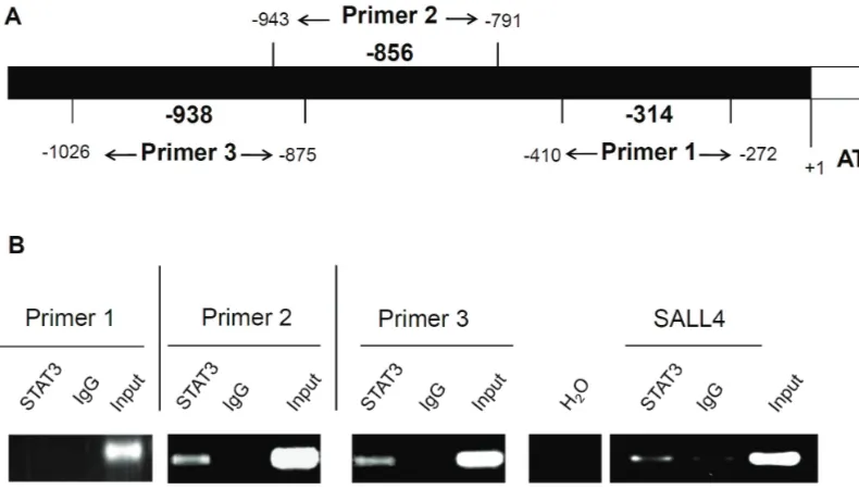
sequences contained the specific STAT3 binding sequence, namely TTMXXXDAA (D: A,G, or T;M:A or C)(summarized in Table 1) [27]. These puta-tive STAT3 binding sites are located at positions -314, -856 and -938, upstream of the ATG tran-scription initiation site. To provide direct evi-dence that STAT3 binds to these three sites, we performed chromatin immunoprecipitation us-ing MCF-7 cells. As shown in Figure 1, both primer 2 (to detect STAT3 binding to the -856 site) and primer 3 (to detect STAT3 binding to the -938 site) showed amplifiable products. In contrast, no detectable amplification was ob-served for primer 1 (to detect STAT3 binding to the -314 site). The input lanes were included as a control for the PCR effectiveness. PCR without the addition of DNA templates was used as a negative control. The SALL4 primer served as the positive control, as published previously [28].
STAT3 regulates the transcriptional activity and protein levels of β-catenin
To determine if the expression of STAT3 affects the transcriptional activity and/or protein level of β-catenin, we subjected two breast cancer cell lines (MCF-7 and BT-474) to STAT3 knock-down using siRNA. As shown in Figure 2A, trans-Table 1. Putative STAT binding sites on human β-catenin gene promoter
Site number Location relative to ATG Consensus sites TTN (4-6)AA
1 -254 TTCCCCAA
2 -314 TTCGGGAAA*
3 -782 TTGTTGAA
4 -856 TTAACCTAA*
5 -938 TTCTCCAAA*
6 -970 TTTCACAAA
7 -1000 TTCTCTATAA
[image:4.612.81.534.99.223.2]fection of STAT3 siRNA resulted in a substantial downregulation in the STAT3 protein levels in both cell lines. In the same blots, the protein levels of β-catenin were also decreased. We also found evidence that STAT3 regulates the transcriptional activity of β-catenin. As shown in Figure 2B, downregulating STAT3 using siRNA in MCF-7 cells resulted in a significant downregu-lation of the β-catenin transcriptional activity, as assessed by the TOPFlash/FOPFlash system (p=0.0006)(Figure 2B). Furthermore, transient transfection of STAT3C (i.e. constitutively active STAT3) in MCF-7 and BT-474 cells led to a significant increase in the transcriptional activity of β-catenin, as compared to transfection of an empty vector (p=0.003 for both cell lines) (Figure 3A). Also, we performed subcellular frac-tionation after STAT3C transfection on MCF-7 but we did not see any change in β-catenin nu-clear translocation (Figure 3B). Lastly, to further support that STAT3 regulates β-catenin, we
em-Figure 1. (A) Schematic representation of the three primers sets specific for three putative STAT3 binding sites in the
[image:5.612.103.498.83.308.2]β-catenin gene promoter region. (B) Chromatin immunoprecipitation was performed using MCF-7 cells. A rabbit poly-clonal antibody against STAT3 was used. Normal rabbit IgG antibody instead of anti-STAT3 served as a negative con-trol. PCR with both primer 2 and primer 3 revealed amplicons. In contrast, no amplicons were detected with primer 1 and in the negative control. SALL4 gene promoter primer served as the positive control.
[image:5.612.82.288.393.654.2]ployed our generated MCF-7 cell clone that has been stably transfected with an inducible (tetracycline-off) STAT3C expression vector (labeled as STAT3Ctet-off MCF-7), as previously
described [22]. As shown in Figure 4A, increas-ing levels of tetracycline added to these cells resulted in a gradual downregulation of the total STAT3 level as well as the FLAG tag. Using the TOPFlash/FOPFlash system, the luciferase level from cells treated with 20 μg/ml and 60 μg/ml tetracycline were significantly lower than that of negative controls (p=0.04 and 0.03 respec-tively) (Figure 4B). These results are derived from triplicate experiments. Also, we performed subcellular fractionation after downregulation of STAT3C levels using tetracycline; however we did not see any change in β-catenin nuclear translocation (data not shown).
Nuclear expression of β-catenin significantly
correlates with pSTAT3 expression in breast cancer samples
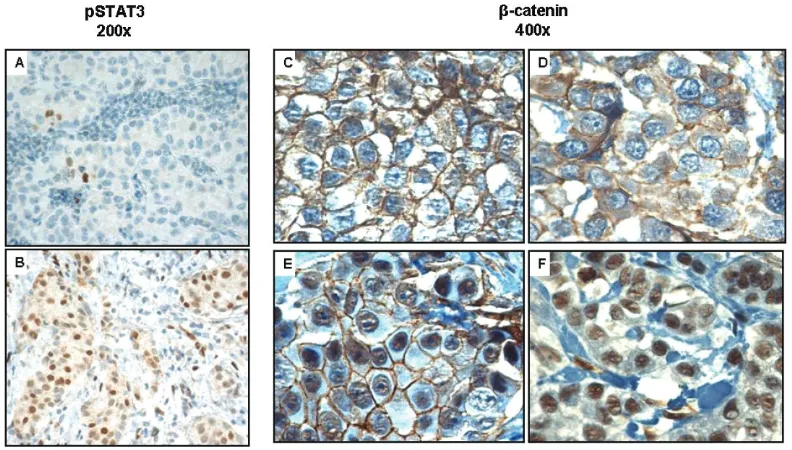
Using an anti-β-catenin antibody and immuno-histochemistry, we surveyed the expression of nuclear β-catenin in a cohort of formalin-fixed, paraffin-embedded breast cancer samples (n=129). Nuclear β-catenin was detected in 24 (19%) cases. Similarly, we surveyed the expres-sion of pSTAT3 using a monoclonal antibody and immunohistochemistry. pSTAT3 was detect-able in 61 (47%) cases (Tdetect-able 2). Importantly, the expression of these two markers signifi-cantly correlated with each other (p=0.003, Fisher exact test). However, the expression of these two markers did not significantly correlate with the overall survival. The staining results for pSTAT3 are illustrated in Figure 5A and 5B. The staining results for β-catenin are illustrated in
[image:6.612.138.477.82.450.2]Figure 5C-F. Cases scored negative for β-catenin showed no detectable nuclear staining, but some cases had staining on the cell membrane
(Figure 5C) whereas other cases showed both membraneous and cytoplasmic staining (Figure 5D). Cases scored positive for β-catenin showed definitive nuclear staining, with some cases also showing staining in the cytoplasm (Figure 5E) whereas other cases showing only nuclear stain-ing (Figure 5F).
β-catenin promotes cell growth in breast cancer
To investigate the biological importance of β -catenin on breast cancer, we downregulated β -catenin levels in MCF-7 using siRNA, and a sig-nificant downregulation of β-catenin was shown on Western blot (Figure 6A). An MTS assay was performed on day 1, 2, and 3 after β-catenin was downregulated. As shown in Figure 6B, there was a significant decrease in cell growth in cells treated with β-catenin siRNA, as com-pared to those treated with scrambled siRNA. Apoptosis, as detected by PARP and caspase 3 cleavages, was not detectable (not shown).
Discussion
Our presented data support the concept that STAT3 is a regulator of β-catenin in breast can-cer. Specifically, we found that the gene pro-moter of β-catenin carries multiple STAT3 con-sensus sequences and our chromatin precipita-tion experiments provided direct evidence of STAT3 binding at two specific sites (856 and -938) in the β-catenin gene promoter region. Furthermore, in two different breast cancer cell lines, we found evidence that the protein level and transcriptional activity of β-catenin can be modulated in response to a change in the ex-pression and/or activity of STAT3, in both tran-sient and stable transfection conditions. In fur-ther support that STAT3 regulates β-catenin in breast cancer, our immunohistochemical stud-ies using a large cohort of breast tumors re-vealed a significant correlation between the expression of pSTAT3 and β-catenin. Our conclu-sion regarding β-catenin being a downstream target of STAT3 echoes the findings described in a recent study of colon cancer, which showed that STAT3 activity regulates the transcriptional activity of β-catenin in colon cancer cells [23]. In contrast with the same study, we did not detect any change in the nuclear translocation of β -catenin following STAT3 knockdown using nu-clear/cytoplasmic fractionation. Based on our overall study results, the regulation of the tran-scriptional activity of β-catenin by STAT3 in
Figure 4. (A) Downregulation of exogenous STAT3C using tetracycline in STAT3Ctet-off MCF-7 cells was
revealed by western blot analysis, as the expression levels of the FLAG tag and total STAT3 were gradually decreased with increasing concentrations of tetracy-cline. Cell lysates were prepared 24 hours after tetra-cycline was added to the cell culture. B) STAT3Ctet-off
[image:7.612.84.282.82.360.2]MCF-7 cells treated with 20 μg/ml and 60 μg/ml tetracycline had a significant decrease in the tran-scriptional activity of β-catenin, as compared to cells without tetracycline (p=0.04 and 0.03 respectively). Results were derived from four independent experi-ments, each performed in triplicate.
Table 2. Immunohistochemistry of pSTAT3 and β-catenin in breast cancer patients’ samples (p=0.003)
β-catenin
positive βnegative-catenin Total
pSTAT3 positive
18 43 61
pSTAT3
negative 6 62 68
[image:7.612.80.290.571.650.2]breast cancer appears to be related to the ob-servations that STAT3 controls the total protein level of β-catenin.
In the present study, we have presented evi-dence that STAT3 can directly regulate the gene
transcription of β-catenin and its protein expres-sion level. We are also aware of other mecha-nisms by which STAT3 can potentially regulate β -catenin through modulating the upstream of the WCP. Specifically, the gene promoter of
[image:8.612.108.504.84.309.2]Wnt3a has been shown to carry the consensus
Figure 5. An immunohistochemical study revealing the correlation between pSTAT3 and β-catenin expression in a cohort of paraffin-embedded breast tumors. A pSTAT3-negative case containing only rare positive cells (i.e. <10%) is shown in A, whereas a pSTAT3-positive case containing ≥10% stained cells is shown in B. pSTAT3 staining was largely nuclear. Staining for β-catenin is illustrated in Figure 5C-F. Figure 5C and 5D show two negative cases in which no definitive nuclear staining was detectable. The case shown in 5C had membranous staining whereas the case in Fig-ure 5D had both membranous and cytoplasmic staining. FigFig-ure 5E and 5F show two positive cases, in which defini-tive nuclear staining was detectable. The case shown in Figure 5E also showed cytoplasmic staining, whereas the case in Figure 5F showed nuclear staining only.
[image:8.612.79.387.419.604.2]binding sequence for STAT3 [29]. STAT3 also has been shown to induce the expression of Wnt5a in rat cardiac myocytes [30, 31]. Taken together, it is possible that STAT3 regulates β -catenin via multiple mechanisms: 1) direct modulation of the gene transcription of β -catenin, and 2) modulate the secretion of differ-ent Wnt's, thereby regulating β-catenin via the WCP.
While we did not observe a prognostic signifi-cance for β-catenin in our cohort of breast can-cer patients, we are aware of the results of a previously published study which showed that β -catenin is prognostically important in breast cancer [14]. In contrast with the study by Lin et al, who scored the β-catenin regardless of the staining pattern [14], we considered β-catenin positivity only when the staining was definitively nuclear. We also would like to point out that, one of the authors (JM) in a paper recently pub-lished that β-catenin is useful in predicting re-lapse in breast cancer patients [12]; however, no significant correlation between β-catenin nuclear staining and the overall survival was found (personal communication).
Although the focus of this manuscript is to docu-ment the functional interaction of STAT3 and β -catenin, we also examined whether β-catenin is biologically important in breast cancer cell lines. As shown in this study, downregulation of β -catenin using siRNA inhibited cell growth in MCF -7. Growth inhibition induced by a downregula-tion of β-catenin has been observed for eso-phageal cancer, colon cancer and glioma [32-34]. β-catenin was found to promote cell migra-tion in a breast cell line [35], but we are not aware of any previous study in which the growth -promoting effect of β-catenin was examined in breast cancer. Thus, to our knowledge, these findings represent the first evidence that β -catenin promotes cell growth in breast cancer.
Both β-catenin and STAT3 have been shown to be oncogenic in various types of human cancer including breast cancer [11, 21]. Our findings described in this manuscript raise the possibility that the oncogenic effects of STAT3 may syner-gize with that of β-catenin. Of interest, it have been reported that β-catenin regulates the STAT3 transcriptional activity in esophageal cancer cell lines, thus potentially creating a positive a feedback loop between these two signaling proteins [36]. Since specific agents inhibiting STAT3 and β-catenin are available
[37, 38], our data provide the rationale for com-bining these inhibitors in treating specific forms of cancer, such as in a subset of breast cancer in which both STAT3 and β-catenin are acti-vated. In-vitro studies evaluating the potential synergistic effects of combining these agents will be of great interest.
Acknowledgement
This work was partly supported by operating research grants from the Canadian Cancer Soci-ety and the Alberta Cancer Foundation awarded to RL.
Please address correspondence to: Raymond Lai, MD, PhD, Department of Laboratory Medicine and Pathology, Cross Cancer Institute, 11560 University Avenue, Edmonton, Alberta, Canada T6G 1Z2, Tel: (780) 432-8338, Fax: (780) 432-8214, E-mail: rlai@ualberta.ca
References
[1] Huber AH, Weis WI. The structure of the beta-catenin/E-cadherin complex and the molecular basis of diverse ligand recognition by beta-catenin. Cell. 2001 May 4;105(3):391-402. [2] Brennan KR, Brown AM. Wnt proteins in
mam-mary development and cancer. J Mammam-mary Gland Biol Neoplasia. 2004 Apr;9(2):119-31. [3] Polakis P. Wnt signaling and cancer. Genes
Dev. 2000 Aug 1;14(15):1837-51.
[4] Clevers H. Wnt/beta-catenin signaling in devel-opment and disease. Cell. 2006 Nov 3;127 (3):469-80.
[5] Gordon MD, Nusse R. Wnt signaling: multiple pathways, multiple receptors, and multiple transcription factors. J Biol Chem. 2006 Aug 11;281(32):22429-33.
[6] He TC, Sparks AB, Rago C, Hermeking H, Zawel L, da Costa LT, Morin PJ, Vogelstein B, Kinzler KW. Identification of c-MYC as a target of the APC pathway. Science. 1998 Sep 4;281 (5382):1509-12.
[7] Mikels AJ, Nusse R. Wnts as ligands: process-ing, secretion and reception. Oncogene. 2006 Dec 4;25(57):7461-8.
[8] Miller JR. The Wnts. Genome Biol. 2002;3 (1):REVIEWS3001.
[9] Shtutman M, Zhurinsky J, Simcha I, Albanese C, D'Amico M, Pestell R, Ben-Ze'ev A. The cyclin D1 gene is a target of the beta-catenin/LEF-1 pathway. Proc Natl Acad Sci U S A. 1999 May 11;96(10):5522-7.
[10] Ewan KB, Dale TC. The potential for targeting oncogenic WNT/beta-catenin signaling in ther-apy. Curr Drug Targets. 2008 Jul;9(7):532-47. [11] Imbert A, Eelkema R, Jordan S, Feiner H, Cowin
mammary gland. J Cell Biol. 2001 Apr 30;153 (3):555-68.
[12] Asgarian N, Hu X, Aktary Z, Chapman KA, Lam L, Chibbar R, Mackey J, Greiner R, Pasdar M. Learning to predict relapse in invasive ductal carcinomas based on the subcellular localiza-tion of junclocaliza-tional proteins. Breast Cancer Res Treat. 2009 Sep 29.
[13] Lim SC, Lee MS. Significance of E-cadherin/ beta-catenin complex and cyclin D1 in breast cancer. Oncol Rep. 2002 Sep-Oct;9(5):915-28. [14] Lin SY, Xia W, Wang JC, Kwong KY, Spohn B,
Wen Y, Pestell RG, Hung MC. Beta-catenin, a novel prognostic marker for breast cancer: its roles in cyclin D1 expression and cancer pro-gression. Proc Natl Acad Sci U S A. 2000 Apr 11;97(8):4262-6.
[15] Benhaj K, Akcali KC, Ozturk M. Redundant ex-pression of canonical Wnt ligands in human breast cancer cell lines. Oncol Rep. 2006 Mar;15(3):701-7.
[16] Schlange T, Matsuda Y, Lienhard S, Huber A, Hynes NE. Autocrine WNT signaling contributes to breast cancer cell proliferation via the ca-nonical WNT pathway and EGFR transactiva-tion. Breast Cancer Res. 2007;9(5):R63. [17] Ryo A, Nakamura M, Wulf G, Liou YC, Lu KP.
Pin1 regulates turnover and subcellular local-ization of beta-catenin by inhibiting its interac-tion with APC. Nat Cell Biol. 2001 Sep;3(9):793 -801.
[18] Levina E, Oren M, Ben-Ze'ev A. Downregulation of beta-catenin by p53 involves changes in the rate of beta-catenin phosphorylation and Axin dynamics. Oncogene. 2004 May 27;23 (25):4444-53.
[19] Liu J, Stevens J, Rote CA, Yost HJ, Hu Y, Neufeld KL, White RL, Matsunami N. Siah-1 mediates a novel beta-catenin degradation pathway linking p53 to the adenomatous polyposis coli protein. Mol Cell. 2001 May;7(5):927-36.
[20] Turkson J. STAT proteins as novel targets for cancer drug discovery. Expert Opin Ther Tar-gets. 2004 Oct;8(5):409-22.
[21] Kunigal S, Lakka SS, Sodadasu PK, Estes N, Rao JS. Stat3-siRNA induces Fas-mediated apoptosis in vitro and in vivo in breast cancer. Int J Oncol. 2009 May;34(5):1209-20.
[22] Dien J, Amin HM, Chiu N, Wong W, Frantz C, Chiu B, Mackey JR, Lai R. Signal transducers and activators of transcription-3 up-regulates tissue inhibitor of metalloproteinase-1 expres-sion and decreases invasiveness of breast can-cer. Am J Pathol. 2006 Aug;169(2):633-42. [23] Kawada M, Seno H, Uenoyama Y, Sawabu T,
Kanda N, Fukui H, Shimahara Y, Chiba T. Signal transducers and activators of transcription 3 activation is involved in nuclear accumulation of beta-catenin in colorectal cancer. Cancer Res. 2006 Mar 15;66(6):2913-7.
[24] Staal FJ, Noort Mv M, Strous GJ, Clevers HC. Wnt signals are transmitted through
N-terminally dephosphorylated beta-catenin. EMBO Rep. 2002 Jan;3(1):63-8.
[25] Sekikawa A, Fukui H, Fujii S, Ichikawa K, Tomita S, Imura J, Chiba T, Fujimori T. REG Ialpha pro-tein mediates an anti-apoptotic effect of STAT3 signaling in gastric cancer cells. Carcinogene-sis. 2008 Jan;29(1):76-83.
[26] Seidel HM, Milocco LH, Lamb P, Darnell JE, Jr., Stein RB, Rosen J. Spacing of palindromic half sites as a determinant of selective STAT (signal transducers and activators of transcription) DNA binding and transcriptional activity. Proc Natl Acad Sci U S A. 1995 Mar 28;92(7):3041-5.
[27] Ehret GB, Reichenbach P, Schindler U, Horvath CM, Fritz S, Nabholz M, Bucher P. DNA binding specificity of different STAT proteins. Compari-son of in vitro specificity with natural target sites. J Biol Chem. 2001 Mar 2;276(9):6675-88.
[28] Bard JD, Gelebart P, Amin HM, Young LC, Ma Y, Lai R. Signal transducer and activator of tran-scription 3 is a trantran-scriptional factor regulating the gene expression of SALL4. FASEB J. 2009 May;23(5):1405-14.
[29] Li X, Placencio V, Iturregui JM, Uwamariya C, Sharif-Afshar AR, Koyama T, Hayward SW, Bhowmick NA. Prostate tumor progression is mediated by a paracrine TGF-beta/Wnt3a sig-naling axis. Oncogene. 2008 Nov 27;27 (56):7118-30.
[30] Fujio Y, Matsuda T, Oshima Y, Maeda M, Mohri T, Ito T, Takatani T, Hirata M, Nakaoka Y, Ki-mura R, Kishimoto T, Azuma J. Signals through gp130 upregulate Wnt5a and contribute to cell adhesion in cardiac myocytes. FEBS Lett. 2004 Aug 27;573(1-3):202-6.
[31] Miyagi C, Yamashita S, Ohba Y, Yoshizaki H, Matsuda M, Hirano T. STAT3 noncell-autonomously controls planar cell polarity dur-ing zebrafish convergence and extension. J Cell Biol. 2004 Sep 27;166(7):975-81.
[32] Pu P, Zhang Z, Kang C, Jiang R, Jia Z, Wang G, Jiang H. Downregulation of Wnt2 and beta-catenin by siRNA suppresses malignant glioma cell growth. Cancer Gene Ther. 2009 Apr;16 (4):351-61.
[33] Wang JS, Zheng CL, Wang YJ, Wen JF, Ren HZ, Liu Y, Jiang HY. Gene silencing of beta-catenin by RNAi inhibits cell proliferation in human eso-phageal cancer cells in vitro and in nude mice. Dis Esophagus. 2009;22(2):151-62.
[34] Huang WS, Wang JP, Wang T, Fang JY, Lan P, Ma JP. ShRNA-mediated gene silencing of beta-catenin inhibits growth of human colon cancer cells. World J Gastroenterol. 2007 Dec 28;13 (48):6581-7.
[36] Yan S, Zhou C, Zhang W, Zhang G, Zhao X, Yang S, Wang Y, Lu N, Zhu H, Xu N. beta-Catenin/TCF pathway upregulates STAT3 expression in hu-man esophageal squamous cell carcinoma. Cancer Lett. 2008 Nov 18;271(1):85-97. [37] Takemaru KI, Ohmitsu M, Li FQ. An oncogenic
hub: beta-catenin as a molecular target for cancer therapeutics. Handb Exp Pharmacol. 2008(186):261-84.
[38] Yue P, Turkson J. Targeting STAT3 in cancer:


