Original Article
Role of far upstream element binding protein 1 in
colonic epithelial disruption during dextran
sulphate sodium-induced murine colitis
Qiyun Tang1*, Weiwei Xia2*, Qianqian Ji2, Runzhou Ni2, Jian’an Bai1, Liren Li2, Yongwei Qin3
1Department of Gastroenterology, The First Affiliated Hospital of Nanjing Medical University, 300 Guangzhou
Road, Nanjing 210000, Jiangsu Province, People’s Republic of China; 2Department of Gastroenterology, Affiliated
Hospital of Nantong University, 20 Xisi Road, Nantong 226001, Jiangsu Province, People’s Republic of China;
3Department of Pathogen Biology, Nantong University Medical College, 19 Qixiu Road, Nantong 226001, Jiangsu
Province, People’s Republic of China. *Equal contributors.
Received March 13, 2014; Accepted April 26, 2014; Epub April 15, 2014; Published May 1, 2014
Abstract: Aim: Intestinal epithelial barrier is essential for maintaining normal intestinal homeostasis; its breakdown
leads to chronic inflammatory pathologies, such as inflammatory bowel diseases. Far upstream element binding protein 1 (FBP1) has been reported to play an important role in cell apoptosis and proliferation. We aimed to inves
-tigate the expression and the role of FBP1 in dextran sodium sulphate (DSS)-induced experimental colitis. Methods: Mice experimental colitis model was established by administration of DSS, and the expression and localization of FBP1 was examined using Western blot and immunohistochemistry. Colon epithelial cell line HT-29 was used to de
-termine the role of FBP1. In vitro study, the expression of FBP1 was determined in HT-29 cells stimulated with tumor necrosis factor α (TNF-α). HT-29 cells were transfected with FBP1 siRNA and then measured for viability. Results: Significant decreasing of FBP1 expression was found in mice colitis. In addition, FBP1 was cleaved and translocated from nucleus to cytoplasm during apoptosis. Downregulated expression of FBP1 induced cell cycle arrest. Conclu
-sions: We demonstrate that apoptosis-mediated cleavage of FBP1 and its decreased expression in epithelial cells
induces cell cycle arrest, which may play an important role in colonic epithelial disruption.
Keywords: Colitis, intestinal epithelial cells, FBP1
Introduction
Inflammatory bowel disease (IBD), including
both colitis and ileitis, is a chronic, relapsing,
and remitting inflammatory dysfunction, which
involving the mucosal layer of the colon and rectum [1]. It has been a worldwide health-care problem with a continually increasing
inci-dence. The etiology of IBD is complex and
incompletely understood. As researchers deep-en their knowledge regarding the mechanisms
underlying the pathogenesis of IBD, emerging
evidence has revealed that the intestinal thelium plays a central role [2]. Intestinal epi-thelial cells (IECs) which are the major composi-tions of intestinal epithelium are the primary cell type coming into contact with the external
environment and act as the host’s first line of
the defense against potential harmful
stimu-lants. IECs produce the mucus layer covering the entire length of the intestinal tract, whose role is to further protect the mucosal surface from harmful molecules and bacteria, and rein-force the overall intestinal barrier. Moreover, IECs also represent a core element of innate immunity within the gut mucosa, displaying a wide array of immune functions [3]. As such, IECs play an important role in maintaining the integrity of gut barrier and normal mucosal immune homeostasis. One form of epithelial
cell injury in the inflamed colonic mucosa has
been reported to involve the apoptosis of these cells [4]. Increased IEC apoptosis has been
detected at the acute inflammatory sites in IBD
increases during acute inflammation, the prolif -eration of epithelial cells is necessary to supply the reduction of intestinal cell library. The fur-ther decrease of the proliferation of intestinal epithelium, along with the increase of apopto-sis, contributes to the injury of the epithelial
barrier. However, molecular mechanisms that regulate intestinal epithelial barrier during IBD
are incompletely understood.
Far upstream element binding protein (FBP)
family includes three members which have been ascribed different functions in gene
regu-lation [8, 9]. The human FBP1 gene, also known as FUBP1, encodes a 644-amino acid protein with three well-defined domains: an amphipa -thic helix N-terminal domain, a tyrosine-rich
C-terminal transactivation domain and a DNA-binding central domain [10]. FBP1 was first identified as a DNA-binding protein that regu -lates c-Myc gene transcription [11]. In recent
years, mounting evidence suggests that FBP1
acts as an RNA-binding protein and regulates mRNA translation or stability of genes [12].
During retroviral infection, FBP1 binds to and
mediates replication of RNA from Enterovirus
71 [13]. In addition, FBP1 is also an important
oncoprotein overexpressed in several human carcinomas, which promotes cell proliferation in a cell cycle-dependent pathway [14, 15].
Undoubtedly, FBP1 is a multifunctional protein.
Previous studies reported that as a nuclear
pro-tein, FBP1 is cleaved by caspase-3 and -7 dur -ing apoptosis induced by various stimuli, which
leads to FBP1 translocation from the nucleus to the cytoplasm [16]. Hypothetically, since that IEC apoptosis increases at the acute inflamma
-tory sites in IBD might cause the change of FBP1 expression, the intestinal epithelial barri -er would be destructed by the alt-ered protein expression.
To clarify the mechanisms responsible for IBD
development, a variety of experimental models
for IBD has been developed, including a spon -taneous colitis model, induced colitis models, as well as transgenic and knockout colitis
mod-els [17, 18]. Dextran sulfate sodium (DSS) has
been used as one model to induce experimen-tal colitis, which is often used to model the
epi-thelial damage and a robust inflammatory response in IBDs. The aim of this study was to investigate the expression and role of FBP1 in colonic epithelial disruption during
DSS-indu-ced mice colitis.
Materials and methods
Animals and experimental colitis
Experiments were performed in accordance
with National Institutes of Health Guidelines for the Care and Use of Laboratory Animals (National Research Council, 1996, USA) and
were approved by the Chinese National
Committee to Use of Experimental Animals for
Medical Purposes, Jiangsu Branch. We obtained seven- to eight-week-old female C57Bl/6 mice (n = 22) weighing 18-20 g from the Experimental Animal Center of Nantong
University. Both control group (n = 9) and exper -imental group (n = 13) of mice were maintained
under specific pathogen-free conditions and
given free access to food. Control mice were given normal drinking water, while 4% dextran
sulfate sodium (4% (w/v) DSS; molecular
weight, 36,000-50,000; MP Biomedicals) was contained in the water of experimental group
from day 0 until day 7. And they were sacrificed on day 7 (n = 5). For recovery studies, DSS was administered for the first 7 days as indicated, then DSS was removed from the drinking water and mice (n = 5) were sacrificed 7 days after cessation of DSS treatment. Additional experi
-mental mice (n = 3) were sacrificed on day 0.
The control mice (n = 3 per time point) were
respectively sacrificed on day 0, 7, 14.
Assessment of colitis activity
To examine the severity of colitis, we evaluated
the body weight, disease activity index (DAI), and colon length. The DAI was determined by
scoring changes in animal weight, occult blood, positivity, gross bleeding, and stool
consisten-cy. We used five grades of weight loss (0, no
loss or weight gain; 1, 1-5% loss; 2, 5-10% loss; 3, 10-20% loss; 4, 20% loss), three grades of stool consistency (0, normal; 2, loose; and 4, diarrhea), and three grades of occult blood (0, negative; 2, occult blood-positive; and 4, gross
bleeding). After determination of the DAI, mice
were euthanized, the entire colon was removed from the cecum to the anus, and the colon length was measured as an indirect marker of
inflammation.
Cell culture and stimulation
The human colon epithelial cell line HT-29 was
purchased from Cell library, China Academy of
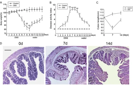
Figure 1. Multiple indicators is used to assess the success of DSS-induced colitis model. The changes of (A) body weight, (B) DAI, (C) colon length, and (D) colonic tissue structure by H&E-staining during the development of DSS-induced colitis in mice. Each value indicates the mean ± SE for 6 mouse. *(**)Significant difference at P < 0.05 (P
< 0.01) compared with control mice receiving water alone. Scale bar column, 200 µm.
BRL, Grand Island, NY, USA) medium containing 10% fetal bovine serum (FBS), 100 U/ml peni
-cillin, and 100 μg/ml streptomycin (GibCo BRL, Grand Island, NY, USA). Cultures were incubat -ed at 37°C in a 95% air/5% CO2 atmosphere. Cells were treated with different
concentra-tions (1, 10, 100 ng/ml) of TNF-α (Sigma-Aldrich, USA). Cultured HT29 cells were collect -ed, washed with phosphate buffer solution (PBS) and suspended in hypotonic buffer to achieve nuclear extracts. The cultured cells were homogenized, meanwhile nuclei were pel-leted. Then we removed the cytoplasmic ex- tracts and re-suspended nuclei in a low-salt buffer. A high-salt buffer was added to release soluble proteins from the nuclei, and the nuclei were removed by centrifugation. The nuclear extracts were dialyzed into a moderate salt solution.
Western blot analysis and antibodies
For Western blot analysis, colon tissues were dissected and flash-frozen at -80°C. To prepare the lysates, frozen samples were weighed and
minced on ice. The samples were homogenized in lysis buffer (1% NP-40, 50 mmol/L Tris, Ph =
7.5, 5 mmol/L EDTA, 1% SDS, 1% sodium deoxy
-cholate, 1% Triton-X100, 1 mmol/L PMSF, 10 μg/mL aprotinin, and 1 μg/mL leupeptin) and
centrifuged at 12,000 rpm and 4°C for 20 min to collect the supernatant. Cell cultures for immunoblot were lysed with sodium lauryl sul-fate loading buffer and stored at -80°C until use. After determining the protein
concentra-tion with the Bradford assay (Bio-Rad, Hercules, CA, USA), protein samples were subjected to SDS-polyacrylamide gel electrophoresis (SDS-PAGE) and transferred to a polyvinylidene difluoride filter (PVDF) membranes (Millipore, Bedford, MA, USA). The membranes were
blocked with 5% fat-free milk in TBST (20 mM Tris, 150 mM NaCl, 0.05% Tween-20) for 2 h at
room temperature, and then, the filters were
washed with TBST for three times and incubat-ed overnight with polyclonal antibody against
using the primary antibodies against anti-FBP1
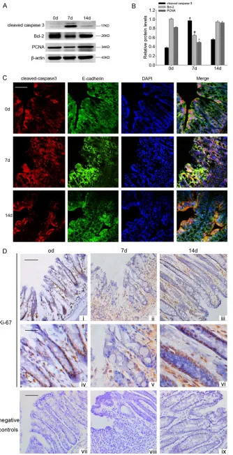
cleaved-cas-Figure 2. (A) Signifi -cant upregulation of cleaved-caspase 3, while downregulation of Bcl-2 and PCNA protein levels were detected
in the mice fed DSS. β-actin served as the
loading control. (B) The bar graph indicated the density of Bcl-2, cleaved-caspase 3 and
PCNA versus β-actin at each time point. Data
are presented as mean ± SEM. (n = 3, *, #, ^P
< 0.05,). (C) Double im
-munofluorescent
analy-sis showed that en-hanced co-localizing of cleaved-caspase 3 with E-cadherin was
observed after DSS administration. (D) Cell
cycle analysis using an anti-Ki-67 antibody in the colonic crypts
of DSS-induced colitis
mice. There were some anti-Ki-67 positive cells in the lower part of the
crypt before DSS ad
-ministration (i, iv). How -ever, the anti-Ki-67 pos-itive cells were reduced
in the mice fed DSS (ii,
v). After stopped
giv-ing DSS, the anti-Ki-67
positive cells were
in-creased significantly
at day 14 (iii, vi). Scale
bar (i-iii), 100 µm; Scale
bar (iv-vi), 50 µm. (vii-ix: negative controls).
pase 3 (anti-rabbit, 1:500; Cell Signali- ng), cleaved PARP (anti-rabbit, 1:500; Cell Signaling), cyclin A (anti-rabbit, 1:10- 00; Santa Cruz), cy-
clin D1 (anti-rabbit,
1:1000; Santa Cruz),
β-actin (anti-mouse,
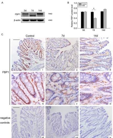
[image:4.612.92.426.71.723.2]Figure 3. Changes of FBP1 expression in colonic tissues during mice experimental colitis. (A) Significant downregula
-tion and cleavage of FBP1 was detected in the mice fed DSS in comparison to control mice. β-actin served as the loading control. (B) Quantitative analysis of the FBP1 protein level in mice colon with and without DSS treatment.
The data are means ± SEM. (n = 3, *, P < 0.05; significantly different from the controlled group). (C) Immunohisto
-chemistry with anti-FBP1 mouse monoclonal antibody on transverse cryosections of colonic tissues to identify the changes of FBP-1 immunoreactivity in DSS-induced mice colitis model at different time points. Scale bar (i-iii), 100 µm; Scale bar (iv-vi), 50 µm. (vii-ix: negative controls).
at-anti-rabbit conjugated horseradish peroxi-dase (1:2000, Southern-Biotech) for 2 h and
visualized using an enhanced
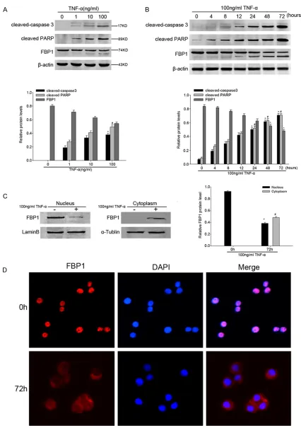
Figure 4. A. Cleaved-caspase 3, cleaved PARP and FBP1 protein in whole-cell lysates derived from different concen
-tration of TNF-α treated (24 h) HT-29 cells shown by representative western blots of FBP1 and the loading control, β-actin. Data are presented as mean ± SEM of 3 independent measurements (*, #P < 0.05). B. A representative
Immunofluorescent and immunohistochemical studies
Mice colon samples were freshly isolated and
frozen in O.C.T (Sakura Finetek, USA) or fixed in
10% neutral buffered formalin and embedded
in paraffin wax. Immunohistochemical studies were performed using paraffin-embedded sec
-tions (4 μm). All sec-tions were deparaffinized,
rehydrated, and thereafter, the sections were
processed in 10 mM citrate buffer (pH 6.0) and
heated to 121°C in an autoclave for 20 minutes to retrieve the antigen. After rinsing in PBS,
sec-tions were first treated with 3% hydrogen perox -ide to block endogenous peroxidase activity, and then blocked by 1.5% normal goat serum
for 20 min. For analysis for FBP1 and ki-67, sec -tions were incubated 2 h at room temperature
with primary antibody against FBP1 (diluted
1:800) and ki-67 (mouse; 1:1000; Santa Cruz
Biotechnology, USA). All slides were processed
using the peroxidase–anti-peroxidase method
(DAKO, Hamburg, Germany). After rinsing in
PBS, the peroxidase reaction was visualized by incubating the sections with the liquid mixture
DAB (0.1% phosphate buffer solution, 0.02%
diaminobenzidine tetrahydrochloride, and 3%
H2O2). After rinsing in water, the sections were counterstained with hematoxylin, dehydrated, and cover slipped.
Additional sets of sections from mice were
used for double immunofluorescence staining. Frozen sections were first blocked with 10%
normal serum-blocking solution species the same as the secondary antibody, containing 3% (w/v) BSA and 0.1% Triton X-100 and 0.05%
Tween-20 2 h at RT in order to avoid unspecific
staining. Then, the sections were double stained with primary antibodies against cleaved-caspase 3 (anti-mouse, 1:200) and anti-E-cadherin (anti-rabbit, a marker of
epithe-lial cells, 1:100, Santa Cruz). Briefly, sections
were incubated with both primary antibodies overnight at 4°C, subsequently with secondary
antibodies (donkey anti-rabbit Alexa Fluor® 488, 1:500; donkey anti-mouse Alexa Fluor®
568, 1:1,000) (both from Life Technologies
Corporation, Paisley, UK) for 2 h at room tem -perature. To show the nucleus of cells, sections were counterstained with 4,
6-diamidino-2-phe-nylindole (DAPI; 0.1 mg/ml; Sigma Chemical Co., St. Louis, MO, USA) for 40 min at 30°C. The
stained sections were examined under a Leica
fluorescence microscope (Leica, DM 5000B; Leica CTR 5000; Germany).
To perform cell immunofluorescent staining, cells were fixed in formaldehyde for 1 h at room
temperature. After washing with 1 × PBS, cells were permeabilized with 0.05% Triton X-100 in 1 × PBS for 15 min and incubated with pre-block buffer (3% BSA, 0.02% Triton X-100 in 1 × PBS) for 15 min before being probed with pri-mary antibodies [19]. And then cells were
stained with anti-FBP1 antibody (anti-mouse,
1:200) at 4°C overnight, followed by secondary
antibody (donkey anti-mouse Alexa Fluor®
568, 1:1000) for 2 h at room temperature.
Nuclei were stained with DAPI (1:1000, Santa
Cruz).
siRNA-mediated reduction of FBP1 expression
HT-29 cells were seeded in 6-well plates at a
density of 400,000 cells per well in 1640 medi-um with glutamax supplemented with 1% fetal bovine serum. After 4 h, medium was replaced
by serum-free medium. For transient transfec
-tion, the FBP1 siRNA vector or the non-specific
vector was transfected using lipofectamine 2,000 and plus reagent in OptiMEM (Invitrogen).
The target of FBP1 siRNA was 5’-AUACAGA-UAGCUCCUGACA-3’, while the control target was 5’-UUCUCCGAACGUGUCACGU-3’.
Trans-fected cells were cultured for 48 h before using. Cell cycle analyses
For cell cycle analysis, HT-29 cells were fixed in
70% ethanol overnight at 4°C and then incu-bated with 1 mg/ml RNase A for 20 min. Subsequently, cells were stained with
propidi-um iodide (PI, 50 lg/ml) (Becton–Dickinson, San Jose, CA, USA) in PBS-Triton 9100 for an
additional 20 min at 4°C and analyzed by a
Becton-Dickinson flow cytometer BD FACScan (San Jose, CA, USA) and Cell Quest acquisition
4, 8, 12, 24, 48, and 72 h. β-actin was used as a control for protein load and integrity. Data are presented as mean
± SEM of 3 independent measurements (*, #, ^P < 0.05). C. Nuclear and cytoplasmic FBP1 protein in TNF-α treated HT-29 cells shown by representative western blots. Lamin B and α-tublin were used as a loading control respectively. All the relative protein level was assessed by densitometric analysis (n = 3). Asterisks denote significant differences
and analysis programs. Gating was set to
exclude cell debris, cell doublets, and cell clumps. The results were gained from three independent experiments.
Cell Counting Kit (CCK) -8 assay
Cell proliferation was measured using the com-mercial CCK-8 kit in accordance with the
manu-facturer’s protocol. Briefly, cells were seeded
onto 96-well cell culture cluster plates (Corning,
Corning, NY, USA) at a concentration of 5 × 10⁴ cells/well in volumes of 100 µl and grew
over-night. CCK-8 reagents (Dojindo, Kumamoto,
[image:8.612.92.515.70.486.2]Japan) were added to a subset of wells under different treatments. Cells were incubated for another 1 h at 37°C, and absorbance was Figure 5. A. Upon TNF-α stimulation, FBP1/cyclin D1/cyclin A down-regulated concomitantly, reached the minimum
at 72 h. The bar chart indicated the density of FBP1/cyclin D1/cyclin A versus β-actin. Data are presented as mean
± SEM of 3 independent measurements (*, #, ^P < 0.05). B. A representative Western blot image showed that
FBP1, cyclin A, and cyclin D1 protein expression in the cells were decreased by treatment of FBP1 siRNA and control
siRNA. A bar chart demonstrated the relative protein expression of FBP1, cyclin A, and cyclin D1 in HT-29 cells by control siRNA or FBP1 siRNA as measured by Western blot analysis. Data are presented as mean ± SEM of 3 inde
-pendent measurements. β-actin was used as internal control for Western blot analysis (*, #, ^P < 0.05). C. Cell cycle
analysis revealed an accumulation of cells in the G0/G1 phase, a decreased population at the S phase from 18.05 to 14.60% and G2/M phase from 19.91 to 9.75% after transfection of FBP1 siRNA into the HT-29 cell line. D. The comparison in growth curves between the cells treated with FBP1 siRNA and control siRNA, as measured by CCK-8
quantified using an automated plate reader.
Absorbency was measured at a test wavelength of 490 nm and a reference wavelength of 630 nm using a microplate reader (Bio-Rad). The experiments were repeated at least three times.
Statistical analysis
All values were expressed as mean ± SEM.
Data were compared by using the Student’s t
test. P < 0.05 was required for statistical signifi -cance. Each experiment consisted of at least three replicates per condition. SEM refers to the standard error of the mean.
Results
Mouse DSS-induced colitis
We investigated the susceptibility of mice to
DSS-induced colitis by analyzing the change of body weight, DAI, and the histology of the colon.
The body weight of mice began to decline at day 3 and reached to the minimum at day 8.
Afterwards, as stopped giving DSS, mice body
weight gradually increased (Figure 1A). The clinical scores for weight loss, bleeding and
diarrhea were added to produce the DAI. After giving DSS, the DAI of mice was gradually
increased. Similarly, after stopping
administra-tion, DAI gradually decreased (Figure 1B). As shown in Figure 1C, colon length was signifi
-cantly reduced after 7 days of DSS administra -tion and was shorter than control mice at day
14. H&E-stained colon sections showed that DSS-induced inflammatory cell infiltration with -in the lam-ina propria, focal loss of crypts,
depletion of epithelial cells, disseminated fibro -sis. At day 14, the intestinal crypts proliferated obviously and intestinal mucosal layer basically repaired (Figure 1D).
Increased apoptosis and decreased prolifera-tion of colonic epithelium in DSS-induced
colitis
As DSS is toxic to mucosal epithelial cells and
can affect the epithelium by inhibiting prolifera-tion and inducing apoptosis in mice [20], and eventual dysfunction of mucosal barrier leads
to mucosal inflammation, we examined the
apoptosis and proliferation of colonic
epitheli-um in DSS-induced colitis. Western blot was
performed to examine the expression of cleaved-caspase 3, Bcl-2 and PCNA. As shown in Figure 2A, the expression of
cleaved-cas-pase 3 increased markedly after DSS adminis -tration, while the expression of anti-apoptosis protein Bcl-2 was lower than normal. In addi-tion, the marker of proliferaaddi-tion, PCNA, was lowly expressed in mice with colitis compared with normal mice. We then explored the apop-tosis and proliferation of colonic epithelium by
immunofluorescent and immunohistochemical analyses. Immunofluorescent analysis showed
that enhanced co-localizing of cleaved-caspase 3 with E-cadherin, a marker of epithelial cells,
was observed after DSS administration (Figure 2C). We observed the cell cycle status of the epithelium using immunostaining with an anti-mouse Ki-67 antibody, which indicates the
acti-vated phases of the cell cycle (G1, S, G2, and M
phases). There were some anti-Ki-67 positive
cells in the lower part of the crypt before DSS
administration (Figure 2Di, 2Div). However, the
anti-Ki-67 positive cells were reduced in the
mice fed DSS (Figure 2Dii, 2Dv). While after
stopped giving DSS, the anti-Ki-67 positive cells were increased significantly at day 14
(Figure 2Diii, 2Dvi). These results suggested
that after DSS administration, the apoptosis of
the epithelial cells increases and cell cycle
arrests. However, the precise mechanisms
remain unknown.
Expression and localization of FBP1 in colonic epithelium
Previous researches have shown that FBP1 is
cleaved by executor caspases during
apopto-sis. The caspase-mediated cleavage of FBP1
leads to its decreased presence in the nucleus [16]. In addition, it had been reported that loss
of FBP1 arrested cellular proliferation [21]. Thus, we hypothesized that FBP1 might be rel -evant to colonic epithelial disruption during
DSS-induced murine experimental colitis. We
investigated the temporal expression patterns
of FBP1 in mice colonic mucosa from
DSS-induced colitis model, western blot analysis and immunohistochemistry were employed. The results of western blot analysis indicated
that the level of FBP1 protein was markedly decreased in DSS-induced colitis model. In
addition, the results showed an obvious cleaved
FBP1. While after stopped giving DSS, the expression of FBP1 increased again at day 14
(Figure 3A, 3B). To identify the changes of FBP1 immunoreactivity in DSS-induced mice colitis
model, we then performed
-body on transverse cryosections of colonic tis-sue. The nucleus of intestinal epithelium of controls showed a strong positive staining for
FBP1. While 7 days after DSS administration, the immunostaining of FBP1 in the nucleus was
practically undetectable, it mainly located in cytoplasm of epithelium. At day 14,the
immu-nostaining of FBP1 located in the nucleus of
epithelium again (Figure 3C). Taken together, these results demonstrated clearly that the
expression of FBP1 in the nucleus of epithelial cells decreased in DSS-induced mice colitis.
TNF-α-mediated epithelial cell apoptosis in-duced cleavage of FBP1 and affected cellular localization of FBP1
Results of mice in vivo studies have demon-strated that apoptosis of colonic epithelium
increased in DSS-induced mice colitis, while the expression of FBP1 decreased and FBP1 translocated to cytoplasm during active inflam -mation. Thus, we hypothesized that the
translo-cation of FBP1 was induced by epithelium apoptosis. To confirm this postulation, we per -formed in vitro experiments using the human
IEC line HT-29 cells incubated with tumor necro
-sis factor α (TNF-α), a key cytokine which was involved in the pathogenesis of IBD and could
stimulate cell apoptotic pathways in IECs [22].
First of all, we incubated HT-29 cells with differ
-ent conc-entration (1, 10, 100 ng/ml) of TNF-α
for 24 h to determine the most appropriate stimulant concentration to induce apoptosis.
When using 100 ng/ml TNF-α, the abundance
of cleaved-caspase 3 and the well-known cas-pase substrate, cleaved PARP reached the peak, consistently, it showed a remarkable
cleavage and decrease of FBP1 expression
(Figure 4A). Based on this observation, we
con-tinued with a concentration of 100 ng/ml TNF-α
in all of our subsequent experiments. We
treat-ed HT-29 cells for 4, 8, 12, 24, 48 and 72 h, and TNF-α decreased FBP1 protein expression levels significantly after 24 h and 72 h incuba -tions, at the same time, Western blot showed the up-regulation of cleaved-caspase 3 and cleaved PARP in a time-dependent manner. In
addition, the cleavage of FBP1 occurred along
with the expression of cleaved-caspase 3 (Figure 4B). We then examined whether FBP1 is
translocated during apoptosis. We collected separate cytoplasmic and nuclear protein
lysates from HT-29 cells after TNF-α treatment
for 72 h. The results showed that in controls,
FBP1 protein expressed in nucleus, while after 72 h of TNF-α stimulation, FBP1 protein levels decreased significantly in nuclear compart -ment, but elevated in the cytoplasmic compart-ment (Figure 4C). Furthermore, immunofluores
-cence exhibited the translocation of FBP1 from
nucleus to cytoplasm after apoptotic stimuli (Figure 4D). All these results demonstrated
that FBP1 was cleaved during apoptosis by TNF-α stimuli, and cleavage of FBP1 resulted in
a decrease in its overall nuclear level. Depletion of FBP1 arrested cell cycle of IECs
Several studies have reported that FBP1 regu -lated the expression of some cell cycle
inhibi-tors, which demonstrated that loss of FBP1
promoted the expression of several cell cycle inhibitors [14]. Thus, we hypothesized that
down-regulation of FBP1 might also affect cell cycle of IECs in DSS-induced colitis. TNF-α
induced epithelial cell apoptosis, which caused
cleavage of FBP1 and a decrease in its overall
nuclear level. In vitro experiments, we found that the expression of cell cycle markers such
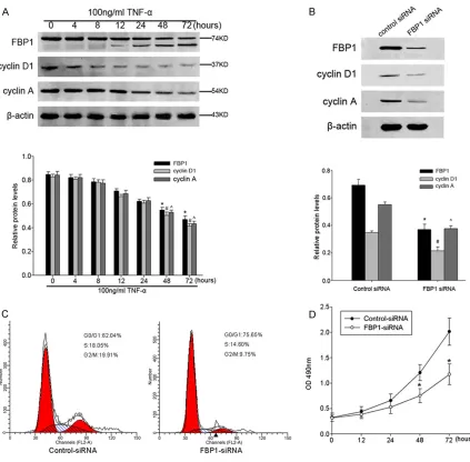
as cyclin A and cyclin D1 down-regulated in a time-dependent manner after TNF-α stimulus, which was similar with FBP1 expression (Figure 5A). To further explore whether the down-regu-lation of these cell cycle marker was relevant to
the FBP1 expression, HT-29 cells were tran
-siently transfected with FBP1 siRNA or control siRNA constructs. Forty-eight hours after trans
-fection, the efficiency of FBP1 siRNA-mediated
down-regulation was assessed by Western blot analysis. It was found that the expression of
FBP1 was significantly reduced in FBP1 siRNA
cells compared with control siRNA group (Figure 5B). The expression of cyclin D1 and cyclin A were correlated positively with FBP1
expression. At the same time, to determine whether the cell cycle distribution was changed
after knockdown of FBP1 in HT-29 cells, we performed flow cytometry 48 h after the FBP1
siRNA transient transfection. Cell cycle analysis
revealed an accumulation of cells in the G0/G1
phase, a decreased population at the S phase
from 18.05 to 14.60% and G2/M phase from 19.91 to 9.75% after transfection of FBP1 siRNA into the HT-29 cell line (Figure 5C),
sug-gesting that loss of FBP1 could induce cell
cycle arrest. In addition, we assessed the
effects of FBP1 knocked down on HT-29 cell
proliferation by CCK-8 assays. The result
exhibited a significant deceleration of cell pro -liferation rate compared with the control siRNA (Figure 5D). Taken together, these results
dem-onstrated that siRNA targeting FBP1 exhibited
inhibition of S-phase and M-phase entry, as
well as a specific inhibitory effect on HT-29 cell
line growth.
Discussion
In our study, 4% DSS induced severe colitis for a short period. The oral administration of DSS
induced several symptoms including diarrhea and weight loss, and histological changes of
the colonic mucosa including inflammation and crypt loss. We detected that DSS administra
-tion certainly induced apoptosis but significant -ly reduced the anti-Ki-67 positive cell ratio as compared to the control mice. These results were accordance with previous studies [23, 24]. The increased apoptosis and cell cycle arrest might lead to a breakdown of the epithe-lial barrier function at a very early phase of coli-tis, which might lead to the mucosal invasion of
intestinal bacterial and their toxins. However,
the precise mechanisms responsible for this apoptosis and cell cycle arrest remain unknown. In this study, we presented new insight into the molecular mechanisms for epithelial barrier
disruption by FBP1 in DSS-induced experimen -tal colitis.
In DSS-induced experimental colitis, we detect
-ed that FBP1 was cleav-ed and translocat-ed
from nucleus to cytoplasm in the colonic muco-sa epithelium. As a transcription regulator,
FBP1 is primarily located in the nucleus where
it exerts multiple biological function. The
nucle-ar localization of FBP1 protein is driven by three
nuclear localization signals (NLSs), one in each of its N-terminal, central and C-terminal
domains. Previous study reported that FBP1
localized in cytoplasm in apoptotic cells [16]. Such localization is related to the fact that
FBP1 is a substrate of caspase-3 and -7, which cleave FBP1 at the DQPD consensus site locat -ed in the N-terminal NLS. When cells suffer-ed from apoptotic stimuli, the caspase-mediated
cleavage of FBP1 led to its decreased presence
in the nucleus, accompanied by the marked downregulation of its various target gene.
According to the above findings, we hypothe
-sized that the cleavage of FBP1 in mice colitis
may be due to the apoptosis of intestinal epi-thelial cells. To test this conjecture, we
con-structed a model of apoptosis in vitro with
TNF-α stimulation. Previous study has con
-firmed that TNF is an important inducer of IEC
apoptosis [25]. Our in vitro results
demonstrat-ed clearly that TNF-α-mdemonstrat-ediatdemonstrat-ed epithelial cell apoptosis induced FBP1 cleavage and which
contributed to its decreasing presence in the nucleus and translocating to the cytoplasm. Since the apoptosis level of intestinal epithelial cells increased, the proliferative epithelial cells is necessary to supply the reduction of
intesti-nal cell library after injury. However, we detect -ed that proliferative epithelial cells were
reduced in the mice fed DSS. The specific
mechanism about this is not yet very clear.
Several studies have reported that loss of FBP1 could arrest cell cycle. FBP1 was first identified as a DNA-binding protein that regulates c-Myc
gene transcription through binding to the far upstream element in the promoter region 1.5 kb upstream of the transcription start site [26]. c-Myc is a master switch of cellular proliferation and this topic has been reviewed extensively elsewhere [27, 28]. Previous study has been
reported that loss of FBP1 extinguished c-Myc expression [21, 29]. Decreased expression of
c-Myc mRNA has been associated with
preven-tion of G1-S phase transipreven-tion and inhibipreven-tion of
cell proliferation [30, 31]. In addition, several
studies reported that FBP1 regulated cell prolif -eration through c-Myc-independent
mecha-nisms. For example, FBP1 downregulated p21 mRNA probably through AREs at the 3’-UTR [32]. p21 mRNA expression was significantly increased following stable FBP1 knockdown
[14]. The p21 gene encodes an inhibitor of cyclin-dependent kinases and inhibits cell pro-liferation in general [33]. Moreover, ectopic
expression of FBP1 may also induce the cell
cycle inhibitor p27Kip by promoting its
transla-tion via IRES in the 5’-UTR [12]. All of these
results demonstrated that downregulated
expression of FBP1 in the nucleus and ectopic expression of FBP1 both promoted cell cycle arrest. In our study, we found that TNF-α-mediated HT-29 cell apoptosis induced cleav
-age of FBP1 which lead to downregulated expression of FBP1 in the nucleus and ectopic expression of FBP1. Moreover, knockdown of FBP1 in HT-29 cell line lead to cell cycle arrest and reduced cell growth. However, the mecha
-nism that FBP1 regulates the cell cycle of IECs
In summary, our study demonstrated for the
first time that intestinal epithelial cells apopto
-sis induced cleavage of FBP1, which could lead
to cell cycle arrest and thus result in a break-down of epithelial barrier function at a very early phase of colitis. Previous studies reported that repeated cycles of the oral administration
of DSS in rats was able to induce an abnormal
increase in the proliferation of colonic epithelial cells [34]. It is reasonable that the repair and/ or proliferation processes might catch up with
the progression of the DSS-induced apoptosis
and cell cycle arrest. If animals were given
repeated cycles of DSS administration, the colonic epithelium might undergo chronic
infla-mmation, including subsequently increased proliferation. It is possible that this chronic coli-tis may lead to an increased incidence of colon cancer. In this study, we just investigated the
expression and biological function of FBP1 at
the early phase of mice colitis, whereas what
functions FBP1 exerts in the pathogenesis of mice chronic colitis and IBD remains to be fur -ther studied.
Acknowledgements
This work was supported by Natural Science
Foundation of China (81201252); Jiangsu
Pro-vince’s Key Provincial Talents Program (grant RC2011079); The project of the six talent sum-mit of Jiangsu Province (grant 2009-B1-002).
Disclosure of conflict of interest
None.
Address correspondence to: Dr. Liren Li, Department of Gastroenterology, Affiliated Hospital of Nantong University, 20 Xisi Road, Nantong 226001, Jiangsu
Province, People’s Republic of China. Tel:
+86-513-85051857; Fax: +86-513-85051896; E-mail: Larry- [email protected]; Dr. Yongwei Qin, Department of Path-ogen Biology, Nantong University Medical College,
19 Qixiu Road, Nantong 226001, Jiangsu Province, People’s Republic of China. Tel: +86-513-85051736;
Fax: +86-513-85052307; E-mail: qinyongwei012- @163.com
References
[1] Gross KJ and Pothoulakis C. Role of neuropep
-tides in inflammatory bowel disease. Inflamm Bowel Dis 2007; 13: 918-932.
[2] Pastorelli L, De Salvo C, Mercado JR, Vecchi M and Pizarro TT. Central Role of the Gut Epithe -lial Barrier in the Pathogenesis of Chronic
In-testinal Inflammation: Lessons Learned from Animal Models and Human Genetics. Front Im -munol 2013; 4: 280.
[3] Maldonado-Contreras AL and McCormick BA. Intestinal epithelial cells and their role in in-nate mucosal immunity. Cell Tissue Res 2011; 343: 5-12.
[4] Iimura M, Nakamura T, Shinozaki S, Iizuka B,
Inoue Y, Suzuki S and Hayashi N. Bax is down
-regulated in inflamed colonic mucosa of ulcer
-ative colitis. Gut 2000; 47: 228-235.
[5] Hagiwara C, Tanaka M and Kudo H. Increase in
colorectal epithelial apoptotic cells in patients with ulcerative colitis ultimately requiring
sur-gery. J Gastroenterol Hepatol 2002; 17:
758-764.
[6] Di Sabatino A, Ciccocioppo R, Luinetti O, Ricev
-uti L, Morera R, Cifone MG, Solcia E and Coraz
-za GR. Increased enterocyte apoptosis in in
-flamed areas of Crohn’s disease. Dis Colon
Rectum 2003; 46: 1498-1507.
[7] Schulzke JD, Bojarski C, Zeissig S, Heller F, Git
-ter AH and Fromm M. Disrupted barrier func -tion through epithelial cell apoptosis. Ann N Y Acad Sci 2006; 1072: 288-299.
[8] Briata P, Forcales SV, Ponassi M, Corte G, Chen CY, Karin M, Puri PL and Gherzi R. p38-depen -dent phosphorylation of the mRNA decay-pro-moting factor KSRP controls the stability of select myogenic transcripts. Mol Cell 2005; 20: 891-903.
[9] Dean JL, Sully G, Clark AR and Saklatvala J. The involvement of AU-rich element-binding
proteins in p38 mitogen-activated protein ki-nase pathway-mediated mRNA stabilisation. Cell Signal 2004; 16: 1113-1121.
[10] Duncan R, Collins I, Tomonaga T, Zhang T and Levens D. A unique transactivation sequence
motif is found in the carboxyl-terminal domain
of the single-strand-binding protein FBP. Mol
Cell Biol 1996; 16: 2274-2282.
[11] Avigan MI, Strober B and Levens D. A far up -stream element stimulates c-myc expression in undifferentiated leukemia cells. J Biol Chem 1990; 265: 18538-18545.
[12] Zheng Y and Miskimins WK. Far upstream ele -ment binding protein 1 activates translation of p27Kip1 mRNA through its internal ribosomal entry site. Int J Biochem Cell Biol 2011; 43: 1641-1648.
[14] Rabenhorst U, Beinoraviciute-Kellner R, Bre-zniceanu ML, Joos S, Devens F, Lichter P, Riek
-er RJ, Trojan J, Chung HJ, Levens DL and Zornig
M. Overexpression of the far upstream ele-ment binding protein 1 in hepatocellular
carci-noma is required for tumor growth. Hepatology
2009; 50: 1121-1129.
[15] Ding Z, Liu X, Liu Y, Zhang J, Huang X, Yang X, Yao L, Cui G and Wang D. Expression of far up
-stream element (FUSE) binding protein 1 in hu -man glioma is correlated with c-Myc and cell proliferation. Mol Carcinog 2013; [Epub ahead of print.
[16] Jang M, Park BC, Kang S, Chi SW, Cho S, Chung
SJ, Lee SC, Bae KH and Park SG. Far upstream
element-binding protein-1, a novel caspase substrate, acts as a cross-talker between apoptosis and the c-myc oncogene. Oncogene 2009; 28: 1529-1536.
[17] Mayer L. The development of animal models of
inflammatory bowel disease. Int Rev Immunol
2000; 19: 77-90.
[18] Mizoguchi A. Animal models of inflammatory
bowel disease. Prog Mol Biol Transl Sci 2012; 105: 263-320.
[19] Liu Y, Chen Y, Lu X, Wang Y, Duan Y, Cheng C
and Shen A. SCYL1BP1 modulates neurite out-growth and regeneration by regulating the Mdm2/p53 pathway. Mol Biol Cell 2012; 23: 4506-4514.
[20] Kim YH, Kwon HS, Kim DH, Shin EK, Kang YH, Park JH, Shin HK and Kim JK. 3,3’-diindolyl
-methane attenuates colonic inflammation and tumorigenesis in mice. Inflamm Bowel Dis
2009; 15: 1164-1173.
[21] He L, Liu J, Collins I, Sanford S, O’Connell B, Benham CJ and Levens D. Loss of FBP function
arrests cellular proliferation and extinguishes c-myc expression. EMBO J 2000; 19: 1034-1044.
[22] Wang J and Fu YX. Tumor necrosis factor family members and inflammatory bowel disease. Im -munol Rev 2005; 204: 144-155.
[23] Araki Y, Mukaisyo K, Sugihara H, Fujiyama Y and Hattori T. Increased apoptosis and de -creased proliferation of colonic epithelium in dextran sulfate sodium-induced colitis in mice. Oncol Rep 2010; 24: 869-874.
[24] Tessner TG, Cohn SM, Schloemann S and Sten
-son WF. Prostaglandins prevent decreased epi -thelial cell proliferation associated with
dex-tran sodium sulfate injury in mice.
Gastroen-terology 1998; 115: 874-882.
[25] Goretsky T, Dirisina R, Sinh P, Mittal N, Mana
-glia E, Williams DB, Posca D, Ryu H, Katzman RB and Barrett TA. p53 mediates TNF-induced epithelial cell apoptosis in IBD. Am J Pathol
2012; 181: 1306-1315.
[26] Wierstra I and Alves J. The c-myc promoter: still MysterY and challenge. Adv Cancer Res 2008; 99: 113-333.
[27] de Nigris F, Sica V, Herrmann J, Condorelli G, Chade AR, Tajana G, Lerman A, Lerman LO and
Napoli C. c-Myc oncoprotein: cell cycle-related events and new therapeutic challenges in can-cer and cardiovascular diseases. Cell Cycle 2003; 2: 325-328.
[28] Morrish F, Isern N, Sadilek M, Jeffrey M and Hockenbery DM. c-Myc activates multiple met -abolic networks to generate substrates for cell-cycle entry. Oncogene 2009; 28: 2485-2491. [29] Chung HJ and Levens D. c-myc expression:
keep the noise down! Mol Cells 2005; 20: 157-166.
[30] Heikkila R, Schwab G, Wickstrom E, Loke SL, Pluznik DH, Watt R and Neckers LM. A c-myc
antisense oligodeoxynucleotide inhibits entry
into S phase but not progress from G0 to G1.
Nature 1987; 328: 445-449.
[31] Holt JT, Redner RL and Nienhuis AW. An oligo -mer complementary to c-myc mRNA inhibits
proliferation of HL-60 promyelocytic cells and
induces differentiation. Mol Cell Biol 1988; 8: 963-973.
[32] Xiao J, Zhang Z, Chen GG, Zhang M, Ding Y, Fu
J, Li M and Yun JP. Nucleophosmin/B23
inter-acts with p21WAF1/CIP1 and contributes to its
stability. Cell Cycle 2009; 8: 889-895.
[33] Abbas T and Dutta A. p21 in cancer: intricate
networks and multiple activities. Nat Rev Can-cer 2009; 9: 400-414.
[34] Inoue T, Murano M, Kuramoto T, Ishida K, Kawakami K, Abe Y, Morita E, Murano N, Toshi-na K, Nishikawa T, Maemura K, Shimamoto C,