www.impactjournals.com/oncotarget/ Oncotarget, Vol. 7, No. 15
Quantification of mutant alleles in circulating tumor DNA can
predict survival in lung cancer
Xue Yang1,*, Minglei Zhuo1,*, Xin Ye2, Hua Bai1, Zhijie Wang1, Yun Sun2, Jun Zhao1, Tongtong An1, Jianchun Duan1, Meina Wu1, Jie Wang1
1 Department of Thoracic Medical Oncology, Key Laboratory of Carcinogenesis and Translational Research (Ministry of
Education), Peking University Cancer Hospital and Institute, Beijing, China
2Asia and Emerging Markets Innovative Medicine of AstraZeneca R & D, Shanghai, China *These authors contributed equally to this work
Correspondence to: Jie Wang, e-mail: zlhuxi@163.com
Keywords: non-small-cell lung cancer, epidermal growth factor receptor, circulating tumor DNA, droplet digital PCR,
next-generation sequencing
Received: August 25, 2015 Accepted: February 15, 2016 Published: March 10, 2016
ABSTRACT
Purpose: We aimed to investigate the feasibility of droplet digital PCR (ddPCR) for the quantitative and dynamic detection of EGFR mutations and next generation sequencing (NGS) for screening EGFR-tyrosine kinase inhibitors (EGFR-TKIs) resistance-relevant mutations in circulating tumor DNA (ctDNA) from advanced lung adenocarcinoma (ADC) patients.
Results: Detection limit of EGFR mutation in ctDNA by ddPCR was 0.04%. Taking the EGFR mutation in tumor tissue as the golden standard, the concordance of EGFR mutations detected in ctDNA was 74% (54/73). Patients with EGFR mutation in ctDNA (n = 54) superior progression-free survival (PFS, median, 12.6 vs. 6.7 months, P < 0.001) and overall survival (OS, median, 35.6 vs. 23.8 months, P = 0.028) compared to those with EGFR wild type in ctDNA (n = 19). Patients with high EGFR-mutated abundance in ctDNA (> 5.15%) showed better PFS compared to those with low EGFR mutated abundance (≤ 5.15%) (PFS, median, 15.4 vs. 11.1 months, P = 0.021). NGS results showed that 66.6% (8/12) total mutational copy number were elevated and 76.5% (26/34) mutual mutation frequency increased after disease progression.
Methods: Seventy-three advanced ADC patients with tumor tissues carrying EGFR mutations and their matched pre- and post-EGFR-TKIs plasma samples were enrolled in this study. Absolute quantities of plasma EGFR mutant and wild-type alleles were measured by ddPCR. Multi-genes testing was performed using NGS in 12 patients. Conclusions: Dynamic and quantitative analysis of EGFR mutation in ctDNA could guide personalized therapy for advanced ADC. NGS shows good performance in multiple genes testing especially novel and uncommon genes.
INTRODUCTION
Great advances have been made in the treatment of non-small cell lung cancer (NSCLC) over the past 40 years. The discovery of oncogenic drivers such as
EGFR (epidermal growth factor receptor) has enabled
a new and increasingly efficacious phase in cancer
biopsy of tumor tissues can be challenging. Moreover, owing to spatial and temporal heterogeneity, molecular detection methods that use initial tissue samples are not appropriate for therapeutic guidance throughout the entire process of treatment, especially after disease progression. Therefore, methods for detecting mutations have been investigated in other specimens that can be acquired easily and dynamically; examples include the use of specimens
like plasma [11–15], pleural fluid [15–17], and sputum [18, 19], among which plasma testing has been identified
as the most promising tool in EGFR mutation analysis [11, 20].
Previous studies have shown the feasibility of investigating EGFR mutation status in ctDNA [21–24]. However, most of these prior studies did not present methods for the dynamic and quantitative evaluation of ctDNA. They simply performed qualitative detection of EGFR mutations in plasma. To date, reported quantitative detection methods for evaluating EGFR mutation status
include BEAMing (beads, emulsion, amplification,
magnetics), ddPCR, and NGS-based methods. Each of these approaches has their own advantages and disadvantages [25–30]. Here, we chose and evaluated the performance of two methods, ddPCR and NGS, to address the commonly encountered problems of low sensitivity and complex multi-gene testing.
This study aimed to assess the concordance and feasibility of ddPCR and NGS for the detection of mutations in plasma samples. Moreover, we also evaluated the clinical outcomes according to the quantity of EGFR mutations in advanced NSCLC.
RESULTS
Patient characteristics
Seventy-three patients from Peking University Cancer Hospital diagnosed with lung adenocarcinoma, including 29 male and 44 female patients, were enrolled in the study. Fifty-three of the patients had never smoked
(72.6%, 53/73) and 20 were former/current smokers. All
of the patients carried EGFR mutations in their TKI-naïve tissue samples: there were 41 cases for exon 19 deletion alone, 30 cases for the L858R mutation alone, and 2 cases harboring concomitant exon 19-del and L858R mutations.
Thirty-seven patients (51.0%, 37/73) received EGFR-TKIs as the first-line therapy, while the rest received
EGFR-TKIs treatment as second (or beyond) line treatment. The patients’ clinical characteristics are listed in Table 1. Survival data were followed till the end of October 2014, and the median follow-up period was 23.8 months (range, 4.3 months–52.8 months). By the end of the last follow-up, sixty-seven patients experienced progressive disease (PD) and thirty-eight of the patients had died. The mortality rate
was 52.1% (38/73).
Matched plasma samples, both pre-EGFR-TKIs therapy and post-PD of EGFR-TKIs, were obtained form
67of 73 patients. The time interval from the diagnosis of
PD to blood sampling for ddPCR was no more than four weeks, with no intervening chemotherapy. The matched
plasma samples for the other 6 patients were obtained
during treatment without disease progression.
Evaluation of the consistency of activating EGFR mutations between TKI-naïve tissue and plasma DNA by ddPCR
Fifty-four of 73 patients were positive for
EGFR mutations in de novo ctDNA (31 cases for exon 19 deletion, 23 cases for L858R). EGFR mutations in
ctDNA were identified in 74% (54/73)of the patients
that had documented EGFR mutations in their tumors. The median absolute and relative EGFR mutant allele
quantities in TKI-naive plasma from 54 patients was 487 copies/reaction and 5.15% respectively. The response
rates (RR) and disease control rates (DCR) were not
significantly different between patients with EGFR mutant
and wild-type alleles.
Qualitative and quantitative analysis of EGFR mutations in de novo plasma by ddPCR predicted survival
OS1 was defined as the first day of the TKIs or
chemotherapy until death from any cause or the date of the last follow-up. OS2 was defined as the time from disease
progression after EGFR-TKIs therapy to death from any cause or the date of the last follow-up. OS1 represented the
overall survival and OS2 stood for the post-TKIs survival.
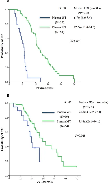
According to the EGFR mutation status of ctDNA
in TKI-naïve patients, all 73 patients were divided into
two subgroups: a group that carried mutations in both specimens (T+/B+, n = 54), and a group that carried
mutations only in tissues rather than in ctDNA (T+/B-, n = 19). The T+/B+ group showed superior PFS (median, 12.6 vs. 6.7 months, P < 0.001, Figure 1A) and OS1 (median, 35.6 vs. 23.8 months, P = 0.028) as compared to the T+/B- group (Figure 1B).
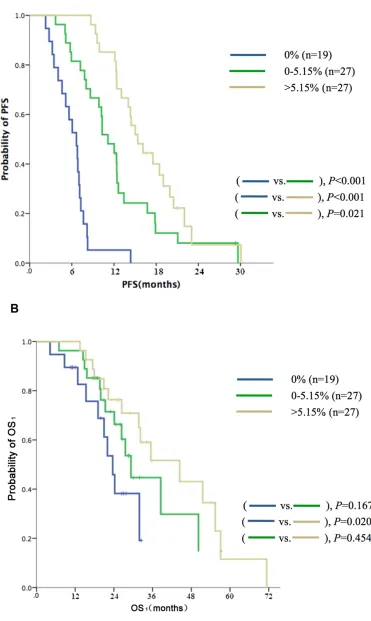
In addition to the qualitative analysis of EGFR mutations, quantitation of EGFR mutant alleles was also
performed. In the cohort of 73 cases, the patients were
subdivided into three groups based on the relative quantity of EGFR mutant alleles (median,5.15%) in TKI-naive plasma samples (high: > 5.15%, n = 27; low: ≤ 5.15% , n = 27; and nil: 0%, n = 19); the respective median PFS
values were 15.4 vs. 11.1 vs. 6.7 months (P < 0.001, Figure 2A); the respective median OS1 values were 44.5
vs. 29.3 vs. 23.8 months (P = 0.072, Figure 2B). Selected
No correlation was found between post-PD EGFR mutation abundance and OS2 (median, 17.2 vs. 16.6 months, P = 0.247). No significant differences were
found between the absolute quantity of in post-PD plasma samples.
Dynamic change in the abundance of EGFR mutations was associated with survival
Analysis of the plasma DNA from the 67 patients with PD, 29 cases (43.3%, 29/67) showed decreasing
EGFR mutation abundance following EGFR-TKIs
treatment, 13 cases (19.4%, 13/67) kept the same EGFR
mutation abundance following EGFR-TKIs treatment,
and 25 (37.3%, 25/67) cases showed increasing EGFR
abundance following EGFR-TKIs treatment.
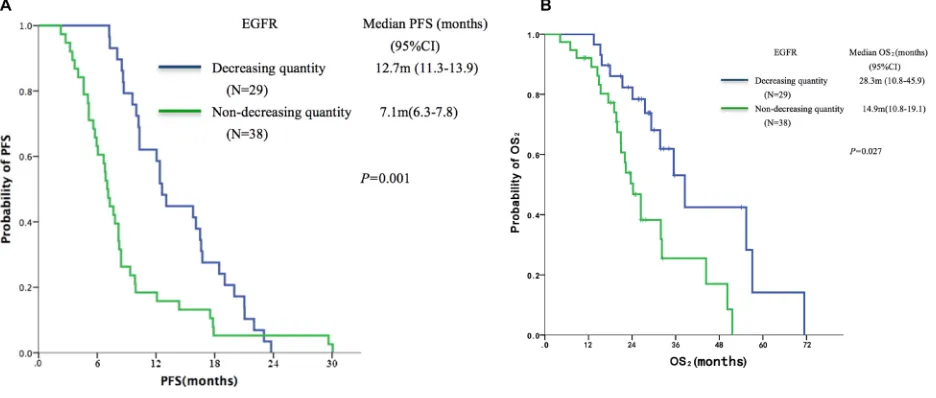
The 67 patients with PD were divided, based on
dynamic changes in EGFR mutation abundance in plasma DNA, into the ‘decreasing quantity group’ (n = 29) and the ‘non-decreasing quantity group’ (n = 38). Patients in the decreasing quantity group showed better PFS (median,
12.7 vs.7.1 months, P = 0.001, Figure 3A) and OS2
(median, 28.3 vs.14.9 months, P = 0.027, Figure 3B) as
compared with the non-decreasing quantity group.
Detection of T790M status in PD plasma samples using ddPCR
The T790M mutation was detected in 29 out of 67 (43.3%) patients when their disease progressed
after EGFR-TKIs therapy. Fourteen of 29
T790M-positive patients expressed the elevation of EGFR mutated abundance after PD, while another 15 patients demonstrated the decline of EGFR mutated abundance
after PD. No correlation was found between the T790M
mutation and survival.
The potential relationships between clinical modes of EGFR-TKI failure (dramatic PD and slow PD) and
T790M status were also analyzed. Dramatic PD was defined as rapid progression of multiple organs or target
lesions or symptom deterioration after disease control
lasting ≥ 3 months with EGFR-TKIs treatment. Slow PD was defined as gradual and local progression excluding
dramatic PD [31]. There were 23 patients with dramatic PD modes and 44 patients with slow PD modes. No
correlation was found between T790M mutation status and
TKI-failure modes (P = 0.975).
The relationship of ctDNA with metastasis site and previous treatment
Considering the potential effects of numbers of metastasis site (tumor burden) and previous treatment on ctDNA content, we investigated the relationships between these factors with ctDNA levels by Kruskal-Wallis Test. The de novo plasma samples had higher ctDNA levels than those obtained after front-line treatment (namely,
[image:3.612.59.556.68.342.2]EGFR-TKIs as first line versus non-first line, P = 0.025). However, no association was found between numbers of metastasis sites and ctDNA levels (P = 0.373). We put
Table 1: Patients’ clinical characteristics (
n =
73)
Characteristics No of Patients % of patients
Age, years
Median 58
Range 34–89
Gender
Male 29 40
Female 44 60
Smoking status
Never smoker 53 73
Former/current smoker 20 27
EGFR-TKIs treatment line
First line 37 51
Second or beyond 36 49
EGFR-TKIs failure modes n = 67
Dramatic PD 23 34
Slow PD 44 66
these two factors (metastasis site and previous treatment) into the Cox regression and found that EGFR mutation status of de novo plasma determined by ddPCR remained to be an independent predictor of PFS of EGFR-TKIs therapy (Table 3).
Frequently mutated genes and altered pathways in ctDNA determined by NGS
To explore other resistance mechanisms beyond
T790M, we randomly selected 12 matched plasma DNA pre-EGFR-TKIs and post-PD from 67 patients for deep
sequencing. Of 12 patients, 10 had matched plasma samples both pre-EGFR-TKIs and post-PD, another two cases (cases 25 and 29) had multi-point plasma samples (Table 4). The total plasma sample number used for the NGS detection experiment was twenty-seven. The sensitivity of the deep sequencing protocol with target regions for 483 genes designed by Agilent SureSelect allowed for the detection of a mutant allele fraction of
5%, with a mean sequencing depth of 609 ×. For each
patient, if the mutant fraction less than 5%, it would not be directly detected by the software. However, if other
samples of the same patient harbored mutant fractions of more than 5%, then the lost information for a given sample could be retrieved.
Using this approach, EGFR sensitive mutations
were identified as the most common somatic variation
in baseline plasma DNA; there were 3 cases with exon 19 deletions, 8 cases with the L858R mutation, and 1 case with overlapping L833V and H835L mutations. The
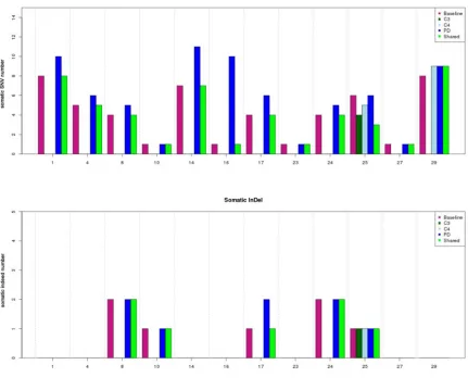
T790M mutation was not identified in baseline plasma DNA. However, it was found in 6 of 12 patients (50%) in PD, and 5 of 6 T790M positive patients coupled with EGFR level increasing (Figure 4). In addition, 66.6% (8/12)of patients presented elevated frequencies of total mutational copy numbers, and 76.5% (26/34) of cases
exhibited increased mutual mutation frequencies after PD (Figure 5).
Other mutations were identified as follows: 4 cases (33.3%, 4/12) with the TP53 mutation, 2 cases (16.7%, 2/12) with the PTEN mutation, and 2 cases (16.7%, 2/12) with the MLL2 (mixed-lineage leukemia 2)
[image:5.612.59.557.76.394.2]mutation. We further performed pathway analysis to identify other signaling cascades in addition to the canonical EGFR pathway. Mutated genes were
Table 2: Selected characteristics of patients with different abundances of EGFR mutations (
n =
54)
Variables EGFR abundance P value
Low abundance
(N = 27) High abundance (N = 27)
N % N %
Age 1.00
< 65 y 21 77.8 21 77.8
≥ 65 y 6 22.2 6 22.2
Gender 0.40
Male 10 37.0 13 48.1
Female 17 63.0 14 51.9
Smoking History 0.36
Former smoker 6 22.2 9 33.3
Never smoker 21 77.8 18 66.7
EGFR-TKIs treatment line 0.10
First line 15 55.6 9 33.3
Second or beyond 12 44.4 18 66.6
DCR 0.58
PR 13 48.1 11 40.7
SD 14 51.9 16 59.3
Notes: factors were tested by χ2test.
remarkably enriched in a DNA damage checkpoint pathway that forms a part of the cell-cycle pathway. We
also observed significant enrichment of mutated genes in TGF-β signaling in two cases that were synchronously accompanied by the T790M mutation (Figure 6).
Most of the mutant genes existed during both baseline readings and during PD. An exception to this was the CREBBP mutation, which emerged in the course of PD. All of the mutation frequencies existing at both baseline and PD that were enriched in the DNA damage
checkpoint pathway and in TGF-β signaling pathway
increased in the course of PD.
Comparison of the performance of ddPCR and NGS in ctDNA testing
We compared ddPCR with NGS methods for the analysis of 12 patients’ plasma samples. As mentioned above, the limit of quantitation for the ddPCR and NGS methods were 0.04% and 5%, respectively. For patients who had mutant frequencies equal to or greater than 5% in at least one plasma sample, both the sensitivity
and the specificity of the NGS method were 100%. For
patients who had mutant frequencies less than 5% in all of the plasma samples, some true positive mutations
were filtered out by Perlscript in order to avoid excessive amounts of false positives. The sensitivity and specificity
for NGS were 89% and 100% respectively. ctDNA levels of the same patient detected by both the ddPCR and NGS
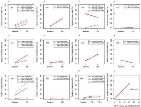
methods showed a similar trend. In general, quantification
of mutant frequencies via the ddPCR and NGS methods showed excellent agreement (Spearman Correlation,
R2 = 0.953, P < 0.001, Figure 7). These results validated
the feasibility of both methods.
DISCUSSION
Due to the existence of intra-tumor heterogeneity and dynamic alterations of EGFR-TKIs-related sensitive and resistant genes that occur in the process of therapy, the real-time, quantitative, and parallel detection of the status of multiple driver genes has value for evaluating suitable therapeutic regimens for EGFR-mutated patients. Here, our results indicated that ddPCR is a highly sensitive method for EGFR mutation analysis with ctDNA for advanced lung adenocarcinoma patients, with a detection limit 0.04%. Patients with high EGFR mutant abundance (> 5.15%) had a longer PFS as compared with those
with low EGFR mutant abundances (≤ 5.15%). Dynamic
changes in the abundance of EGFR mutations were associated with the survival of EGFR-TKIs treatment. NGS shows good performance for the testing of multiple genes simultaneously.
Previous studies have compared the coincidence rate between for the detection of EGFR mutations from ctDNA and tissue samples. ctDNA test sensitivity was
46–82%, specificity was 90–99%, and concordance was 78–88% in assessing EGFR mutation [11, 21–23, 32, 33].
Taking the detection of EGFR mutations in tumor tissue as the golden standard, the concordance of the detection
of EGFR mutations from ctDNA was 74% (54/73)in our study. There was 26% (19/73) discordance between the
tissue and plasma samples in our study with regard to the detection of EGFR mutations. A possible reason for
this discrepancy may be that nearly 50% (36/73) of the
[image:7.612.67.535.477.676.2]patients in our study received EGFR-TKIs as the second (or beyond) line therapy, and the time-points for the collection of their blood specimens was different from that of the tissues samples, which were sampled before
initial therapy. Chemotherapy prior to EGFR-TKIs may reduce EGFR mutation frequency in ctDNA of patients with advanced NSCLC [34].
In accordance with previous reports, our study showed that patients with EGFR mutations in TKI-naïve plasma had longer PFS and OS as compared to patients without EGFR mutations in plasma [11, 22, 23].
Further, we realized that, in addition to mutation status,
the abundance of mutant alleles also predicted survival. In a previous study, Zhou et al. [35] demonstrated that the relative abundances of EGFR mutations in tumor
samples could predict the potential benefit from
EGFR-TKIs treatment for advanced NSCLC. However, the underlying biology of the quantitative analysis of EGFR mutations in plasma is not yet fully understood. Yung
et al. [20] were the first to use digital PCR to quantify
EGFR mutations in plasma and proved the relationship between the response rate and the abundance of EGFR
mutations, but they did not analyze the predictive and/
or prognostic value of ctDNA. Our results indicate that patients with highly abundant EGFR mutations (> 5.15%) at baseline had a longer PFS compared with those carrying low abundance EGFR mutations
(≤ 5.15%). The relatively high frequency of intratumor
EGFR mutant alleles might imply that TKIs-sensitive mutant clones accounted for the major part of whole tumor clones, and needed longer time to develop drug resistance compared with the those with low frequency
of intratumor EGFR mutant alleles. Our previous [36]
and other studies [35] have demonstrated that high frequency of EGFR mutant alleles yielded superior
efficacy and survival of EGFR-TKIs compared with
low frequency ones. This intra-tumor heterogeneity of
EGFR mutation can be reflected by the quantification
of EGFR mutant alleles in ctDNA detected by ddPCR. Therefore, we speculated that the superior survival of EGFR-TKIs therapy for patients with high quantities of EGFR mutations at baseline might be attributed to the presence of high percentage of EGFR mutant clones in the tumor entities.
Moreover, we found that patients undergoing decreasing EGFR mutation alleles had better survival compared with those without decreasing. The possible
reason included that 1) the significant reduction of EGFR
mutation in peripheral blood during therapy meant higher sensitivity of tumor to EGFR-TKIs therapy compared to those without decrease 2) EGFR mutant clones might also carry other gene aberrances, causing the activation of related pathways correspondingly and leading to resistance to EGFR-TKIs subsequently.
Using the NGS technique, somatic mutations such as PTEN, MLL2, and TP53 were also detected. Previous studies have suggested that PTEN loss contributes to erlotinib resistance in EGFR-mutant lung cancer via the
activation of AKT and EGFR [37]. In this study, somatic
mutations in PTEN were found in two patients. According to previous studies, the incidence of PTEN mutations in NSCLC is 4%–4.5%, and is more common in smokers and squamous carcinoma [38–40]. However, the PTEN mutation
rate was 16.7% (2/12) in our study, and these were both
detected in non-smoking women with adenocarcinoma. This result is similar to a study that reported a PTEN mutation
rate of 16.1% in EGFR-mutant NSCLC [41]. Reasons for the differences might include the limited sample size or
variations in different human sub-populations. We also found frequent mutation of the MLL2 gene in 2 EGFR-mutant patients. This gene encodes a histone lysine methyltransferase that plays an important role in the regulation of transcription. Our results were in agreement with a previous report in which deleterious mutations of MLL2 occurred in 11.4% of Chinese NSCLC patients [42]. In addition to the canonical EGFR pathway, we also
found significant enrichment of gene mutations related with cell cycle pathway and the TGF-β pathways. TGF-β signaling pathway has been justified being associated
with EGFR-TKIs resistance, which may be induced by epithelial-mesenchymal transition (EMT), the activation
of MAPK pathway and IL-6 axis [43–46]. During the
[image:8.612.56.560.68.217.2]disease progression along with the enlarged tumor burden, rapid replication of cancer cell DNA produced more errors
Table 3: Association between clinical features and PFS using multivariate analysis
P HR 95% CI
Previous treatment EGFR-TKI first line
0.016 0.514 0.298–0.885
Number of metastatic sites 1 0.009
2 0.359 1.351 0.710–2.572
3+ 0.002 2.634 1.420–4.884
EGFR mutation status Negative 0.000
Low abundance 0.000 0.122 0.057–0.258
High abundance 0.000 0.066 0.030–0.149
in the process of cell cycle, causing the activation of DNA damage checkpoint pathway especially in the present of dysfunctional mutations in TP53 and ATM. Previous studies have showed multiple mechanisms of resistance can occur synchronously in the same patient [43]. In this
study, two cases harbored T790M together with the TGF-β
pathway related genes variants, a situation that might indicate multiple mechanisms of resistance.
We used both ddPCR and NGS to monitor dynamic changes of EGFR mutation in ctDNA. Levels of
EGFR-sensitive mutations and the T790M mutation analyzed via the two separate methods were significantly correlated
with each other. The detection limit of ddPCR is about 0.04%, which is more sensitive and with lower cost than regular NGS analyses. However, one-time ddPCR assay can detect only one known gene mutation, while NGS can monitor multiple gene mutation simultaneously and identify novel functional gene aberrances in whole-genomic scope. So, as a quantitative detection method, ddPCR can be well applied to testing a known gene mutation, especially with low frequency.
There were several limitations to out study. This was a retrospective and single-institution study. Repeat biopsies were not performed to obtain matched tissue samples with
blood after disease progression to confirm the presence of
resistance related gene mutations. However, based on the positive and meaningful results in this study, we launched a multicenter clinical trial using ddPCR as a screening tool to select EGFR mutation patients in ctDNA to
receive EGFR-TKI therapy (BENEFIT, NCT02282267), which had finished enrollment to date. This prospective
multicenter study will further validate the conclusion of the present study.
In summary, plasma DNA testing is promising and should be ready for use in the near future for the quantitative and qualitative analysis of multi-gene variations using ddPCR and NGS. Highly abundant EGFR mutations in de novo plasma samples predicted longer PFS on EGFR-TKIs. Compared to the non-decreasing quantity group, patients with decreasing quantities of EGFR mutated alleles after PD showed better PFS and OS. Our results warrant further investigation in a prospective study.
MATERIALS AND METHODS
Sample collection and processingSeventy-three patients diagnosed with advanced (stage IV) NSCLC admitted to the Peking University Cancer Hospital were recruited from May 1st, 2010, to April 1st, 2013. Enrollment Criteria included: 1) histological type must be adenocarcinoma; 2) patients were undergoing EGFR-TKIs treatment. 3) Patients’ tumor tissues carried EGFR mutations as detected by denaturing high-performance liquid chromatography (DHPLC).
DNA was extracted from 2 mL of plasma using QIAamp Circulating Nucleic Acid Kits (Qiagen) and
eluted with 55 μl of Elution Buffer.
[image:9.612.57.561.59.254.2]The study was approved by the Institutional Review Board of the Peking University Cancer Hospital. All of the patients signed informed consent before the collection and use of their tumor specimens and blood samples.
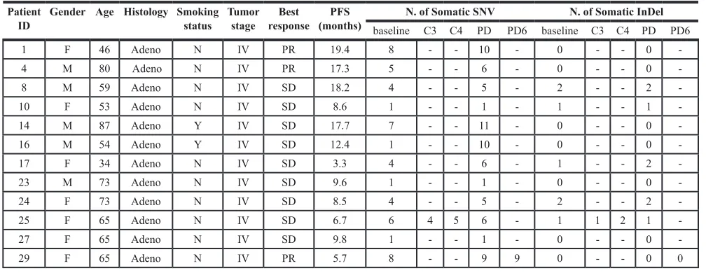
Table 4: Patient characteristics and NGS monitoring results in 12 cases
Patient
ID Gender Age Histology Smoking status Tumor stage responseBest (months)PFS baseline C3 C4 PDN. of Somatic SNV PD6 baseline C3 C4 PDN. of Somatic InDel PD6
1 F 46 Adeno N IV PR 19.4 8 - - 10 - 0 - - 0
-4 M 80 Adeno N IV PR 17.3 5 - - 6 - 0 - - 0
-8 M 59 Adeno N IV SD 18.2 4 - - 5 - 2 - - 2
-10 F 53 Adeno N IV SD 8.6 1 - - 1 - 1 - - 1
-14 M 87 Adeno Y IV SD 17.7 7 - - 11 - 0 - - 0
-16 M 54 Adeno Y IV SD 12.4 1 - - 10 - 0 - - 0
-17 F 34 Adeno N IV SD 3.3 4 - - 6 - 1 - - 2
-23 M 73 Adeno N IV SD 9.6 1 - - 1 - 0 - - 0
-24 F 73 Adeno N IV SD 8.5 4 - - 5 - 2 - - 2
-25 F 65 Adeno N IV SD 6.7 6 4 5 6 - 1 1 2 1
-27 F 65 Adeno N IV SD 9.8 1 - - 1 - 0 - - 0
-29 F 65 Adeno N IV PR 5.7 8 - - 9 9 0 - - 0 0
Dashes indicate that plasma was not available.
Abbreviations: F, female; M, male; Adeno, adenocarcinoma; Y, Yes; N, No; PR, partial remission; SD, stable disease;
C3,taking during the third months in TKI treatment;
C4, taking during the fourth months in TKI treatment; PD, progression of disease;
Droplet digital PCR analysis for plasma samples
Droplet digital PCR assays were done using a QX100 Droplet Digital PCR System (BIO-RAD). The QX100 droplet generator was used to create an emulsion of ~20,000 droplets, with mutant and wild type DNA distributed among the droplets following the Poisson distribution. After the PCR reaction, each
droplet produced a positive or negative fluorescence
signal indicating whether or not the EGFR mutant gene was present, respectively, in a given droplet. We then measured the absolute quantities of circulating EGFR mutant and wild-type alleles. The ratio of the concentration of mutant EGFR ctDNA to wild-type ctDNA was determined by ddPCR.
To evaluate the sensitivity of the ddPCR assays for mutant sequence detection, DNA extracted from cell
lines carrying the L858R mutation, the T790M mutation, and exon 19 deletion (ATCC NCI-H1975, NCI-H1975 and NCI-H1650, respectively) were mixed with human
reference genomic DNA (Promega) to achieve decreasing ratios (1:1 to 1:10,000) of the mutant allele versus the
wild-type allele. Genomic DNA from the H1975 cell line expresses both the EGFR L858R and the T790M
mutations. The three assays were able to detect the mutant alleles in samples containing 2,500 alleles, and the limit of quantitation was approximately 0.04%. Human
[image:10.612.71.545.265.703.2]reference genomic DNA and deionized water were used as negative controls. Mutant alleles from the NCI-H1975 and NCI-H1650 cell lines were used as positive controls [47].
Figure 6: Signaling pathways detected by NGS. Mutations involved in DNA damage checkpoint were shown in red, and mutations
involved with TGF-β pathway were shown in blue.
Figure 7: Comparison between NGS and ddPCR in measuring relative abundance of EGFR mutation in ctDNA. (A–J) Allele frequency (AF) of EGFR mutations in each patient. The dashed line indicates AF measured by ddPCR, and the solid line indicates AF
measured by NGS. EGFR mutation was shown in red line, while T790M was shown in blue line. PD, progressive disease; (K)Concordance
[image:12.612.80.532.327.669.2]NGS-Library preparation, hybrid capture, and sequencing
DNA libraries with ~150 nucleotide base pair insert
size were prepared using a NEBNext DNA Library Prep
Reagent Set (New England BioLabs), according to the manufacturer’s protocol. Next, the libraries were hybrid-captured with custom biotinylated DNA oligo pools (custom SureSelect kit, Agilent) that contained all of the exons of 483 cancer-related genes (driver genes from the
Catalogue of Somatic Mutations in Cancer, COSMIC V68)
and 88 introns from 14 genes that are frequently rearranged in cancer. All samples were sequenced with the Illumina HiSeq2500 platform in a 150 bp paired-end pattern.
Calculation of absolute and relative EGFR mutant allele quantities by ddPCR
As described in a previous paper [47], the absolute
and relative quantities of EGFR mutated alleles were
determined by analyzing the number of positive and negative fluorescence signals in droplets. Plasma sample
DNA input, as a unit of ‘genome copies per reaction’ represented the absolute abundance of EGFR mutations. The percentage of EGFR mutated alleles represented the relative abundance of EGFR mutations.
Statistical analysis
We used SPSS statistical software, version 17.0 (SPSS), to analyze the data. We used χ2 tests to assess
the relationships between EGFR mutation status and clinicopathologic features. Median PFS (progression free survival) and OS (overall survival) were calculated by Kaplan-Meier estimation. Log-rank tests were performed to explore the cut-off values of the quantity of EGFR
mutated alleles that could predict efficacy outcomes. P < 0.05 was considered significant. Relationship between metastatic sites (reflecting tumor burden) and previous
treatment with ctDNA levels by Kruskal-Wallis Test.
DAVID software (Database for Annotation, Visualization, and Integrated Discovery) was used to analyze cancer
genes in pathway enrichment.
ACKNOWLEDGMENTS AND SUPPORT
This study was supported by 2015 scientific activity prioritized funding for scholars study abroad
from the ministry of human resources and social security of the People’s Republic of China, and from Beijing Municipal Administration of hospitals Clinical medicine Development of special funding support, code: ZYLX201509. We thank John Hugh Snyder for manuscript proofreading.
CONFLICTS OF INTEREST
The authors declare that there are no conflicts of
interest.
REFERENCES
1. Lynch TJ, Bell DW, Sordella R, Gurubhagavatula S, Okimoto RA, Brannigan BW, Harris PL, Haserlat SM, Supko JG, Haluska FG, Louis DN, Christiani DC, Settleman J, et al. Activating mutations in the epidermal growth factor receptor underlying responsiveness of
non-small-cell lung cancer to gefitinib. N Engl J Med. 2004;
350:2129–2139.
2. Paez JG, Janne PA, Lee JC, Tracy S, Greulich H,
Gabriel S, Herman P, Kaye FJ, Lindeman N, Boggon TJ, Naoki K, Sasaki H, Fujii Y, et al. EGFR mutations in lung cancer: correlation with clinical
response to gefitinib therapy. Science. 2004; 304: 1497–1500.
3. Dienstmann R, Martinez P, Felip E. Personalizing therapy
with targeted agents in non-small cell lung cancer.
Oncotarget. 2011; 2:165–177. doi: 10.18632/oncotarget.245.
4. Shtivelman E, Hensing T, Simon GR, Dennis PA, Otterson GA, Bueno R, Salgia R. Molecular pathways and therapeutic targets in lung cancer. Oncotarget. 2014;
5:1392–1433. doi: 10.18632/oncotarget.1891.
5. Mok TS, Wu YL, Thongprasert S, Yang CH, Chu DT, Saijo N, Sunpaweravong P, Han B, Margono B, Ichinose Y,
Nishiwaki Y, Ohe Y, Yang JJ, et al. Gefitinib or
carboplatin-paclitaxel in pulmonary adenocarcinoma. N Engl J Med.
2009; 361:947–957.
6. Maemondo M, Inoue A, Kobayashi K, Sugawara S, Oizumi S, Isobe H, Gemma A, Harada M, Yoshizawa H, Kinoshita I, Fujita Y, Okinaga S, Hirano H, et al. Gefitinib
or chemotherapy for non-small-cell lung cancer with
mutated EGFR. N Engl J Med. 2010; 362:2380–2388. 7. Mitsudomi T, Morita S, Yatabe Y, Negoro S, Okamoto I,
Tsurutani J, Seto T, Satouchi M, Tada H, Hirashima T,
Asami K, Katakami N, Takada M, et al. Gefitinib versus
cisplatin plus docetaxel in patients with non-small-cell lung cancer harbouring mutations of the epidermal growth factor receptor (WJTOG3405): an open label, randomised phase 3 trial. Lancet Oncol. 2010; 11:121–128.
8. Zhou C, Wu YL, Chen G, Feng J, Liu XQ, Wang C, Zhang S, Wang J, Zhou S, Ren S, Lu S, Zhang L, Hu C,
et al. Erlotinib versus chemotherapy as first-line treatment
for patients with advanced EGFR mutation-positive non-small-cell lung cancer (OPTIMAL, CTONG-0802): a multicentre, open-label, randomised, phase 3 study. Lancet
Oncol. 2011; 12:735–742.
9. Rosell R, Carcereny E, Gervais R, Vergnenegre A,
treatment for European patients with advanced EGFR mutation-positive non-small-cell lung cancer (EURTAC): a multicentre, open-label, randomised phase 3 trial. Lancet
Oncol. 2012; 13:239–246.
10. Russo A, Franchina T, Ricciardi GR, Picone A, Ferraro G, Zanghi M, Toscano G, Giordano A, Adamo V. A decade of EGFR inhibition in EGFR-mutated non small cell lung cancer (NSCLC): Old successes and future perspectives. Oncotarget.
2015; 6:26814–25. doi: 10.18632/oncotarget.4254.
11. Bai H, Mao L, Wang HS, Zhao J, Yang L, An TT, Wang X, Duan CJ, Wu NM, Guo ZQ, Liu YX, Liu HN, Wang YY, et al. Epidermal growth factor receptor mutations in plasma DNA samples predict tumor response in Chinese patients with stages IIIB to IV non-small-cell lung cancer. J Clin
Oncol. 2009; 27:2653–2659.
12. Schwarzenbach H, Hoon DS, Pantel K. Cell-free nucleic
acids as biomarkers in cancer patients. Nature reviews
Cancer. 2011; 11:426–437.
13. Punnoose EA, Atwal S, Liu W, Raja R, Fine BM,
Hughes BG, Hicks RJ, Hampton GM, Amler LC, Pirzkall A,
Lackner MR. Evaluation of circulating tumor cells and circulating tumor DNA in non-small cell lung cancer: association with clinical endpoints in a phase II clinical
trial of pertuzumab and erlotinib. Clin Cancer Res. 2012;
18:2391–2401.
14. Akca H, Demiray A, Yaren A, Bir F, Koseler A, Iwakawa R, Bagci G, Yokota J. Utility of serum DNA and pyrosequencing for the detection of EGFR mutations in
non-small cell lung cancer. Cancer Genet. 2013; 206:73–80.
15. Yeo CD, Kim JW, Kim KH, Ha JH, Rhee CK, Kim SJ, Kim YK, Park CK, Lee SH, Park MS, Yim HW. Detection and comparison of EGFR mutations in matched tumor tissues, cell blocks, pleural effusions, and sera from patients with NSCLC with malignant pleural effusion, by PNA clamping and direct sequencing. Lung cancer. 2013;
81:207–212.
16. Buttitta F, Felicioni L, Del Grammastro M, Filice G, Di
Lorito A, Malatesta S, Viola P, Centi I, D’Antuono T, Zappacosta R, Rosini S, Cuccurullo F, Marchetti A. Effective assessment of egfr mutation status in
bronchoalveolar lavage and pleural fluids by next-generation sequencing. Clin Cancer Res. 2013; 19:691–698. 17. Liu D, Lu Y, Hu Z, Wu N, Nie X, Xia Y, Han Y, Li Q,
Zhu G, Bai C. Malignant pleural effusion supernatants are substitutes for metastatic pleural tumor tissues in EGFR mutation test in patients with advanced lung
adenocarcinoma. PloS one. 2014; 9:e89946.
18. Kim CE, Tchou-Wong KM, Rom WN. Sputum-based molecular biomarkers for the early detection of lung cancer:
limitations and promise. Cancers. 2011; 3:2975–2989.
19. Hubers AJ, Heideman DA, Yatabe Y, Wood MD, Tull J, Taron M, Molina MA, Mayo C, Bertran-Alamillo J, Herder GJ, Koning R, Sie D, Ylstra B, et al. EGFR mutation analysis in sputum of lung cancer patients: a multitechnique study. Lung cancer. 2013; 82:38–43.
20. Yung TK, Chan KC, Mok TS, Tong J, To KF, Lo YM. Single-molecule detection of epidermal growth factor
receptor mutations in plasma by microfluidics digital PCR
in non-small cell lung cancer patients. Clin Cancer Res.
2009; 15:2076–2084.
21. Karachaliou N, Casas CM-dl, Queralt C, Aguirre Id. Association of EGFR L858R Mutation in Circulating Free DNA With Survival in the EURTAC Trial. JAMA Oncol.
2015; 257:2374–2437.
22. Mok TS, Wu YL, Soo Lee J, Yu CJ, Sriuranpong V, Sandoval-Tan J, Ladrera G, Thongprasert S, Srimuninnimit V, Liao M, Zhu Y, Zhou C, Fuerte F, et al. Detection and Dynamic Changes of EGFR Mutations from Circulating Tumor DNA as a Predictor of Survival Outcomes in NSCLC Patients Treated with First-line Intercalated Erlotinib and Chemotherapy. Clin Cancer Res.
2015; 21:3196–203. doi: 10.1158/1078-0432.CCR-14-2594.
23. Douillard JY, Ostoros G, Cobo M, Ciuleanu T, Cole R, McWalter G, Walker J, Dearden S, Webster A, Milenkova T,
McCormack R. Gefitinib treatment in EGFR mutated
caucasian NSCLC: circulating-free tumor DNA as a surrogate for determination of EGFR status. J Thorac Oncol. 2014; 9:1345–1353.
24. Sorensen BS, Wu L, Wei W, Tsai J, Weber B, Nexo E, Meldgaard P. Monitoring of epidermal growth factor
receptor tyrosine kinase inhibitor-sensitizing and resistance
mutations in the plasma DNA of patients with advanced non-small cell lung cancer during treatment with erlotinib.
Cancer. 2014; 120:3896–3901.
25. Taniguchi K, Uchida J, Nishino K, Kumagai T, Okuyama T, Okami J, Higashiyama M, Kodama K, Imamura F, Kato K. Quantitative detection of EGFR mutations in circulating tumor DNA derived from lung adenocarcinomas. Clin
Cancer Res. 2011; 17:7808–7815.
26. Forshew T, Murtaza M, Parkinson C, Gale D, Tsui DW, Kaper F, Dawson SJ, Piskorz AM, Jimenez-Linan M, Bentley D, Hadfield J, May AP, Caldas C, et al. Noninvasive identification and monitoring of cancer mutations by
targeted deep sequencing of plasma DNA. Sci Transl Med.
2012; 4:136ra168.
27. Murtaza M, Dawson SJ, Tsui DW, Gale D, Forshew T, Piskorz AM, Parkinson C, Chin SF, Kingsbury Z, Wong AS, Marass F, Humphray S, Hadfield J, et al. Non-invasive
analysis of acquired resistance to cancer therapy by
sequencing of plasma DNA. Nature. 2013; 497:108–112. 28. Oxnard GR, Paweletz CP, Kuang Y, Mach SL, O’Connell A,
Messineo MM, Luke JJ, Butaney M, Kirschmeier P, Jackman DM, Janne PA. Noninvasive detection of response and resistance in EGFR-mutant lung cancer using quantitative next-generation genotyping of cell-free plasma
DNA. Clin Cancer Res. 2014; 20:1698–1705.
patients with advanced cancers referred for experimental
targeted therapies. Oncotarget. 2015; 6:12809–12821. doi: 10.18632/oncotarget.3373.
30. Bria E, Pilotto S, Amato E, Fassan M, Novello S, Peretti U, Vavala T, Kinspergher S, Righi L, Santo A, Brunelli M, Corbo V, Giglioli E, et al. Molecular heterogeneity assessment
by next-generation sequencing and response to gefitinib of
EGFR mutant advanced lung adenocarcinoma. Oncotarget.
2015; 6:12783–12795. doi: 10.18632/oncotarget.3727.
31. Yang JJ, Chen HJ, Yan HH, Zhang XC, Zhou Q, Su J, Wang Z, Xu CR, Huang YS, Wang BC, Yang XN, Zhong WZ, Nie Q, et al. Clinical modes of EGFR tyrosine kinase inhibitor failure and subsequent management in advanced non-small cell lung cancer. Lung cancer. 2013;
79:33–39.
32. Han B. Determining the prevalence of EGFR mutations in Asian and Russian patients (pts) with advanced non-small-cell lung cancer (aNSCLC) of adenocarcinoma (ADC) and non-ADC histology: IGNITE study. European Lung Cancer Conference (ELCC),abstract. 2015.
33. Reck M. Investigating the utility of circulating-free tumour-derived DNA (ctDNA) in plasma for the detection of epidermal growth factor receptor (EGFR) mutation status in European and Japanese patients (pts) with advanced non-small-cell lung cancer (NSCLC): ASSESS study. European Lung Cancer Conference (ELCC), abstract. 2015.
34. Bai H, Wang Z, Chen K, Zhao J, Lee JJ, Wang S, Zhou Q, Zhuo M, Mao L, An T, Duan J, Yang L, Wu M, et al.
Influence of chemotherapy on EGFR mutation status among
patients with non-small-cell lung cancer. J Clin Oncol.
2012; 30:3077–3083.
35. Zhou Q, Zhang XC, Chen ZH, Yin XL, Yang JJ, Xu CR, Yan HH, Chen HJ, Su J, Zhong WZ, Yang XN, An SJ, Wang BC, et al. Relative abundance of EGFR mutations
predicts benefit from gefitinib treatment for advanced non-small-cell lung cancer. J Clin Oncol. 2011; 29:3316–3321. 36. Bai H, Wang Z, Wang Y, Zhuo M, Zhou Q, Duan J, Yang L,
Wu M, An T, Zhao J, Wang J. Detection and clinical
significance of intratumoral EGFR mutational heterogeneity
in Chinese patients with advanced non-small cell lung
cancer. PloS one. 2013; 8:e54170.
37. Sos ML, Koker M, Weir BA, Heynck S, Rabinovsky R,
Zander T, Seeger JM, Weiss J, Fischer F, Frommolt P, Michel K, Peifer M, Mermel C, et al. PTEN loss contributes to erlotinib resistance in EGFR-mutant lung cancer by activation of Akt and EGFR. Cancer Res. 2009;
69:3256–3261.
38. Leary RJ, Kinde I, Diehl F, Schmidt K, Clouser C, Duncan C, Antipova A, Lee C, McKernan K, De La
Vega FM, Kinzler KW, Vogelstein B, Diaz LA, Jr. et al. Development of personalized tumor biomarkers using
massively parallel sequencing. Sci Transl Med. 2010; 2:20ra14.
39. Lee SY, Kim MJ, Jin G, Yoo SS, Park JY, Choi JE, Jeon HS, Cho S, Lee EB, Cha SI, Park TI, Kim CH, Jung TH, et al. Somatic mutations in epidermal growth factor receptor signaling pathway genes in non-small cell lung cancers.
J Thorac Oncol. 2010; 5:1734–1740.
40. Lopez-Chavez A, Thomas A, Rajan A, Raffeld M,
Morrow B, Kelly R, Carter CA, Guha U, Killian K, Lau CC,
Abdullaev Z, Xi L, Pack S, et al. Molecular Profiling and
Targeted Therapy for Advanced Thoracic Malignancies: A Biomarker-Derived, Multiarm, Multihistology Phase II
Basket Trial. J Clin Oncol. 2015; 33:1000–1007.
41. Kim HR, Cho BC, Shim HS, Lim SM, Kim SK, Chang J, Kim DJ, Kim JH. Prediction for response duration to epidermal growth factor receptor-tyrosine kinase inhibitors in EGFR mutated never smoker lung adenocarcinoma. Lung
cancer. 2014; 83:374–382.
42. Yin S, Yang J, Lin B, Deng W, Zhang Y, Yi X, Shi Y, Tao Y, Cai J, Wu CI, Zhao G, Hurst LD, Zhang J, et al. Exome
sequencing identifies frequent mutation of MLL2 in
non-small cell lung carcinoma from Chinese patients. Sci Rep.
2014; 4:6036.
43. Bean J, Brennan C, Shih JY, Riely G, Viale A, Wang L,
Chitale D, Motoi N, Szoke J, Broderick S, Balak M, Chang WC, Yu CJ, et al. MET amplification occurs with or without T790M mutations in EGFR mutant lung tumors with acquired resistance to gefitinib or erlotinib. Proc Natl Acad Sci U S A. 2007; 104:20932–20937.
44. Sequist LV, Waltman BA, Dias-Santagata D, Digumarthy S, Turke AB, Fidias P, Bergethon K, Shaw AT, Gettinger S, Cosper AK, Akhavanfard S, Heist RS, Temel J, et al. Genotypic and histological evolution of lung cancers acquiring resistance to EGFR inhibitors. Sci Transl Med.
2011; 3:75ra26.
45. Yao Z, Fenoglio S, Gao DC, Camiolo M, Stiles B, Lindsted T, Schlederer M, Johns C, Altorki N, Mittal V,
Kenner L, Sordella R. TGF-beta IL-6 axis mediates
selective and adaptive mechanisms of resistance to molecular targeted therapy in lung cancer. Proc Natl Acad
Sci U S A. 2010; 107:15535–15540.
46. Brunen D, Willems SM, Kellner U, Midgley R,
Simon I, Bernards R. TGF-beta: an emerging player in drug resistance. Cell cycle (Georgetown, Tex). 2013;
12:2960–2968.
47. Zhu G, Ye X, Dong Z, Lu YC, Sun Y, Liu Y, McCormack R,
Gu Y, Liu X. Highly Sensitive Droplet Digital PCR Method for Detection of EGFR Activating Mutations in Plasma Cell-Free DNA from Patients with Advanced Non-Small