Original Article
B7-H4 expression is correlated with tumor progression
and clinical outcome in urothelial cell carcinoma
Min Fan1*, Qianfeng Zhuang1*, Yiming Chen1*, Tao Ding1, Hongwei Yao1, Lujun Chen2, Xiaozhou He1, Xianlin Xu1
1Department of Urology, The Third Affiliated Hospital of Soochow University, Changzhou 213000, Jiangsu, China; 2Department of Tumor Biological Treatment, The Third Affiliated Hospital of Soochow University, Changzhou
213000, Jiangsu, China. *Equal contributors.
Received August 11, 2014; Accepted September 13, 2014; Epub September 15, 2014; Published October 1, 2014
Abstract: Objective: To investigate the mRNA and protein levels of B7-H4, a B7 family molecule, in human urothelial cell carcinoma (UCC), to analyze the relationship between B7-H4 protein expression level and pathological stage of UCC, and to examine the potential of B7-H4 as a prognostic factor in UCC. Methods: mRNA and protein levels of B7-H4 were measured in pairs of tumor tissues and matched adjacent nontumor tissue obtained from patients with UCC by quantitative reverse transcription-polymerase chain reaction (qRT-PCR) and immunohistochemical staining, respectively. Association of the protein level of B7-H4 with pathological tumor stage and the overall survival of UCC
patients were also analyzed. Results: B7-H4 mRNA and protein level were significantly higher in UCC tumor tissues
compared with adjacent nontumor tissues as assessed by qRT-PCR and immunohistochemical staining, respec-tively. Higher B7-H4 protein levels were observed in patients with more advanced pathological stage of UCC and
were also associated with decreased overall survival of patients with UCC. Conclusions: The findings from this study
indicate that B7-H4 has the potential to be an independent prognostic indicator for UCC.
Keywords: Bladder cancer, B7 family, B7-H4, prognosis, gene expression
Introduction
Bladder cancer, with 383,000 new cases diag-nosed worldwide in 2008 [World Cancer Research Fund International (WCRF Inter national)], is the ninth most common cancer in the world and the second most common genito-urinary malignancy [1, 2]. About 95% of bladder cancers are urothelial cell carcinoma [UCC, also known as transitional cell carcinoma (TCC)] [3]. UCC is a type of malignant tumour originat-ing from the urothelium linoriginat-ing the urinary tract
from the renal calyces to the ureteral orifice.
UCC is a clinically heterogeneous disease, with 70% of total cases presenting non-invasive tumors and 30% presenting muscle-invasive tumors [4-6]. Muscle muscle-invasive tumors usually implies metastases and poor prognosis [4-6]. Surgery is the option for most people with
UCC. However, a significant number of patients
suffer from disease recurrence and progres-sion after radical cystectomy [6]. For example, a group from the University of Texas MD
Anderson Cancer Center found that metasta-ses developed in 97 of the 382 patients (25%) with transitional cell carcinoma of the bladder a median of 12 months after cystectomy [7]. Therefore, there is an urgent need to identify
prognositc biomarkers with high specificity and
sensitivity for UCC in order to distinguish tumors with the potential to progress and metastasize.
T cell-mediated immunity depends on specific
recognition of antigen-major histocompatibility complex (MHC) by T cell receptor (TCR) and co-regulatory signals [8]. The co-co-regulatory signals come from the B7 family molecules, which are a group of structurally related, peripheral mem-brane proteins mainly located on activated anti-gen presenting cells (APCs) [8]. B7 molecules function as co-regulatory ligands by binding to corresponding receptors on T cell surfaces, pro-ducing co-regulatory signals to either enhance
or decrease T cell-mediated, antigen-specific immune responses [9]. The newly identified B7
B7S1), negatively regulate in T cell-mediated immunity by arresting T cell cell-cycle progres-sion and thus inhibiting T cell proliferation, cyto-kine secretion, and cytotoxic activity [10, 11]. B7-H4 has been reported to be highly expressed in different tumors, including ovarian, breast, non-small-cell lung cancers, etc, however, there is a little or no B7-H4 expression in normal tis-sues [12, 13]. It has also been reported that B7-H4 promotes malignant transformation and lymph node metastasis [12, 14, 15].
Up to date, however, no reports have
investi-gated the clinical significance of B7-H4 expres -sion in patients with bladder cancer. In this study, we analyzed B7-H4 expression in UCC tissues and normal urothelium tissues using both immunohistochemical method and real time RT-PCR. Additionally, we investigated the relationship between B7-H4 expression level and clinicopathological variables and evaluat-ed the prognostic values of B7-H4 using log-rank survival analysis.
Materials and methods
Patient identification
The study protocol was approved by the ethics
committee of the Third Affiliated Hospital of
Soochow University, and all tissue samples were collected from patients and donors with appropriate informed consent. The criteria for study enrollment were histopathological diag-nosis of UCC of the bladder, no history of other tumour, no chemotherapy before surgery,
avail-ability of sufficient tumour sample, and the
potential to follow-up. By applying these crite-ria, sixty two patients who underwent surgeries for bladder cancer (between July 2006 and July 2012) at the Department of Urology, the Third
Affiliated Hospital of Soochow, Changzhou,
Jiangsu, China, were included in this study. The patients were followed for 3-6 months after surgery with cystoscopic examination at the
outpatient clinic. The tumors were classified
according to the 2010 Union for International
Cancer Control (UICC) TNM classification for
pathologic staging and the 2004 World Health
Organization classification for the pathological grading based on the findings of clinical, radio -logical, or histological examinations [16, 17]. The hematoxylin and eosin staining was evalu-ated by two independent pathologists or urolo-gists without knowledge of patient outcome.
Immunohistochemical staining
The bladder cancer specimens were fixed in
10% neutral buffered formalin, embedded in
paraffin, cut into 5 µm serial sections, and then
mounted on glass slides. The slides were
depa-raffinized with xylene and rehydrated in graded
alcohol. Antigen retrieval was performed tissue sections in a citrate buffer (10 mmol/l, pH 6.0) at 100°C for 30 min. After cooling, slides were incubated in 0.3% H2O2 solution for 30 min to block endogenous peroxidase. After washing three times with PBS (pH 7.4) for 5 min each, slides were incubated with the primary antibod-ies (mouse anti-B7-H4 polyclonal antibody, USCNLIFE, USA) diluted 1:400 in PBS with 1.0% bovine serum albumin (BSA, Sigma) in a humid chamber at 4°C overnight followed by three times 5 min washes in PBS. After washing three times with PBS (pH 7.4) for 5 min each, sec-tions were incubated with the secondary anti-body (mouse/rabbit general second antianti-body, Maixin Biotechnology Co. Ltd, Fuzhou) at room temperature for 30 min. After washing again with PBS, the sections were visualized by incu-bation with diaminobenzidine (Dako Cytoma- tion) substrate for 8 min. Finally, slides were counterstained with for 1 min with hematocylin, and coverslips were applied. Stained sections were photographed using the BX50 microscope (Olympus America, Center Valley, PA) with an attached QImaging Retiga 2000R Digital Camera (Quantitative Imaging, Surrey, BC, Canada).
Evaluation of B7-H4 staining
The slides were examined by two pathologists, and the sections were evaluated according to the immunohistochemical scores (IHS) [18, 19]. The staining intensity the proportion of positive cells was semiquantitatively evaluated. The staining intensity was scored as 0, no stain-ing; 1, weak stainstain-ing; 2, moderate stainstain-ing; and 3, intense staining. The proportion of positive cells was scored as 0 (< 5% positive cells), 1 (6-25% positive cells), 2 (26-50% positive cells), 3 (51-75% positive cells), and 4 (> 75% positive
cells). The final B7-H4 staining score was calcu -lated using the percent of positive cell score × staining intensity score ranging 0-12. In this
study, the B7-H4 expression is defined as weak
Table 1. Primers and probes for human B7-H4 and β-actin Gene Primers and probes Sequence (5’ to 3’)
B7-H4 Forward primer CACCAGGATAACATCTCTCAGTGAA Reverse primer TGGCTTGCAGGGTAGAATGA
Probe FAM-AAGCTGAAGATAATCCCATCAGGCAT-TAMRA
β-Actin Forward primer GGAAGGTGAAGGTCGGAGTC
Reverse primer CGTTCTCAGCCTTGACGGT
Probe FAM-TTTGGTCGTATTGGGCGCCTG-TAMRA
Real-time reverse transcription-polymerase chain reaction (RT-PCR)
Tumor tissues were frozen in liquid nitrogen immediately until RNA extraction. Total RNA was extracted from tissues using a total RNA
purification kit (Shenergy Biocolor BioScience
and Technology Co., Shanghai, China) accord-ing to the manufacturer’s instructions. One microgram of total RNA was reversely tran-scribed to cDNA with 100 units of Moloney murine leukemia virus (M-MLV) reverse tran-scriptase (USB, Cleveland, OH, USA) according to the manufacturer’s protocol. TaqMan® gene expression assays (Applied Biosystems, Foster City, CA, USA) were used to quantify mRNA
expression of human B7-H4 and β-actin (inter -nal control) genes. Primers and probes in the TaqMan assay are presented in Table 1. PCR reactions were performed on a CFX96 Real-Time PCR Detection System (Bio-Rad, Hercules,
CA, USA) in duplicate in a 10 µl volume contain
-ing 5 µl Universal PCR Master Mix (Applied Biosystems), 0.5 µl TaqMan® assay and 4.5 µl
diluted cDNA (50 ng reverse-transcribed RNA). PCR cycling conditions were 50°C for 2 min, 95°C for 10 min and 40 cycles of 95°C for 15 s and 60°C for 1 min. PCR products were
visual-ized on 1.2% agarose, purified and then verified
by sequencing. The relative expression level of B7-H4 mRNA was normalized with β-actin
expression level and calculated using the 2-ΔΔCt method [20].
Statistical analysis
Statistical analyses were performed using the GraphPad Prism version 5.0 software package (GraphPad Software, San Diego, CA, USA). Data are presented as the mean standard error (SE) from at least three independent experiments. Paired samples t-test was performed to analyze
significant differences between the UCC and
adjacent nontumor tissue. The relationship between B7-H4 expression and clinical
param-eters was evaluated
using Pearson χ2 test. The overall survival ra- tes were calculated by the Kaplan-Meier meth-od, and the difference in survival was compared with the log-rank test. The Cox proportional hazards regression mo- del was used for univari-ate and multivariunivari-ate analyses to assess the effects of the clinicopathological variables and B7-H4 expression on overall survival. Two-tailed
P values < 0.05 were considered to be
statisti-cally significant.
Results
B7-H4 expression in human UCC tissues
B7-H4 expression in 62 tissue specimens obtained from patients with bladder cancer was assessed by immunohistochemical stain-ing. Interobserver agreement in the
assess-ment of immunohistochemical findings was
excellent. Positive B7-H4 immunohistochemi-cal staining was predominantly observed on the membrane and in cytoplasm of the urothe-lial cancer cells (Figure 1), while weak staining was found in normal bladder tissues. Forty seven out of 62 specimens of bladder cancer tissues showed positive B7-H4 staining. Therefore, our result indicated that higher B7-
H4 expression was identified in 75.8% bladder
cancer specimens. Immunohistochemical anal-ysis demonstrated that B7-H4 was highly expressed in bladder cancer tissues (Figure 1B and 1C), whereas there was no or very weak B7-H4 staining in the or adjacent normal tis-sues (Figure 1A).
B7-H4 mRNA expression in tumour tissues and adjacent non-tumour tissues from 20 UCC patients was assessed by real-time RT-PCR. As shown in Figure 2, the mRNA levels of B7-H4 in
UCC tumour samples was significantly higher
than those in non-tumour tissue samples (P = 0.012).
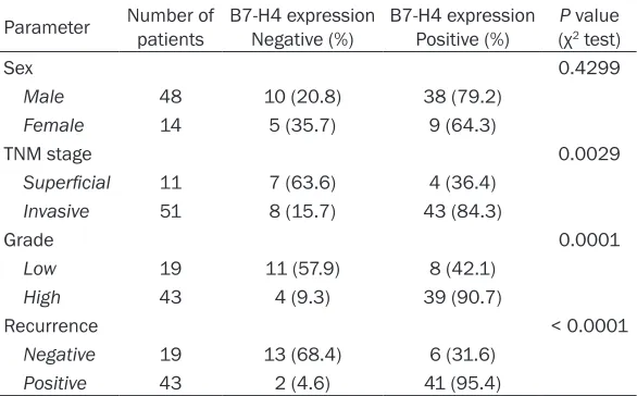
Correlations of B7-H4 expression with clinico-pathological parameters for UCC patients
2. Gender was not found to be significantly
associated with B7-H4 expression (P = 0.4299).
There was a significant association between
B7-H4 expression and the cancer grade: [low grade (G1/2: 8 out of 19: 42.1%) and high grade (G3/4: 39 out of 43: 90.7%)]. In addition,
B7-H4 expression was significantly higher in
patients with muscle invasive tumors (43 out of
51: 84.3%) than in those with superficial tumors
(4 out of 11: 36.4%). The rate of B7-H4 higher expressing specimens in patients with
recur-rence (41 out of 43: 95.4%) was also signifi -cantly higher than that for B7-H4 lower express-ing patients without recurrence (6 out of 19: 31.6%).
Correlation between B7-H4 expression and bladder cancer recurrence-free rate
The recurrence-free rate of UCC was analyzed by the Kaplan-Meier method. A time period of
60 months was defined to investigate the
recurrence-free rate. The recurrence-free rate was determined from the date of the operation to the time of the detection of bladder cancer recurrence or the last follow-up. The impacts of B7-H4 staining, tumour stage, and cancer grade on the recurrence free rate were investi-gated. A log-rank test revealed that positive
B7-H4 expression was significantly associated
[image:4.612.83.527.68.596.2]Figure 1. Immunohistochemical staining of B7-H4. The B7-H4 immunohistochemical staining in bladder cancer. A: Adjacent normal tissues with negative Expression. B: Noninvasive blad-der cancer with lower expression. C: High-grade invasive bladder cancer with higher expression.
with an increased incidence of the cancer recurrence (Figure 3A). Additionally, the higher tumour stage (Figure 3B) and the higher cancer grade (Figure 3C) showed significant associa -tion the poor recurrence free rate.
Cox proportional hazards model univariate and multivariate analyses of individual parameters for correlations with overall survival
Univariate analysis using a Cox proportional hazards model to evaluate the potential of using B7-H4 mRNA expression level as a prog-nostic marker for UCC patients after surgery showed that B7-H4 overexpression (P = 0.006), tumour grade (P = 0.013), and primary tumour stage (P < 0.001) were the prime variables for UCC prognosis (Table 3). After adjusting for clinicopathologic variables, B7-H4 overexpres-sion (P = 0.011), tumour state (P = 0.013), and primary tumour stage (P = 0.023) remained
sig-nificantly correlated with the prognosis of UCC
patients (Table 3). Discussion
The co-regulatory B7 family members are cell-surface protein ligands, binding to receptors on lymphocytes to regulate immune responses [9]. They can provide either positive or negative signal to stimulate or inhibit T-cell activation
[9]. B7-H4 is a recently identified member of
the B7 family [10]. In this study, we investigated the mRNA and protein levels of B7-H4 in human UCC and analyzed the relationship between B7-H4 protein expression level and clinicopath-ological parameters of UCC.
poor prognosis of the patients suffering from gastric cancer [23]. B7-H4 expression in human esophageal squamous cell cancer was shown to be associated with cancer progression, reduced tumour immune surveillance and worse patient outcomes [24]. In this study, B7-H4 mRNA and protein level were found to be
significantly higher in UCC tumour tissues com -pared with adjacent nontumor tissues as assessed by qRT-PCR and immunohistochemi-cal staining, respectively. Higher B7-H4 protein levels were observed in patients with more advanced pathological stage of UCC and asso-ciated with decreased overall survival of patients with UCC. Thus, these previous
stud-ies along with our findings suggest that B7-H4
expression may serve as a universal prognostic indicator for various cancers.
Although B7-H4 overexpression has been found various human tumors, the exact role of B7-H4 in tumourigenesis is still under active investiga-tions. Currently, it is believed that B7-H4 may protect cancer cells by inhibiting tumour-target-ed T cell-mtumour-target-ediattumour-target-ed immune surveillance. In
vitro studies suggest that B7-H4 may deliver an inhibitory signal to T cells, thereby inhibiting CD4+ and CD8+ T cell proliferation, progres-sion, and cytokine production [25-27]. Blockade of B7-H4 has also been shown to enhance the activity of cytotoxic T lymphocytes [28]. Thus, these results imply that B7-H4 may be involved in the evasion mechanism of cancer cells from tumour-targeted T cell-mediated immune sur-Table 2. Correlation between B7-H4 protein expression and
clinico-pathologic parameters of the patients with urothelial cell carcinoma (UCC) (n = 62)
Parameter Number of patients B7-H4 expression Negative (%) B7-H4 expression Positive (%) P(χ value 2 test)
Sex 0.4299
Male 48 10 (20.8) 38 (79.2)
Female 14 5 (35.7) 9 (64.3)
TNM stage 0.0029
Superficial 11 7 (63.6) 4 (36.4)
Invasive 51 8 (15.7) 43 (84.3)
Grade 0.0001
Low 19 11 (57.9) 8 (42.1)
High 43 4 (9.3) 39 (90.7)
Recurrence < 0.0001
Negative 19 13 (68.4) 6 (31.6)
Positive 43 2 (4.6) 41 (95.4)
An association between tu- mour-associated B7-H4 ex- pression and clinicopatho-logical features has been recently found in prostate, renal cell, and esophageal cancers. In prostate cancer, strong expression of B7-H4 is positively correlated with extra capsular extension, seminal vesicle invasion, and distant metastasis [21]. In clear-cell renal cell can-cer, patients with B7-H4-positive tumors showed a poorer survival rate than those with B7-H4-negative tumors [22]. Higher B7-H4 expression was found to be
[image:5.612.91.384.108.290.2]Figure 3. Kaplan-Meir curves demonstrating overall survival of 62 patients with urothelial cell carcinoma following surgery according to mRNA levels of B7-H4 (A), tumour stage (B), and tumour grade (C). P-value was deter-mined by paired samples t-test.
Table 3. Univariate and multivariate analyses of different clinicopathological variables and B7-H4 expres-sion status as predictors for overall survival of urothelial cell carcinoma (UCC)
Variable Univariate analysis Multivariate analysis HR (95% CI) P-value HR (95% CI) P-value Sex (male vs. female) 1.341 (0.689-2.678) 0.354 1.571 (0.759-3.210) 0.215 TNM stage (high vs. low) 2.818 (1.545-5.032) < 0.001 2.033 (0.122-3.688) 0.013 Grade (G3/4 vs. G1/2) 2.215 (1.310-4.925) 0.013 1.523 (1.183-4.125) 0.023 B7-H4 expression (high vs. low) 1.561 (1.125-2.358) 0.006 1.365 (1.115-2.521) 0.011
CI, confidence interval; HR, hazard ratio.
veillance. In addition, B7-H4 may promote tumourigenesis by rendering tumour cells refractory to apoptosis. For example, knock-down of B7-H4 mRNA and protein expression in the SKBR3 breast cancer cell line enhanced intracellular caspase activity, leading to accel-eration of tumour cell apoptosis [29].
In conclusion, the present study has shown for
the first time that B7-H4 mRNA and protein are
increased in UCC tissues and that higher B7-H4 levels are associated with advanced clinical tumour stage and shorter overall survival. The precise role of B7-H4 in UCC development and progression, however, remains to be elucidated
and further investigations in cell and animal models are in progress.
Acknowledgements
Preparation of this manuscript was supported by Changzhou Health Research Program, grant no. CE20125025.
Disclosure of conflict of interest
None.
[image:6.612.93.525.441.521.2]Affiliated Hospital of Soochow University, Changzhou
213000, Jiangsu, China. E-mail: xianlinxu@126.com (Xianlin Xu); fnmong@hotmail.com (Xiaozhou He)
References
[1] Badar F, Sattar A, Meerza F, Irfan N, Siddiqui N. Carcinoma of the urinary bladder in a tertiary care setting in a developing country. Asian Pac J Cancer Prev 2009; 10: 449-452.
[2] Rogers CG, Palapattu GS, Shariat SF, Karakie-wicz PI, Bastian PJ, Lotan Y, Gupta A, Vazina A, Gilad A, Sagalowsky AI, Lerner SP, Schoenberg MP. Clinical outcomes following radical cystec-tomy for primary nontransitional cell carcino-ma of the bladder compared to transitional cell carcinoma of the bladder. J Urol 2006; 175: 2048-2053.
[3] Abel PD. Prognostic indices in transitional cell carcinoma of the bladder. Brit J Urol 1988; 62: 103-109.
[4] Kaufman DS, Shipley WU and Feldman AS. Bladder cancer. Lancet 2009; 374: 239-249. [5] Catto JW, Yates DR, Rehman I, Azzouzi AR,
Pat-terson J, Sibony M, Cussenot O, Hamdy FC. Be-havior of urothelial carcinoma with respect to anatomical location. J Urol 2007; 177: 1715-1720.
[6] Stein JP, Lieskovsky G, Cote R, Groshen S, Feng AC, Boyd S, Skinner E, Bochner B, Thangathu-rai D, Mikhail M, Raghavan D, Skinner DG. Radical cystectomy in the treatment of inva-sive bladder cancer: long-term results in 1,054 patients. J Clin Oncol 2001; 19: 666-675. [7] Slaton JW, Swanson DA, Grossman HB, Dinney
CP. A stage specific approach to tumor surveil -lance after radical cystectomy for transitional cell carcinoma of the bladder. J Urol 1999; 162: 710-714.
[8] Sharpe AH, Freeman GJ. The B7-CD28 super-family. Nat Rev Immunol 2002; 2: 116-126. [9] Greenwald RJ, Freeman GJ, Sharpe AH. The B7
family revisited. Annu Rev Immunol 2005; 23: 515-548.
[10] Sica GL, Choi IH, Zhu G, Tamada K, Wang SD, Tamura H, Chapoval AI, Flies DB, Bajorath J, Chen L. B7-H4, a molecule of the B7 family, negatively regulates T cell immunity. Immunity 2003; 18: 849-861.
[11] Choi IH, Zhu G, Sica GL, Strome SE, Cheville JC, Lau JS, Zhu Y, Flies DB, Tamada K, Chen L. Ge-nomic organization and expression analysis of B7-H4, an immune inhibitory molecule of the B7 family. J Immunol 2003; 171: 4650-4654. [12] Salceda S, Tang T, Kmet M, Munteanu A,
Ghosh M, Macina R, Liu W, Pilkington G, Pap-koff J. The immunomodulatory protein B7-H4 is overexpressed in breast and ovarian
can-cers and promotes epithelial cell transforma-tion. Exp Cell Res 2005; 306: 128-141. [13] Sun Y, Wang Y, Zhao J, Gu M, Giscombe R,
Lef-vert AK, Wang X. B7-H3 and B7-H4 expression in non-small-cell lung cancer. Lung Cancer 2006; 53: 143-151.
[14] Tan DS, Agarwal R, Kaye SB. Mechanisms of transcoelomic metastasis in ovarian cancer. Lancet Oncol 2006; 7: 925-934.
[15] Tringler B, Liu W, Corral L, Torkko KC, Enomoto T, Davidson S, Lucia MS, Heinz DE, Papkoff J, Shroyer KR. B7-H4 overexpression in ovarian tumors. Gynecol Oncol 2006; 100: 44-52. [16] In: Sobin LH, Gospodarowicz MK and Wittekind
CH, editors. TNM classification of malignant
tumors. 7th edition. Oxford: Blackwell Publish-ing Ltd; 2010.
[17] Seitz M, Zaak D, Knüchel-Clarke R, Stief C. [Uri-nary bladder tumors. The new 2004 WHO
clas-sification]. Urologe A 2005; 44: 1073-1086.
[18] Soslow RA, Dannenberg AJ, Rush D, Woerner BM, Khan KN, Masferrer J, Koki AT. COX-2 is expressed in human pulmonary, colonic, and mammary tumors. Cancer 2000; 89: 2637-2645.
[19] Hao L, Zhang C, Qiu Y, Wang L, Luo Y, Jin M, Zhang Y, Guo TB, Matsushima K, Zhang Y. Re-combination of CXCR4, VEGF, and MMP-9 pre-dicting lymph node metastasis in human breast cancer. Cancer Lett 2007; 253: 34-42. [20] Livak KJ and Schmittgen TD. Analysis of
rela-tive gene expression data using real-time
quantitative PCR and the 2-ΔΔCT method.
Methods 2001; 25: 402-408.
[21] Zang X, Thompson RH, Al-Ahmadie HA, Serio AM, Reuter VE, Eastham JA, Scardino PT, Shar-ma P, Allison JP. B7-H3 and B7x are highly ex-pressed in human prostate cancer and associ-ated with disease spread and poor outcome. Proc Natl Acad Sci U S A 2007; 104: 19458-19463.
[22] Krambeck AE, Thompson RH, Dong H, Lohse CM, Park ES, Kuntz SM, Leibovich BC, Blute ML, Cheville JC, Kwon ED. B7-H4 expression in renal cell carcinoma and tumor vasculature: associations with cancer progression and sur-vival. Proc Natl Acad Sci U S A 2006; 103: 10391-10396.
[23] Jiang J, Zhu Y, Wu C, Shen Y, Wei W, Chen L, Zheng X, Sun J, Lu B, Zhang X. Tumor expres-sion of B7-H4 predicts poor survival of patients suffering from gastric cancer. Cancer Immunol Immunother 2010; 59: 1707-1714.
[25] Glaspy JA. Therapeutic options in the manage-ment of renal cell carcinoma. Semin Oncol 2002; 29: 41-46.
[26] Bromwich EJ, McArdle PA, Canna K, McMillan DC, McNicol AM, Brown M, Aitchison M. The
relationship between T-lymphocyte infiltration,
stage, tumour grade and survival in patients undergoing curative surgery for renal cell can-cer. Br J Cancer 2003; 89: 1906-1908. [27] Nakano O, Sato M, Naito Y, Suzuki K, Orikasa
S, Aizawa M, Suzuki Y, Shintaku I, Nagura H, Ohtani H. Proliferative activity of intratumoral CD8+ T-lymphocytes as a prognostic factor in human renal cell carcinoma clinicopathologic demonstration of antitumor immunity. Cancer Res 2001; 61: 5132-5136.
[28] Sica GL, Choi IH, Zhu G, Tamada K, Wang SD, Tamura H, Chapoval AI, Flies DB, Bajorath J, Chen L. B7-H4, a molecule of the B7 family, negatively regulates T cell immunity. Immunity 2003; 18: 849-861.