ISSN Online: 2160-8806 ISSN Print: 2160-8792
DOI: 10.4236/ojog.2018.814153 Dec. 18, 2018 1520 Open Journal of Obstetrics and Gynecology
Use of Basal Serum Testosterone Level as
Predictor for Poor Ovarian Response in Women
with Unexplained Infertility Undergoing
In
Vitro
Fertilization Cycle: Prospective Study
Waleed M. Khalaf, Hayam Fathy, Sarah Safwat
*Faculty of Medicine, Ain Shams University, Cairo, Egypt
Abstract
Background: Delayed pregnancy in women and marked increase in the numbers of older women who fail to respond to ovarian stimulation had been a significant issue. This study aims to assess the value of basal serum testos-terone level as a predictor of ovarian response for induction of ovulation in women with unexplained infertility undergoing IVF (in vitro fertilization) cycle. Patients and Methods: A prospective study was conducted in Ain Shams University Maternity hospital Infertility Center during a period of time from October 2016 to June 2017. This study recruited 89 women. On day 2 or 3 of a spontaneous menstrual cycle of the included women within 3 months before fresh IVF cycle, basal hormonal (FSH, LH, estradiol, total tes-tosterone) concentrations, AFC (antral follicle count) were performed. Using the Long-protocol for induction of ovulation, serial monitoring of ovarian response was assessed by transvaginal ultrasound. When the expected ovarian response was reached (at least three oocytes ≥ 17 mm), we gave trigger dose of HCG. Ultrasound guided oocyte aspiration was performed 34 - 36 hours later. Two to three days after oocyte aspiration, we transferred the embryos according to the patient’s age and the condition of embryos available. Bio-chemical pregnancy was considered if serum B-hCG test was positive at day 14 from embryo transfer, where all the data were correlated with serum tes-tosterone level and ovarian response as 1 ry outcome. Results: There were significant positive correlations between testosterone and LH, Prolactin, AFC, Number of oocytes & Number of Embryos (0.014, 0.032, 0.023, 0.004, 0.033, p < 0.001 respectively). Poor responders versus good responders as regards tes-tosterone level (0.81 ± 0.47 versus 1.08 ± 0.45) Fertilized & pregnant cases had significantly higher testosterone than non-fertilized & non pregnant had (1.20 ± 0.45, 0.92 ± 0.47 p value 0.035, 0.021 respectively). Yet, testosterone
How to cite this paper: Khalaf, W.M., Fathy, H. and Safwat, S. (2018) Use of Basal Serum Testosterone Level as Predictor for Poor Ovarian Response in Women with Unexplained Infertility Undergoing In Vitro Fertilization Cycle: Prospective Study. Open Journal of Obstetrics and Gynecology, 8, 1520-1531.
https://doi.org/10.4236/ojog.2018.814153
Received: November 22, 2018 Accepted: December 15, 2018 Published: December 18, 2018
Copyright © 2018 by authors and Scientific Research Publishing Inc. This work is licensed under the Creative Commons Attribution International License (CC BY 4.0).
http://creativecommons.org/licenses/by/4.0/
DOI: 10.4236/ojog.2018.814153 1521 Open Journal of Obstetrics and Gynecology had significant low diagnostic performance in prediction of poor response and pregnancy (AUC 0.654, 0.676 respectively), (p value 0.015, 0.022 respec-tively). Conclusion: Basal T levels are helpful for predicting ovarian re-sponse, hence the dosage of gonadotropins used in induction. But it can’t be used as single marker for prediction of ovarian response.
Keywords
Testosterone, Induction, Ovarian Reserve, Pregnancy
1. Introduction
Two to thirty percent of women undergoing induction of ovulation experience poor response which results in cycle cancellation and reduced pregnancy rate [1]. In recent years, several studies showed that response to induction in poor responders might become better with androgen supplementation. These prod-ucts are cheap and available, yet their use remains controversial. This is due to lack of sufficient studies for their efficacy and safety [2] [3].
Androgen receptor mRNA is correlated with FSH receptor mRNA expression in granulose cells. Thus androgens augment FSH receptor expression in the granulose cells amplifying FSH effects on follicular growth [4] [5].
At the other extreme, androgen excess affect oocyte quality, so the current study excluded patients with hyperandrogenemia as in PCOS (polycystic ovarian syndrome)[6].
A previous study by Guo et al. suggested that basal testosterone, instead of DHEAS concentration, couldn’t be used as single predictor, for POR (poor ova-rian reserve). However multifactors as age, AFC, basal FSH, basal FSH/LH and basal testosterone are better predictors for POR and clinical pregnancy than AFC alone. They also observed that as testosterone levels increase, total gonado-tropins dosage decrease while numbers of oocytes retrieved, cleavage-stage em-bryos, frozen embryos and pregnancy rates all increase significantly [7].
Assessment of serum androgen levels prior to controlled ovarian stimulation might be useful to predict the ovarian response and, thus adjust the starting dose of exogenous gonadotrophins or even pretreatment with transdermal testoste-rone before ovarian stimulation may be a useful approach for women known to have poor ovarian response.
This study aims to assess the value of basal serum testosterone level as a predictor of ovarian response for induction of ovulation in women undergoing IVF cycle.
2. Patients and Methods
conti-DOI: 10.4236/ojog.2018.814153 1522 Open Journal of Obstetrics and Gynecology nuous marital life for at least two years after marriage, all patients were ovulatory and had regular menstrual cycles, BMI: 20 - 30, normal pelvic transvaginal ultraso-nography concerning exclusion of pelvic pathology, normal hysterosalpingogram (confirming tubal patency and showing absence of uterine anomalies or filling de-fects, or tubal anomalies as hydrosalpinx), normal hormonal profile regarding es-tradiol, FSH (Day 2 < 10 mIU/ml), LH (Day 2 < 8 mIU/ml), TSH (0.5 - 4.5 mIU/L), serum prolactin (2 - 29 ng/ml).All husbands of the selected patients showed normal semen analysis parameters. Women with endocrinal problems such as DM, thyroid disorders or hyperprolactinemia were excluded. Women having AFC < 5 follicles (Antral Follicular Count in both ovaries) were also excluded.
Ethics: The study was approved from the Ethical Committee of the Depart-ment of Obstetrics and Gynecology, Faculty of Medicine, Ain Shams University. Written informed consents were obtained from all participants.
All participants were subjected to careful history taking and clinical examina-tion including BMI to ensure fulfillment of selecexamina-tion criteria. Compleexamina-tion of the series of investigations to fulfill the selection criteria on day 2 or 3 of a sponta-neous menstrual cycle within 3 months before fresh IVF cycle, a blood sample was taken in the morning to evaluate basal hormonal (FSH, LH, estradiol, total testosterone): Complete Blood Picture, Thyroid Profile, prolactin, AMH (anti-mullerian hormone). On the same day, trans-vaginal sonography was performed to obtain AFC (the follicles visualized and counted were 2 - 10 mm in size, and the numbers of follicles in both ovaries were added to obtain the total AFC) [8], at the same time will be used to identify uterine fibroids, PCO, polyps. Recent hysterosalpingogram & semen analysis of husband should be availabe.
Using the Long-protocol for induction of ovulation:
This study used controlled-release of long-acting GnRH analogue (Decapep-tyl®; 0.1 mg/ampoule Tryptorelin, Ferring, Germany) daily from mid-luteal day of the previous cycle till the day of hCG administration. When a satisfactory pi-tuitary desensitization is achieved (Estradiol level below 40 pg/ml), human
me-nopausal gonadotrophins (Menogon®; 75 mg/ampoule HMG, Ferring GmbH
DOI: 10.4236/ojog.2018.814153 1523 Open Journal of Obstetrics and Gynecology Biochemical pregnancy was considered if serum B-hCG test was positive at day 14 from embryo transfer, where all the data were correlated with serum tes-tosterone level, with ovarian responders(the 1ry outcome) and pregnancy out-come (the 2ry outcome).
Hormonal Assay:
All blood samples were immediately processed to separate serum. Serum samples were stored at −20˚C and hormonal assays were performed in the docrine Laboratory of Ain Shams University Hospital, Cairo, Egypt using En-zyme-Linked Immunosorbent Assay for the Quantitative Determination in Hu-man Serum (ELISA Technique) by kits named Immunospec Quantitative Assay (Immunospec Corporation, 7018 Owensmouth Ave. Suite 103, Canoga Park, CA, 91303) according to the manufacture instructions to Testosterone, FSH, LH and Estradiol with analytical sensitivity is <0.1 ng/ml, 2.5 mIU/ml, 2 mIU/ml, 5 pg/ml respectively.
Statistical methods:
The collected data were coded, tabulated, and statistically analyzed using IBM SPSS statistics (Statistical Package for Social Sciences) software version 22.0, IBM Corp., Chicago, USA, 2013.
Descriptive statistics were done for quantitative data as minimum& maximum of the range as well as mean ± SD (standard deviation) for quantitative parame-tric data, while it was done for qualitative data as number and percentage.
Inferential analyses were done for quantitative variables using independent t-test in cases of two independent groups with parametric data. In qualitative data, inferential analyses for independent variables were done using Chi square test for differences between proportions. While correlations were done using Pearson correlation for numerical parametric data. Logistic regression model was used to find out independent factors affecting response. The level of signi-ficance was taken at P value < 0.050 is significant, otherwise is non-significant.
3. Results
1) Basal characteristics of the studied women were listed in Table 1.
2) Fertilization characteristics of the studied women. The number of fertilized women in the study 77 out of 89 which represents 88.5% yielding mean number of embryos of 5.5 (Table 2).
3) Pregnancy rate among the studied women was 20.2% (18 out of 89) (Table 3).
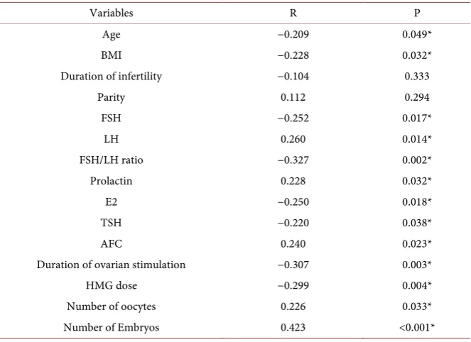
4. Correlation between Testosterone and Other Variables
There were significant positive correlations between testosterone and LH, Pro-lactin, AFC, Number of oocytes & Number of Embryos (P value 0.014, 0.032, 0.023, 0.033, <0.001 respectively) (Table 4).DOI: 10.4236/ojog.2018.814153 1524 Open Journal of Obstetrics and Gynecology
Table 1. Basal characteristics of the studied women.
Characteristics Mean ± SD Range Age (years) 28.5 ± 3.6 21.0 - 35.0 BMI (kg/m2) 27.9 ± 1.2 25.1 - 31.5
Duration of infertility (years) 4.6 ± 1.4 2.0 - 8.0
Parity 1.0 ± 0.8 0.0 - 2.0
FSH (mIU/mL) 7.45 ± 2.1 4.2 - 10.7 LH (mIU/mL) 5.0 ± 1.3 2.1 - 8.3
FSH/LH ratio 1.9 ± 0.9 0.7 - 5.6 Prolactin (ng/mL) 19.3 ± 4.6 6.8 - 30.4
[image:5.595.207.539.303.362.2]E2 (pg/mL) 53.2 ± 13.5 22.0 - 85.4 TSH (mIU/L) 3.1 ± 0.2 2.6 - 3.6 Testosterone (nmol/L) 0.98 ± 0.48 0.02 - 2.13 Total = 89.
Table 2. Fertilization characteristics of the studied women.
Characteristics Mean ± SD Range Number of embryos 5.5 ± 2.9 0.0 - 13.0
N %
Fertilization 77 88.5
total = 89.
Table 3. Pregnancy among the studied women.
Characteristics N %
Among all cases (Total = 89) 18 20.2 Among fertilized cases (Total = 77) 18 23.4
Table 4. Correlation between testosterone and other variables.
Variables R P
Age −0.209 0.049*
BMI −0.228 0.032*
Duration of infertility −0.104 0.333
Parity 0.112 0.294
FSH −0.252 0.017*
LH 0.260 0.014*
FSH/LH ratio −0.327 0.002*
Prolactin 0.228 0.032*
E2 −0.250 0.018*
TSH −0.220 0.038*
AFC 0.240 0.023*
Duration of ovarian stimulation −0.307 0.003*
HMG dose −0.299 0.004*
[image:5.595.207.538.406.448.2] [image:5.595.207.539.474.715.2]DOI: 10.4236/ojog.2018.814153 1525 Open Journal of Obstetrics and Gynecology
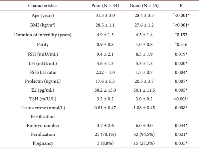
6. Comparison between Poor and Good Responses
regarding Basal Characteristics
Poor responders versus good responders as regards testosterone level (0.81 ± 0.47 versus1.08 ± 0.45). Poor responders had significantly higher age and BMI than good responders had. Poor responders had significantly higher FSH, FSH/LH ratio, E2 and TSH and significantly lower LH, prolactin and Testoste-rone than good responders had (Table 6).
7. Comparison between Pregnancy Conditions regarding
Basal Characteristics
Pregnant cases had significantly higher testosterone than non pregnant had (0.92 ± 0.47 p value, 0.026) according to Table 7.
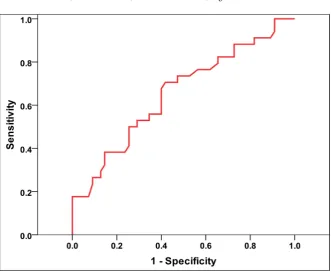
8. Diagnostic Performance of Testosterone
in Prediction of Outcomes
[image:6.595.207.538.378.421.2]Testosterone had significant low diagnostic performance in prediction of poor response and pregnancy, so it can’t be used as marker alone for ovarian response (Table 8 and Figure 1).
Table 5. Relation of fertilization with testosterone (nmol/L).
N Mean ± SD P value
Fertilization Fertilized 77 1.04 ± 0.46 0.002* Not 12 0.56 ± 0.34
^Independent t-test, *significant.
Table 6. Comparison between poor and good responses regarding basal characteristics.
Characteristics Poor (N = 34) Good (N = 55) P Age (years) 31.3 ± 3.0 28.4 ± 3.5 ^<0.001*
BMI (kg/m2) 28.5 ± 1.1 27.6 ± 1.2 ^<0.001*
Duration of infertility (years) 4.9 ± 1.3 4.5 ± 1.4 ^0.153
Parity 0.9 ± 0.8 1.0 ± 0.8 ^0.516
FSH (mIU/mL) 9.4 ± 2.1 8.3 ± 1.9 0.019* LH (mIU/mL) 4.6 ± 1.3 5.3 ± 1.3 0.020* FSH/LH ratio 2.22 = 1.0 1.7 ± 0.7 0.004* Prolactin (ng/mL) 17.6 ± 5.3 20.3 ± 3.7 0.007* E2 (pg/mL) 58.2 ± 15.0 50.1 ± 11.5 0.005* TSH (mIU/L) 3.2 ± 0.2 3.0 ± 0.2 <0.001* Testosterone (nmol/L) 0.81 ± 0.47 1.08 ± 0.45 0.008*
Fertilization
Embryo number 4.7 ± 2.6 6.0 ± 3.0 0.044* Fertilization 25 (78.1%) 52 (94.5%) 0.021* Pregnancy 3 (8.8%) 15 (27.3%) 0.035*
[image:6.595.207.539.464.712.2]DOI: 10.4236/ojog.2018.814153 1526 Open Journal of Obstetrics and Gynecology
Table 7. Comparison between pregnancy conditions regarding basal characteristics.
Characteristics Pregnant (N = 18) Not (N = 71) P Age (years) 28.1 ± 3.3 29.8 ± 3.6 0.073 BMI (kg/m2) 28.0 ± 1.0 27.9 ± 1.3 0.870
Duration of infertility (years) 4.6 ± 1.6 4.6 ± 1.3 0.830 Parity 1.3 ± 0.8 0.9 ± 0.8 0.063 FSH (mIU/mL) 8.1 ± 2.2 8.9 ± 2.0 0.149 LH (mIU/mL) 5.5 ± 1.5 4.9 ± 1.3 0.113 FSH/LH ratio 1.6 ± 0.8 2.0 ± 0.9 0.165 Prolactin (ng/mL) 19.5 ± 2.9 19.2 ± 4.9 0.816 E2 (pg/mL) 50.3 ± 10.5 53.9 ± 14.1 0.312 TSH (mIU/L) 3.0 ± 0.2 3.1 ± 0.2 0.324 Testosterone (nmol/L) 1.20 ± 0.45 0.92 ± 0.47 0.026*
[image:7.595.207.540.355.408.2]^Independent t-test, #Chi square test, *Significant.
Table 8. Diagnostic performance of Testosterone in prediction of outcomes.
Condition AUC SE P 95% CI
Poor response 0.654 0.061 0.015* 0.535 - 0.773 Pregnancy 0.676 0.071 0.022* 0.538 - 0.815 AUC: Area under curve, SE: Standard error, CI: Confidence interval, *Significant.
[image:7.595.209.541.420.691.2]DOI: 10.4236/ojog.2018.814153 1527 Open Journal of Obstetrics and Gynecology
9. Discussion
Several reviews have showed the predictive value of many tests for ovarian re-serve, antral follicle count (AFC) and anti-Mullerianhormone (AMH) were the best. However, even the best marker had a false positive rate of 10% - 20%, might falsely prevent these women from undergoing IVF [9] [10].
In women undergoing IVF cycle who attended Ain shams university materni-ty hospital during October 2015 to June 2016, this study compared basal serum testosterone level in relation to number of follicles in both ovaries(AFC),number of actual oocyte retrieved, number of embryos fertilized, positive pregnancy test to predict PORs (poor ovarian responders).
The current study suggested that serum testosterone level may help in prediction of the type of response for induction of ovulation in women undergoing IVF cycle.
89 women underwent the ovarian stimulation protocol & oocyte aspiration, where 55 women (61.8%) have been good responders, while 34 women (38.2%) have been poor responders. Testosterone level among PORs was 0.81 ± 0.47 nmol/L where it was 1.08 ± 0.45 nmol/L among good responders. Also its level among pregnant women was1.20 ± 0.45 nmol/L, while among non-pregnant women was 0.92 ± 0.47 nmol/L.
Correlation between testosterone and other variables showed that: There were significant positive correlations between testosterone and BMI, LH, Prolactin, AFC, Number of oocytes & Number of Embryos, while there were significant negative correlations between testosterone and age, FSH, FSH/LH ratio, E2, TSH, Duration of ovarian stimulation & HMG dose.
These results were concordant with Jing Guo et al. (2014) who found that bas-al T positively related to body mass index (BMI), AFC, numbers of oocytes re-trieved, mature oocytes and pregnancy outcome, but negatively with age, basal FSH/LH, peak E2, and total gonadotropins dosage. Also these results were con-firmed by Bo Sun et al. (2014), with the exception of falling of basal E2 levels in the positively correlated parameters. That may be due to different magnitude of populations between the studies or the applied tense whether prospectively or retrospectively [6] [11].
Also these results went with that of Fraterelli and Gerber (2006) and Qin et al., 2011) confirming the above data. Colakoglu (1986) found that Testosterone and DHEAs level decline with age, which may be related to POR predicted by FSH and age [9] [12] [13].
As regards relation of fertilization with testosterone level showed that Ferti-lized cases had significantly higher testosterone than non-fertiFerti-lized had.
Comparison between poor and good responses regarding demographic cha-racteristics and hormonal chacha-racteristics showed that poor responders had sig-nificantly higher age, BMI, FSH, FSH/LH ratio, E2 and TSH than good respond-ers had. Poor respondrespond-ers had significantly lower LH, prolactin and Testosterone than good responders had.
DOI: 10.4236/ojog.2018.814153 1528 Open Journal of Obstetrics and Gynecology significantly older than normal ovarian responders (P < 0.001), with higher body mass index (BMI; P = 0.001), basal FSH (P < 0.001), FSH/LH (P < 0.001) and es-tradiol (P < 0.001) and lower AFC (P < 0.001), mean ovarian volume (P < 0.001) and testosterone (P = 0.022). Significantly higher total gonadotropins dosage was consumed and lower peak estradiol, number of mature oocytes, fertilization rate, cleavage rate, implantation rate and clinical pregnancy rate (all P < 0.001) were achieved by poor ovarian responders. Jayaprakasan et al., 2009 confirmed the above data [6] [14].
On the other hand, Bo Sun et al. (2014) found that the good responder women had more oocytes retrieved, good quality embryos, and embryos cryopreserved. However, the pregnancy outcomes were not significantly different between the two groups [11].
Comparison between pregnancy conditions regarding demographic charac-teristics showed no significant difference. While comparison between nancy conditions regarding basal hormonal characteristics showed that preg-nant women had significantly higher testosterone (T) than non-pregpreg-nant women had.
Also comparison between pregnancy conditions regarding stimulation, re-trieval and fertilization showed that pregnant women had significantly higher good response, oocyte and embryo numbers and significantly lower duration and dose than non-pregnant women had.
Also Jing Guo et al. (2014) observed that Testosterone levels of pregnant women were significantly higher than those who were not (1.3 ± 0.57 vs 1.23 ± 0.6 nmolL; P = 0.026) [6].
Testosterone had significant low diagnostic performance in prediction of poor response and pregnancy, in other words can’t be used as a marker alone for pre-diction of ovarian response.
Yingying Qin’s group (2011) suggested that basal T level was a predictor for ovarian response and pregnancy outcome in women with diminished ovarian reserve; but not in those with normal serum FSH. Also, Fratharelli and Peter-son’s study (2004) showed that women with Day 3 testosterone level lesser than 20 ng/dl were five time less likely to achieve pregnancy approved by Fouany and Sharara, 2013.This may be attributed to the theory of ovarian aging suggested by Glechier and Barad (2011) [9] [12] [15] [16].
Disagreement with this opinion Frattarelli and Gerber (2006) adopted the negative relation between basal Testosterone level and pregnancy outcome. Many patients who have poor gonadotropin responsiveness and low-quality oo-cytes and embryos having normal screening results Thus, the tests for dimi-nished ovarian reserve are specific but not sensitive [9].
DOI: 10.4236/ojog.2018.814153 1529 Open Journal of Obstetrics and Gynecology and achieved a clinical pregnancy rate of 30% per oocyte retrieval. There were 20% cancelled cycles [17].
Most studies of serum T levels in infertile women have focused on women with irregular cycles and PCOS. In this study, women with irregular menstrual cycles or a known diagnosis of PCOS were excluded.
The multivariate model composed of age, AFC, basal FSH, basal FSH/LH and basal testosterone performed better than AFC for predicting both POR and pregnancy outcome. This might be meaningful for most of reproductive medi-cine, where AMH measurement is costly and not in routine use, however testos-terone is available, cheap & can be used as preinduction treatment to enhance ovarian response in those with previous poor response.
Identification of women at increased risk for POR prior to IVF could be use-ful, help to modify the gonadotropins dose for induction in order to maximize ovarian response [18].
10. Limitation of the Study
Measuring only basal serum T level, which is an ovarian androgen,. DHEA and other adrenal androgens are also very important.
Another limitation of study, it didn’t study the role and the effect of giving androgens to those of low levels of testosterone and POR & study the change in ovarian response, another drop out that it didn’t mention the relation between the degree of severity of low testosterone level and ovarian response. Another research may be needed to assess the role of basal testosterone level and prein-duction treatment with androgens in those with poor ovarian reserve according to bologna criteria two of those criteria: Advanced maternal age (≥40 years) or any other risk factor for POR, previous POR (≤three oocytes with a conventional stimulation protocol).An abnormal ovarian reserve tests (i.e. AFC, 5 - 7 follicles or AMH, 0.5 - 1.1 ng/ml).
Also this research not including obese women and relation of obesity to basal testosterone level and if preinduction treatment with androgens can be of benefit for those obese infertile women with poor ovarian response. This question need to be put in mind in future studies.
11. Conclusion
Basal T levels are helpful for predicting ovarian response, hence the dosage of gonadotropins used in induction. Yet, testosterone had significant low diagnos-tic performance in prediction of poor response and pregnancy, so can’t be used as single marker for prediction of ovarian response.
Acknowledgements
Materni-DOI: 10.4236/ojog.2018.814153 1530 Open Journal of Obstetrics and Gynecology ty Hospital for the outstanding support during the entire study.
Compliance with Ethical Standards
Disclosure Statement
No potential conflict of interest was reported by the authors.
Funding
This research did not receive any specific grant from funding agencies in the public, commercial, or not-for-profit sectors.
References
[1] Hendriks, D.J., Mol, B.W., Bancsi, L.F., Te, V.E. and Broekmans, F.J. (2005) Antral Follicle Count in the Prediction of Poor Ovarian Response and Pregnancy after In Vitro Fertilization: A Meta-Analysis and Comparison with Basal Follicle-Stimulating Hormone Level. Fertility and Sterility, 83, 291-301.
https://doi.org/10.1016/j.fertnstert.2004.10.011
[2] Kim, C.H., Howles, C.M. and Lee, H.A. (2011) The Effect of Transdermal Testoste-rone Gel Pretreatment on Controlled Ovarian Stimulation and IVF Outcome in Low Responders. Fertility and Sterility, 95, 679-683.
https://doi.org/10.1016/j.fertnstert.2010.07.1077
[3] Nagels, H.E., Rishworth, J.R., Siristatidis, C.S., Kroon, B., et al. (2012) Androgens (Dehydroepiandrosterone or Testosterone) in Women Undergoing Assisted Re-production (Protocol), Copyright © 2012 The Cochrane Collaboration. John Wiley & Sons, Ltd.
[4] Weil, S., Vendola, K., Zhou, J. and Bondy, C.A. (1999) Androgen and Fol-licle-Stimulating Hormone Interactions in Primate Ovarian Follicle Development.
The Journal of Clinical Endocrinology & Metabolism, 84, 2951-2956.
https://doi.org/10.1210/jcem.84.8.5929
[5] Hugues, J.N. and Durnerin, I.C. (2005) Impact of Androgens on Fertili-ty—Physiological, Clinical and Therapeutic Aspects. Reproductive BioMedicine Online, 11, 570-580. https://doi.org/10.1016/S1472-6483(10)61165-0
[6] Guo, J., Zhang, Q., Li, Y., Huang, J., Wang, W., Huang, L., Zhao, X. and Yang, D. (2014) Predictive Value of Androgens and Multivariate Model for Poor Ovarian Response. Reproductive BioMedicine Online, 28, 723-732.
https://doi.org/10.1016/j.rbmo.2014.02.009
[7] Nielsen, M.E., Rasmussen, I.A., Kristensen, S.G., Christensen, S.T., Mollgard, K., Wreford, A.E., Byskov, A.G. and Yding, A.C. (2011) In Human Granulosa Cells from Small Antral Follicles, Androgen Receptor mRNA and Androgen Levels in Follicular Fluid Correlate with FSH Receptor mRNA. Molecular Human Reproduc-tion, 17, 63-70. https://doi.org/10.1093/molehr/gaq073
[8] Qin, Y., Zhao, Z., Sun, M., Geng, L., Che, L. and Chen, Z. (2011) Association of Basal Serum Testosterone Levels with Ovarian Response and In Vitro Fertilization Outcome. Reproductive Biology and Endocrinology, 9, 9.
https://doi.org/10.1186/1477-7827-9-9
DOI: 10.4236/ojog.2018.814153 1531 Open Journal of Obstetrics and Gynecology https://doi.org/10.1016/j.fertnstert.2009.04.040
[10] Colakoglu, M. (1986) The Effect of Dehydroepiandrosterone Sulfate Prolactin and Testosterone Hormones in Female Fertility and Hirsutism. Clinical and Experi-mental Obstetrics and Gynecology, 13, 32-34.
[11] Ferraretti, A.P., La Marca, A., Fauser, B.C., Tarlatzis, B., Nargund, G. and Gianaroli, L. (2011) ESHRE Consensuson the Definition of “Poor Response” to Ovarian Sti-mulation for In Vitro Fertilization: The Bologna Criteria. Human Reproduction, 26, 1616-1624. https://doi.org/10.1093/humrep/der092
[12] La Marca, A., Sighinolfi, G., Radi, D., Argento, C., Baraldi, E., Artenisio, A.C., Sta-bile, G. and Volpe, A. (2010) Anti-Mullerian Hormone (AMH) as a Predictive Marker in Assisted Reproductive Technology (ART). Human Reproduction Update, 16, 113-130. https://doi.org/10.1093/humupd/dmp036
[13] Jayaprakasan, K., Al-Hasie, H., Jayaprakasan, R., Campbell, B., Hopkisson, J., John-son, I. and Raine-Fenning, N. (2009) The Three-Dimensional Ultrasonographic Ovarian Vascularity of Women Developing Poor Ovarian Response during Assisted Reproduction Treatment and Its Predictive Value. Fertility and Sterility, 92, 1862-1869. https://doi.org/10.1016/j.fertnstert.2008.09.031
[14] Fouany, M.R. and Sharara, F.I. (2013) Is There a Role for DHEA Supplementation in Women with Diminished Ovarian Reserve? Journal of Assisted Reproduction and Genetics, 30, 1239-1244. https://doi.org/10.1007/s10815-013-0018-x
[15] Gleicher, N. and Barad, D.H. (2011) Dehydroepiandrosterone (DHEA) Supple-mentation in Diminished Ovarian Reserve (DOR). Reproductive Biology and En-docrinology, 9.
[16] Balasch, J., Fábregues, F., Peñarrubia, J., Carmona, F., Casamitjana, R., Creus, M., Manau, D., Casals, G. and Vanrell, J.A. (2006) Pretreatment with Transdermal Tes-tosterone May Improve Ovarian Response to Gonadotrophins in Poor-Responder IVF Patients with Normal Basal Concentrations of FSH. Human Reproduction, 21, 1884-1893. https://doi.org/10.1093/humrep/del052
[17] Sun, B., Wang, F., Sun, J., Yu, W.Z. and Sun, Y.P. (2014) Basal Serum Testosterone Levels Correlate with Ovarian Response But Do Not Predict Pregnancy Outcome in Non-PCOS Women Undergoing IVF. Journal of Assisted Reproduction and Genet-ics, 31, 829-835. https://doi.org/10.1007/s10815-014-0246-8
[18] Klinkert, E.R., Broekmans, F.J., Looman, C.W., Habbema, J.D. and Te, V.E. (2005) Expected Poor Responders on the Basis of an Antral Follicle Count Do Not Benefit from a Higher Starting Dose of Gonadotrophins in IVF Treatment: A Randomized Controlled Trial. Human Reproduction, 20, 611-615.