EFFECT OF NANO CLAY AND SILVER NANO PARTICLES ON THE FLEXURAL STRENGTH AND
ANTIMICROBIAL PROPERTY OF ACRYLIC DENTURE BASE RESIN
*Dr. Hamim Rahman Hass
ARTICLE INFO ABSTRACT
Polymethyl methacrylate (PMMA) has been commonly used as denture base material. However, repeated stress from masticatory action d
base. Thus, further improvements in the mechanical property of PMMA denture base material are still needed. This study aimed to
property and antimicrobial activity by addition of nano clay and silver nano particles. are to evaluate the flexural strength and antimicrobial property of conventional PMMA incorporation of 3% nano clay and varied concentrations of silver nano particles. Materials and
were fabricated for testing the flexural strength and antimicrobial property. Test specimens
silver nano particles in heat cure acrylic resin. These included a control group and two test groups with 3% nano clay and varied concentrations of silver nano particles.
Results:
clay and different percentage of silver nano particles in conventional PMMA denture base resin. The increasing order of flexural strength was as follows: Group
flexural strength followed by Group C (3% nano clay and 1% silver nano particles) and Group A (Control) showed the highest flexural strength
(Control) showed the least antimicrobial property followed by Group B (3% nano clay and O.5% silver nano particles) and Group C (3% nano clay and 1% silver nano particles) showed the highest antimicrobial property. Conclusion:
and 0.5% silver nano particles showed the least flexural strength. Incorporation of 3% nano clay and 1% silver nano particles showed higher flexural strength but less
property of
particles showed lower antimicrobial property but higher compared to the control group. Incorporat clay and 1% silver nano particles showed the highest antimicrobial property.
Clinical implications
resin having sufficient flexural strength and possessin
shown by the synthesized material can be considered to be an interesting material for scientific research of antimicrobial acrylic denture base resins.
Copyright©2016, Dr. Hamim Rahman Hassan et al. This
unrestricted use, distribution, and reproduction in any medium, provided the original work is properly cited.
INTRODUCTION
Dentures are made of poly methyl methacrylate (PMMA)
acrylic resin.1 PMMA acrylic resin has proven to be the most
satisfactory denture base material currently available
their popularity, this material is far from ideal in fulfilling the mechanical requirements. Recently the use of nanoparticles as fillers or additives in polymers for various desired effects are receiving an increased interest for research and development.
*Corresponding author: Dr. Hamim Rahman Hassan
JSS University, India.
ISSN: 0975-833X
Vol.
Article History: Received 23rd June, 2016 Received in revised form 20th July, 2016
Accepted 09th August, 2016
Published online 20th September,2016
Key words:
Denture base resin, Nano clay, Silver nano particles, Heat polymerization, Antimicrobial property, Flexural strength.
Citation: Dr. Hamim Rahman Hassan, Dr. Anil Kumar Gujjari, Dr. Dhakshaini, M. R. and Dr. Sandeip Kumar, Nano particles on the flexural strength and antimicrobial property of acrylic denture base resin
Research, 8, (09), 37911-37915.
RESEARCH ARTICLE
EFFECT OF NANO CLAY AND SILVER NANO PARTICLES ON THE FLEXURAL STRENGTH AND
ANTIMICROBIAL PROPERTY OF ACRYLIC DENTURE BASE RESIN– AN INVITRO STUDY
Dr. Hamim Rahman Hassan, Dr. Anil Kumar Gujjari, Dr. Dhakshaini, M.
Dr. Sandeip kumar
JSS University, India
ABSTRACT
Polymethyl methacrylate (PMMA) has been commonly used as denture base material. However, repeated stress from masticatory action during function and impact force from accidental drop can result in fracture of the denture base. Thus, further improvements in the mechanical property of PMMA denture base material are still needed. This study aimed to provide us with an improved acrylic denture base resin which possesses better mechanical property and antimicrobial activity by addition of nano clay and silver nano particles.
to evaluate the flexural strength and antimicrobial property of conventional PMMA incorporation of 3% nano clay and varied concentrations of silver nano particles. Materials and Methods: A total number of 60 specimens were included in the study. were fabricated for testing the flexural strength and 30 disc shaped specimens
antimicrobial property. Test specimens were divided into 3 groups based on the concentration of nano clay and silver nano particles in heat cure acrylic resin. These included a control group and two test groups with 3% nano clay and varied concentrations of silver nano particles.
Results: One way ANOVA descriptive analysis showed significant difference after the incorporation of 3% nano clay and different percentage of silver nano particles in conventional PMMA denture base resin. The increasing order of flexural strength was as follows: Group B (3% nano clay and 0.5% silver nano particles) showed the least flexural strength followed by Group C (3% nano clay and 1% silver nano particles) and Group A (Control) showed the highest flexural strength. The increasing order of antimicrobial property is
(Control) showed the least antimicrobial property followed by Group B (3% nano clay and O.5% silver nano particles) and Group C (3% nano clay and 1% silver nano particles) showed the highest antimicrobial property. Conclusion: The flexural strength of conventional PMMA denture base resin after incorporation of 3% nano clay and 0.5% silver nano particles showed the least flexural strength. Incorporation of 3% nano clay and 1% silver nano particles showed higher flexural strength but lesser compared to the control group.
property of conventional PMMA denture base resin after incorporation of 3% nano clay and 0.5% silver nano particles showed lower antimicrobial property but higher compared to the control group. Incorporat
clay and 1% silver nano particles showed the highest antimicrobial property.
Clinical implications: This study provided a feasible approach of developing a modified acrylic denture base resin having sufficient flexural strength and possessing antimicrobial properties. The optimum microbial inhibition shown by the synthesized material can be considered to be an interesting material for scientific research of antimicrobial acrylic denture base resins.
This is an open access article distributed under the Creative Commons Att use, distribution, and reproduction in any medium, provided the original work is properly cited.
Dentures are made of poly methyl methacrylate (PMMA) PMMA acrylic resin has proven to be the most satisfactory denture base material currently available. Despite their popularity, this material is far from ideal in fulfilling the mechanical requirements. Recently the use of nanoparticles as ives in polymers for various desired effects are receiving an increased interest for research and development.
*Corresponding author: Dr. Hamim Rahman Hassan
Various types of nanoparticles, including nanocarbon, carbon nanotubes, nanoclays, and metal oxides, are currently used to modify the polymer’s performance.
critical parameter for selecting denture base material, as the denture bases must endure various types of stress during function. Decreased flexural strength of the denture base resin will lead to its fracture when subjected to flexural stress. Polymer nanocomposites exhibit excellent biocompatibility as denture base material and have potential application in dental
materials.3 Nano clay dispersed into the PMMA matrix
improves the structure, flexural strength and thermal stability
International Journal of Current Research
Vol. 8, Issue, 09, pp.37911-37915, September, 2016
san, Dr. Anil Kumar Gujjari, Dr. Dhakshaini, M. R. and Dr. Sandeip Kumar, particles on the flexural strength and antimicrobial property of acrylic denture base resin– An In vitro Study
EFFECT OF NANO CLAY AND SILVER NANO PARTICLES ON THE FLEXURAL STRENGTH AND
AN INVITRO STUDY
Dr. Dhakshaini, M. R. and
Polymethyl methacrylate (PMMA) has been commonly used as denture base material. However, repeated stress uring function and impact force from accidental drop can result in fracture of the denture base. Thus, further improvements in the mechanical property of PMMA denture base material are still needed. enture base resin which possesses better mechanical property and antimicrobial activity by addition of nano clay and silver nano particles. The objectives of the study to evaluate the flexural strength and antimicrobial property of conventional PMMA denture base resin after incorporation of 3% nano clay and varied concentrations of silver nano particles.
A total number of 60 specimens were included in the study. 30 rectangular specimens 30 disc shaped specimens were fabricated for testing the were divided into 3 groups based on the concentration of nano clay and silver nano particles in heat cure acrylic resin. These included a control group and two test groups with 3% nano
way ANOVA descriptive analysis showed significant difference after the incorporation of 3% nano clay and different percentage of silver nano particles in conventional PMMA denture base resin. The increasing B (3% nano clay and 0.5% silver nano particles) showed the least flexural strength followed by Group C (3% nano clay and 1% silver nano particles) and Group A (Control) The increasing order of antimicrobial property is as follows: Group A (Control) showed the least antimicrobial property followed by Group B (3% nano clay and O.5% silver nano particles) and Group C (3% nano clay and 1% silver nano particles) showed the highest antimicrobial property.
ural strength of conventional PMMA denture base resin after incorporation of 3% nano clay and 0.5% silver nano particles showed the least flexural strength. Incorporation of 3% nano clay and 1% silver er compared to the control group. The antimicrobial conventional PMMA denture base resin after incorporation of 3% nano clay and 0.5% silver nano particles showed lower antimicrobial property but higher compared to the control group. Incorporation of 3% nano clay and 1% silver nano particles showed the highest antimicrobial property.
: This study provided a feasible approach of developing a modified acrylic denture base g antimicrobial properties. The optimum microbial inhibition shown by the synthesized material can be considered to be an interesting material for scientific research of
is an open access article distributed under the Creative Commons Attribution License, which permits
Various types of nanoparticles, including nanocarbon, carbon nanotubes, nanoclays, and metal oxides, are currently used to
modify the polymer’s performance.2 Flexural strength is a
critical parameter for selecting denture base material, as the denture bases must endure various types of stress during ased flexural strength of the denture base resin will lead to its fracture when subjected to flexural stress. Polymer nanocomposites exhibit excellent biocompatibility as denture base material and have potential application in dental dispersed into the PMMA matrix improves the structure, flexural strength and thermal stability
OF CURRENT RESEARCH
of PMMA, 4 and may decrease the possibility of fracture of the denture base resins. As nano clay is organic in nature, chances of microbial colonization may increase. To compensate for this, incorporation of an antibiotic agent is necessary. As the denture base resin is subjected to heat curing cycles, finishing and polishing procedures which generate huge amount of heat, it may lead to instability of the antibiotic agent within the acrylic denture base resin. Hence we require an antimicrobial agent that overcomes this drawback and is stable within the PMMA matrix when incorporated into the acrylic denture base resin. Silver (Ag) has been well known for its antimicrobial characteristics and has a long history of its application in medicine with a well-tolerated tissue response and low toxicity
profile.5 Due to their small size, nano particles present a large
surface area and therefore larger available area for oxidation.6
These silver nano particles can be used to overcome the drawbacks of an antibiotic agent and also are stable within PMMA matrix of the acrylic denture base resin.
Hence addition of nano clay and silver nano particles may provide us with an improved acrylic denture base resin which possesses better mechanical property and antimicrobial activity. Chronic atrophic Candidiasis, also known as denture stomatitis is a common form of oral Candidiasis involving oral mucosal tissues in complete denture wearers, associated with
the adherence of Candida albicans to denture base surfaces.7
Oral mucosal infection with Staphylococcus aureus has recently been incriminated in a severe form of mucositis in
some group of elderly patients with systemic diseases.8 In the
present study, antimicrobial activity against Candida Albicans and Staphylococcus Aureus were evaluated. The purpose of this in-vitro study was to evaluate the flexural strength and antimicrobial property of conventional PMMA denture base resin after incorporating 3% nano clay and varied concentrations of silver nano particles.
MATERIALS AND METHODS
Fabrication of specimens for assessment of flexural strength and antimicrobial property
A total number of 60 specimens were included in the study. 30 rectangular specimens were fabricated for testing the flexural strength and 30 disc shaped specimens were fabricated for testing the antimicrobial property against test strains of Candida albicans and Staphylococcus aureus. Test specimens were divided into 3 groups based on the concentration of nano clay and silver nano particles in heat cure acrylic resin.
Group A: Heat cure acrylic resin with 0% nano clay and silver nano particles (control)
Group B: Heat cure acrylic resin with 3% nano clay and 0.5% silver nano particles (by Wt)
Group C: Heat cure acrylic resin with 3% nano clay and 1% silver nano particles (by Wt)
Uniform size wax patterns (65×10×2.5 mm rectangle shaped for flexural strength and 6×1 mm disc shaped for antimicrobial property) were fabricated using standardized stainless steel mould for making acrylic resin test specimens to test flexural strength and antimicrobial property.
Preparations of heat cure acrylic resin without Nano clay and Silver nano particles (control)
DPI heat cure denture base resin was mixed in a ceramic mixing jar according to the manufacturer’s recommended ratio of polymer and monomer (3:1 by volume). The resin was left in the mixing jar until it reached the dough stage, and then the mix was kneaded thoroughly to obtain a homogenous dough. The dough was packed into the molds with a thin cellophane sheet placed over it. The flask lid was closed properly and was placed in the hydraulic bench press. The pressure was gradually raised up to 1500 psi pressure to compress the material into the mold. Trial closure was carried out, till no flash was present. The cellophane sheet was removed and the final closure was done. The flasks were bench cured for 1/2 hour at room temperature.
Preparations of heat cure acrylic resin with Nano clay and Silver nano particles
Materials were weighed in an analytical balance, nano clay in (Sigma Aldrich, Nanomer® PGV) and followed by silver nano particles (Ag Flex Technologies Private Ltd, Flexsil ™) were incorporated in pre-defined percentages to the polymer powder of the denture base acrylic resin. These were mixed using an ultrasonic stirrer to obtain a uniform particle distribution. This mixing of nano clay and silver nano particles with acrylic resin polymer resulted in even distribution of filler within polymer matrix. Denture base resin was mixed in a ceramic mixing jar according to the manufacturer recommended ratio of polymer and monomer (3:1 by volume). The resin was left in the mixing jar until it reached the dough stage, and then the mix was kneaded thoroughly to obtain homogenous dough. The dough was packed into the molds with a thin cellophane sheet placed over it. . The flask lid was closed properly and was placed in the hydraulic bench press. The pressure was gradually raised up to 1500 psi pressure to compress the material into the mold. Trial closure was carried out, till no flash was present. The cellophane sheet was removed and the final closure was done. The flasks were bench cured for 1/2 hour at room temperature.
After processing, the specimens for testing flexural strength were retrieved, finished, marked and stored in distilled water at 37°C ± 1°C for 48 hrs prior to subjecting them to flexural strength testing. After processing, the specimens for testing antimicrobial property were retrieved, finished, marked, autoclaved and stored in artificial saliva at 37°C ± 1°C for 7 days prior to subjecting them to antimicrobial property testing.
Assessment of Flexural Strength
were loaded until fracture occurred. The peak load (fracture load) was recorded in chart recorder.
The Peak load is converted to flexural strength by the formula:
S = 3PL/2bd2
Where S= Flexural strength (N/mm2)
P = load at fracture
L = distance between jig supports b = specimen width
d = specimen thickness
The results were tabulated, subjected to statistical analysis and compared.
Assessment of Antimicrobial property
The artificial saliva was discarded and the specimens were subjected to the antifungal and antibacterial activity assays using Agar disc diffusion method.
Antimicrobial property against Candida Albicans
For obtaining the inoculum, the reference strains (Candida albicans ATCC 28366) were inoculated in Mueller Hinton Broth. After incubation at 37°C for 24 hours, the turbidity of Candida albicans inoculum was adjusted to tubes 1 McFarland standard. Mueller Hinton Agar plates were swabbed using sterile cotton swab dipped in 1 McFarland matched Candida albicans culture in Mueller Hinton broth. Specimens measuring 6mm in diameter and 1mm in thickness were placed in 6mm diameter wells prepared on surface of Mueller Hinton Agar plates. The plates were kept at room temperature for 120 min for diffusion of the antimicrobial agent and then incubated in aerobiosis at 37° C for 24 hours. The zone of inhibition diameters were measured with zone measuring scale (in mm) and this measurement indicated the microbial susceptibility to the material.
Antimicrobial property against Staphylococcus Aureus
For obtaining the inoculum, the reference strains
(Staphylococcus aureus ATCC 25293) were inoculated in Mueller Hinton Broth. After incubation at 37°C for 24 hours,
the turbidity of Staphylococcus aureus inoculum was adjusted to tubes 0.5 McFarland standard. Mueller Hinton Agar plates were swabbed using sterile cotton swab dipped in 0.5 McFarland matched Staphylococcus aureus culture in Mueller Hinton broth. Specimens measuring 6mm in diameter and 1mm in thickness were placed in 6mm diameter wells prepared on surface of Mueller Hinton Agar plates. The plates were kept at room temperature for 120 min for diffusion of the antimicrobial agent and then incubated in aerobiosis at 37° C for 24 hours. The zone of inhibition diameters were measured with zone measuring scale (in mm) and this measurement indicated the microbial susceptibility to the material. The results were tabulated, subjected to statistical analysis and compared.
Statistical Analysis
Following statistical methods were applied in analyzing the results of the variables considered in the study.
Descriptive statistics
Independent sample‘t’ test
Scheffe’s Post Hoc test
Analysis of variance
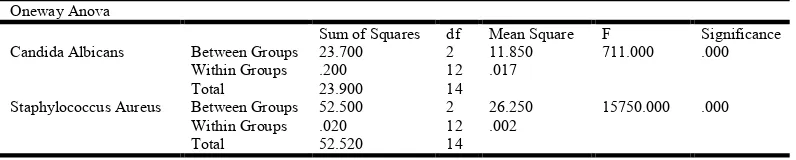
Statistical analysis were conducted with SPSS (version 16). Table 1 shows the One- way ANOVA analysis which demonstrates a highly significant difference (p=.000) between the control and the test groups which were fabricated using different percentages of nano clay and silver nano particles. Table 2 shows the One-way ANOVA analysis which demonstrates a highly significant difference (p=.000) between the control and the test groups which were fabricated with 3% nano clay and different percentages of silver nano particles.
RESULTS
The results of the study showed statistically significant decrease of flexural strength in comparison to the control
group on addition of 3% nano clay and 0.5% silver nano particles. It was followed by statistically significant increase of flexural strength on addition of 3% nano clay and 1% silver nano particles, but lesser when compared to the control group.
Table 1. One way Anova- Group
Oneway Anova flexural strength
Sum of Squares df Mean Square F Significance Between groups 2301.388 2 1150.694 21434.138 .000 Within groups 1.449 27 .054
[image:3.595.104.502.612.692.2]Total 2302.837 29
Table 2. One way anova- group (antimicrobial property)
Oneway Anova
Sum of Squares df Mean Square F Significance Candida Albicans Between Groups 23.700 2 11.850 711.000 .000
Within Groups .200 12 .017
Total 23.900 14
Staphylococcus Aureus Between Groups 52.500 2 26.250 15750.000 .000 Within Groups .020 12 .002
Statistically significant increase in antimicrobial property was observed with the addition of 0.5% and 1% of silver nano particles along with 3% nano clay to the conventional PMMA denture base resin. The antimicrobial property increases as the concentration of silver nano particles increases within the denture base resin.
DISCUSSION
The flexural properties of denture base materials are of importance for predicting their clinical success during function. Dentures are commonly subjected to flexures during masticatory function. Repeated flexures can lead to failure in well fitting denture occluding against natural teeth. Flexural strength is important because it reflects the rigidity of the material, which in turn is important for the integrity of the supporting ridges and tissues, along with the fitting accuracy of
the denture.9 Decreased flexural strength of the denture base
resin will lead to its fracture when subjected to flexural stress. Nano clay dispersed into the PMMA matrix improves the
structure, flexural strength and thermal stability of PMMA, 4
and may decrease the possibility of fracture of the denture base resins. Nano clay being organic in nature may increase the chances of microbial colonization after its incorporation into the denture base resin. To compensate for this, incorporation of an antibiotic agent is necessary.
Silver nano particles have been used for their antimicrobial
effect in different biomedical applications, 10 also in modifying
commercial acrylic resins but not in the formulation of an experimental acrylic for denture bases. Silver nano particles are also reported to be nontoxic to humans and very effective against bacteria, viruses, and other eukaryotic micro-organisms
at very low concentrations and without side effects.11These
silver nano particles can be used as antimicrobial control systems and are also stable within PMMA matrix of the acrylic denture base resin. Addition of nano clay and silver nano particles may provide us with an improved acrylic denture base resin which possesses better mechanical property and antimicrobial activity. In the present study, 3% nano clay and varied concentrations of silver nano particles were added in to the heat polymerized acrylic resin to evaluate the flexural strength and antimicrobial property of the conventional PMMA denture base resin.
Evaluation of flexural strength
A statistically significant decrease of flexural strength in comparison to the control group was observed with the addition of 3% nano clay and 0.5% silver nano particles. This was followed by increase in flexural strength with addition of 3% nano clay and 1% silver nano particles, to the acrylic resin, but lesser when compared to the control group, which is consistent with the findings of previous investigations done using AgNps as antimicrobial agents (A. sodagar et al 2012). The decrease of flexural strength values in this study followed by increase, in agreement with the results of the study done by
Sodagar et al, who reported that probably, at low
concentration, particle dispersion and chemical interactions between PMMA and AgNps are low which result in flexural strength reduction. We can say that here, at the 0.5%
concentration, AgNps act as impurities which usually decrease mechanical strength in the composites. Along with increase in concentration of the nano particles, the more chemical interaction between the C=O groups and the silver overcome the aforementioned negative effects, leading to the flexural
strength improvement.12 However, it is important to realize that
the least mean flexural strength value (84.29 MPa) obtained for the samples in which the percentage of nano clay and silver nano particles was 3% and 0.5 % is more than the minimum flexural strength value of 65 MPa set forth by the ADA specification no.12. Hence this slight reduction may not have
any clinical relevance.13.
Evaluation of antimicrobial property
It was observed that the control specimens (fabricated without incorporation of nano clay and silver nano particles), did not have antimicrobial activity, which agrees with the results of previous studies that report little or no antimicrobial activity of the tested materials. The addition of 0.5% of silver nano particles with 3% nano clay to heat polymerized acrylic resin was enough to provide antimicrobial activity against the test strains. A significant increase in antimicrobial property was observed with the increase in the concentration of silver nano particles along with nano clay at constant concentration. The higher, the percentage of silver nano particles, the greater the antimicrobial property. The smallest inhibition halos were observed for the heat polymerized acrylic resin specimens with 0.5% of silver nano particles with 3% nano clay against Candida albicans and intermediary for Staphylococcus aureus isolate, while the largest inhibition halos were observed for heat polymerized acrylic resin specimens with 1% of silver nano particles with 3% nano clay against Staphylococcus Aureus isolate followed by Candida Albicans isolate for the same. These results may be attributed to silver nano particles
present in the acrylic resin. The major advantage of this
addition could be for the elderly people with restricted manual dexterity or cognitive disturbances. Also, since the benefits of silver nano particles along with nano clay outweigh the toxicity risks, the use of these nano particles in denture base resin can be recommended. An antimicrobial biocompatible PMMA was developed as a denture base.
Conclusion
patients who do not follow a sufficient denture cleaning protocol. The major advantage of this addition could be for the elderly people with restricted manual dexterity or cognitive disturbances.
REFERENCES
1. Diaz-Arnold AM, Vargas MA, Shaull KL. Flexural and
fatigue strengths of denture base resin. J Prosthet Dent. 2008; 100:47–51.
2. Faheem Uddin, Clays, Nanoclays and Montmorillonite
Minerals, Metallurgical and Materials Transactions A, 2804— December 2008, Volume 39A.
3. Xiang Wang, Qiang Su, Yumei Hu, Chuanzeng Wang,
Junping Zheng. Structure and thermal stability of PMMA/MMT nanocomposites as denture base material.
Journal of Thermal Analysis and Calorimetry, February
2014, Volume 115, Issue 2, pp 1143-1151.
4. Vajihesedar Mortazavi, Mohammad Atai,
Mohammadhossein Fathi, Solmaz Keshavarzi, Navid Khalighinejad and Hamid Badrian. The effect of nanoclay filler loading on the flexural strength of fiber-reinforced composites. Dental Research Journal, 2012; 9:273-280.
5. Suganya, et al., Evaluation and comparison of anti-Candida
effect of heat cure polymethylmethacrylate resin enforced with silver nanoparticles and conventional heat cure resins: An in vitro study. Indian Journal of Dental Research, 2014;25:204-7.
6. Monteiro D R, Gorup L F, Takamiya A S, De Camargo E
R, Filho A C R, Barbosa D B, Silver Distribution and Release from an Antimicrobial Denture Base Resin Containing Silver Colloidal Nanoparticles, J Prosthodont., 2012, 21:7-15.
7. Sang E. Park, R Blissett, Candida-albicans adherence to
surface- modified denture resin surface, Journal of
Prosthodontics, 2008:17;365-369
8. A. J. Smith, M. S. Jackson and J. Bagg, The ecology of
Staphylococcus species in the oral cavity, Journal of
Medical Microbiology, November 2001, 50: 940-946.
9. Brooks Se CP, Stafford GD. Comparison of three and four
point flexural testing of denture base polymers. Dental Mat
J., 1989; 51: 2-5
10.Damm C, Münstedt H. Kinetic aspects of the silver ion
release from antimicrobial polyamide/silver
nanocomposites. Appl Phys A Mater Sci Process, 2008; 91:479–486.
11.Jeong SH, Yeo SY, Yi CS. The effect of filler particle size
on the antibacterial properties of compounded
polymer/silver fibers. J Mater Sci., 2005;40:5407–5411.
12.A. Sodagar et al, Effect of silver nano particles on flexural
strength of acrylic resins, Journal of Prosthodontic
Research, 56 (2012) 120–124.
13.Amanda Peracini, L R Davi, Effect of denture cleansers on
physical properties of heat polymerized acrylic resin,
Journal of Prosthodontic Research, 2010;54:78-83.