ASSESSMENT OF ANTIOXIDANT POTENTIAL OF
AS A THERAPEUTIC STRATEGY
*Dr.
Department of Biochemistry, Kongunadu Arts and Science College, Coimbatore
ARTICLE INFO ABSTRACT
Free radicals are substances normally produced by the human body as one of the defense mechanisms against harmful substances. When the rate of their production exceeds the antioxidant capacity of the body, oxidative stress occurs. Oxidative stress carries
implicated in the pathogenesis of various diseases including hypertension, atherosclerosis, diabetes mellitus and cancer. Enzymatic and non
of the bo
directed towards finding natural antioxidants of plant origins. In this study, we assessed enzymatic and non
Tinospora
used to determine the enzymatic antioxidants (Superoxide Dismutase, Catalase, Glutathione Peroxidase, Glutathione S transfer
Vitamin
enzymatic and non
free radicals and protect against oxidative stress causing diseases. In future stem of cordifolia
Copyright©2016, Dr. Krishnakumari and Amudha. This
unrestricted use, distribution, and reproduction in any medium, provided the original work is properly cited.
INTRODUCTION
Free radicals are atoms, molecules or ions with unpaired electrons that are highly unstable and active towards chemical reactions with other molecules. These highly reactive molecules attack the nearest stable molecule to obtain an electron. Subsequently, the targeted molecule becomes a free radical itself and initiates a cascade of events that can ultimately lead to cellular damage (Agarwal
However, at physiological levels, free radicals also help to preserve hemostatic by acting as signal tranducers
al., 2010). Free radicals are the new “buzz” word in
pathophysiology today. They have special affinity for lipids, proteins and nucleic acids (DNA). Most of the molecules have all their electrons in pairs and are therefore not free radicals. Molecules are held together by pair of electrons forming stable bonds, but breaking a bond forms highly reactive free radical (Cheeseman and Slater, 1993). Antioxidants may protect the body against ROS toxicity either by preventing the formation
*Corresponding author: Dr. S. Krishnakumari
Department of Biochemistry, Kongunadu Arts and Science College, Coimbatore- 641 029, Tamilnadu, India.
ISSN: 0975-833X
Article History: Received 03rd May, 2016 Received in revised form 19th June, 2016
Accepted 09th July, 2016 Published online 31st August,2016
Key words:
Oxidativestress,
Enzymatic and Non-enzymatic antioxidants,
Tinospora cordifolia.
Citation: Dr. S. Krishnakumari and Amudha, M. strategy”, International Journal of Current Research, 8, (0
RESEARCH ARTICLE
ASSESSMENT OF ANTIOXIDANT POTENTIAL OF TINOSPORA CORDIFOLIA
AS A THERAPEUTIC STRATEGY
S. Krishnakumari and Amudha, M.
Department of Biochemistry, Kongunadu Arts and Science College, Coimbatore- 641 029
ABSTRACT
Free radicals are substances normally produced by the human body as one of the defense mechanisms against harmful substances. When the rate of their production exceeds the antioxidant capacity of the body, oxidative stress occurs. Oxidative stress carries harmful effects to all the body systems and is implicated in the pathogenesis of various diseases including hypertension, atherosclerosis, diabetes mellitus and cancer. Enzymatic and non-enzymatic antioxidants play an important role in protection of the body against the harmful effects of free radicals. A lot of researchers are going on worldwide directed towards finding natural antioxidants of plant origins. In this study, we assessed enzymatic and non-enzymatic antioxidant properties of stem of Tinospora
Tinospora cordifolia (stem) was carried out by using Soxhlet apparatus. The resultant extraction was used to determine the enzymatic antioxidants (Superoxide Dismutase, Catalase, Glutathione Peroxidase, Glutathione S transferase) and Non-enzymatic antioxidants (Total reduced glutathione, Vitamin – C,) The present study revealed that stem of Tinospora cordifolia
enzymatic and non – enzymatic antioxidants. The present study, reveals the capability to s free radicals and protect against oxidative stress causing diseases. In future stem of cordifolia may serve as a good pharmacotherapeutic agent.
This is an open access article distributed under the Creative Commons Att use, distribution, and reproduction in any medium, provided the original work is properly cited.
atoms, molecules or ions with unpaired electrons that are highly unstable and active towards chemical These highly reactive molecules attack the nearest stable molecule to obtain an ule becomes a free radical itself and initiates a cascade of events that can Agarwal et al., 2008). However, at physiological levels, free radicals also help to preserve hemostatic by acting as signal tranducers (Kothari et . Free radicals are the new “buzz” word in pathophysiology today. They have special affinity for lipids, proteins and nucleic acids (DNA). Most of the molecules have all their electrons in pairs and are therefore not free radicals. are held together by pair of electrons forming stable bonds, but breaking a bond forms highly reactive free radical Antioxidants may protect the body against ROS toxicity either by preventing the formation
Department of Biochemistry, Kongunadu Arts and Science College,
of ROS, by bringing interruption in ROS attack, by scavenging the reactive metabolites or by converting them to less reactive molecules. The antioxidant capacity gives information about the duration while the activity describes the starting dynamics of antioxidant action. Therefore the uses of antioxidants, both natural and synthetic are gaining wide importance in prevention of diseases (Hegde and Joshi
commonly named as “Guduchi” in Sanskrit belonging to family Menispermaceae is a genetically diverse, large, deciduous climbing shrub with greenish yellow typical flowers, found in all plants (Rana et al., 2012
Ayurvedic Pharmacopoeia of India
has been traditionally used for treatment of anti diabetic, anti – inflammatory, anti
antimalarial, hepatoprotective and anti
The main objective of this study is,
enzymatic and non – enzymatic antioxidants of
cordifolia.
International Journal of Current Research
Vol. 8, Issue, 08, pp.36823-36827, August, 2016
INTERNATIONAL
Dr. S. Krishnakumari and Amudha, M. 2016. “Assessment of antioxidant potential of Tinospora cordifolia , 8, (08), 36823-36827.
TINOSPORA CORDIFOLIA (STEM EXTRACT)
641 029, Tamilnadu, India
Free radicals are substances normally produced by the human body as one of the defense mechanisms against harmful substances. When the rate of their production exceeds the antioxidant capacity of the harmful effects to all the body systems and is implicated in the pathogenesis of various diseases including hypertension, atherosclerosis, diabetes enzymatic antioxidants play an important role in protection dy against the harmful effects of free radicals. A lot of researchers are going on worldwide directed towards finding natural antioxidants of plant origins. In this study, we assessed enzymatic Tinospora cordifolia. Aqueous extract of (stem) was carried out by using Soxhlet apparatus. The resultant extraction was used to determine the enzymatic antioxidants (Superoxide Dismutase, Catalase, Glutathione enzymatic antioxidants (Total reduced glutathione, Tinospora cordifolia has an excellent source of enzymatic antioxidants. The present study, reveals the capability to scavenge the free radicals and protect against oxidative stress causing diseases. In future stem of Tinospora
ted under the Creative Commons Attribution License, which permits
of ROS, by bringing interruption in ROS attack, by scavenging the reactive metabolites or by converting them to less reactive molecules. The antioxidant capacity gives information about the duration while the activity describes the starting dynamics ioxidant action. Therefore the uses of antioxidants, both natural and synthetic are gaining wide importance in prevention Joshi, 2009). Tinospora cordifolia commonly named as “Guduchi” in Sanskrit belonging to family s a genetically diverse, large, deciduous climbing shrub with greenish yellow typical flowers, found in 2012; Parthipan et al., 2011; The Ayurvedic Pharmacopoeia of India, 2001). Generally the plant has been traditionally used for treatment of anti - peroidic,
anti-inflammatory, anti – stress, anti-arthritic, antimalarial, hepatoprotective and anti – neoplastics activities.
The main objective of this study is, to evaluate the level of enzymatic antioxidants of Tinospora
INTERNATIONAL JOURNAL OF CURRENT RESEARCH
MATERIALS AND METHODS
Plant collection
The stem of Tinospora cordifolia was collected from Coimbatore district in Tamilnadu during the month of August 2015. The plant was identified and authenticated by Dr. M. Palanisamy, Botanical Survey of India, Southern Circle, Coimbatore – 641 003, (BSI/SRC/5/23/2015/Tech 1820).
Plant sample extraction
The fresh samples were prepared by grinding 1g of Tinospora
cordifolia (stem) in 10ml of water by using motar and pestle
and the extract were centrifuged at 10,000rpm for 15minutes. The supernatant thus obtained were used within four hours for various enzymatic and non-enzymatic antioxidant assays.
Assay of Superoxide Dismutase
The assay of superoxide dismutase was done according to the method of Das et al. 2000. In this method, 1.4ml aliquots of reaction mixture (comprising 1.11ml of 50mM phosphate buffer of pH 7.4, 0.075 ml of 20mM L-Methionine, 0.04 ml of 1% (v/v) Triton X-100, 0.075 ml of 10 mM Hydroxylamine hydrochloride and 0.1ml of 50 mM EDTA) was added to 100µl of the sample extract and incubated at 37ºC for 5 minutes. 80 µl of 50 µM riboflavin was added and the tubes were exposed for 10 min to 200 W-philips fluorescent lamps. After the exposure time, 1ml of Greiss reagent (mixture of equal volume of 1% sulphanilamide in 5% phosphoric acid) was added and the absorbance of the color formed was measured at 543 nm. One unit of enzyme activity was measured as the amount of SOD capable of inhibiting 50% of nitrite formation under assay condition.
Assay of Catalase
Catalase activity was assayed by the method of Sinha 1972. The enzyme extract (0.5 ml) was added to the reaction mixture containing 1ml of 0.01 M phosphate buffer (pH 7.0), 0.5 ml of
0.2M H2O2, 0.4 ml H2O and incubated for different time
period. The reaction was terminated by the addition of 2 ml of acid reagent (dichromate/acetic acid mixture) which was prepared by mixing 5% potassium dichromate with glacial acetic acid (1:3 by volume). To the control, the enzyme was added after the addition of acid reagent. All the tubes were heated for 10 minutes and the absorbance was read at 610 nm.
Catalase activity was expressed in terms of µmoles of H2O2
consumed/min/mg protein.
Assay of Glutathione Peroxidase
Glutathione peroxidase was assayed according to the method of Rotruck et al., 1973 with slight modifications. The reaction mixture consisting of 0.4 ml of 0.4 M sodium phosphate buffer (pH 7.0), 0.1 ml of 10mM sodium azide, 0.2 ml of 4 mM
reduced glutathione, 0.1 ml of 2.5 mM H2O2, 0.2 ml of water
and 0.5 ml of plant extract was incubated at 0, 30, 60, 90 seconds respectively. The reaction was terminated with 0.5 ml of 10% TCA and after centrifugation; 2 ml of the supernatant
was added to 3 ml of phosphate buffer and 1ml of DTNB reagent (0.04% DTNB in 1% sodium citrate). The color developed was read at 412 nm and the enzyme activity is expressed as µg of glutathione utilized / min / mg protein.
Assay of Glutathionine - S –Transferase
Glutathione transferase activity using 2,4 dichloronitrobenzene as substrate was assayed spectrophotometrically as described by Habig et al. (1974). The cuvettes (final volume of 3.0 ml) contained 0.1 Μ phosphate buffer (pH 6.5), 1 mM GSH and 1 mM of chlorodinitrobenzene and 20 μl of appropriately diluted plant extract from the different sources. Change in absorbance at 340 nm was followed against a blank containing all reactants excepting enzyme protein, Specific activity was expressed as µmol conjugate formed/min/mg protein
Assay of Peroxidase
The assay was carried out by the method of Addy and Goodman 1972. The reaction mixture consisted of 3ml of buffered pyrogallol (0.05 M pyrogallol in 0.1 M phosphate
buffer (pH 7.0) and 0.5 ml of 1% H2O2. To this added 0.1 ml
plant extract and O.D. change was measured at 430 nm for every 30 seconds for 2 minutes. The peroxidase activity was calculated using an extinction coefficient of oxidized pyrogallol.
Estimation of Reduced Glutathione
The amount of reduced glutathione in the sample was estimated by the method of Moron et al., 1979 . 1.0 ml of 10% tissue homogenate was precipitated with 4.0 ml of metaphosphoric acid. The precipitate was removed by centrifugation. To 2.0 ml of the supernatant, 2.0 ml of disodium hydrogen phosphate and 1.0 ml of DTNB reagent was added. The absorbance was read at 412 nm against a reagent blank. The amount of glutathione is expressed as µg /mg protein.
Estimation of Vitamin C
1.0 ml of 10 % homogenate was precipitated with 5 % ice-cold TCA and centrifuged for 20mins at 3,500 rev / min.1.0 ml of the supernatant was mixed with 0.2 ml of DTCS reagent and incubated for 3 hours at 37◦C. then 1.5 ml of ice-cold 65 % sulphuric acid was added, mixed well and the solutions were allowed to stand at room temperature for an additional 30 mins. Absorbance was determined at 520 nm. The results are expressed as µg/mg protein.
Statistical Analysis
The results obtained were expressed as mean ± SD (n=3).
RESULTS
Estimation of Enzymatic Antioxidants
The level of enzymatic antioxidants such as SOD, CAT, GPX,
found to be 20.70 ± 0.65 units /mg protein and CAT
35.87±0.50µmole of H2O2 consumed / minute / mg proteins
respectively. The activity of glutathione peroxidase and glutathione -S- Transferase in the stem of Tinospora cordifolia was found to be 10.80 ± 0.18 µg of glutathione utilized /min/mg protein and 02.10 ± 0.07 µmol conjugate formed /min/mg protein respectively. In our study the peroxidase was
[image:3.595.38.284.188.252.2]found to be 26.20 ± 0.35µmoles/g tissue.
Table 1. Levels of enzymatic antioxidants present in fresh sample of Tinospora cordifolia (stem)
S.No Parameters Values
1. Superoxide Dismutase 20.70 ± 0.65
2. Catalase 35.87 ± 0.50
3. Glutathione Peroxidase 10.80 ± 0.18 4. Glutathione -S- Transferase 02.10 ± 0.07
5. Peroxidase 26.20 ± 0.35
Values are expressed as mean ± SD (n=3)
Units:
Superoxide Dismutase: units/mg protein
Catalase : µmole of H2O2 consumed/min/mg protein
Glutathione peroxidase : µg of glutathione utilized /min/mg protein
Glutathione -S- Transferase:µmol conjugate formed /min/mg protein
Peroxidase:µmoles/g tissue
Estimation of Non-enzymatic antioxidants
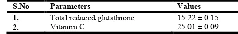
The level of non-enzymatic antioxidants such as Total reduced glutathione and Vitamic C was showed in table 2. The activity of total reduced glutathione and Vitamin C was found to be 15.22 ± 0.15 µg/mg plant tissue and 25.01± 0.09 µg/mg plant tissue respectively.
Table 2. Levels of non-enzymatic antioxidants present in fresh sample of Tinospora cordifolia (stem)
S.No Parameters Values
1. Total reduced glutathione 15.22 ± 0.15 2. Vitamin C 25.01 ± 0.09 Values are expressed as mean ± SD (n=3)
Units:
Total reduced glutathione: µg/mg protein Vitamin C: µg/mg protein
DISCUSSION
The term “antioxidant” refers to any molecule capable of stabilizing or deactivating free radicals before they attack cells. Humans have evolved highly complex antioxidant systems (enzymatic and nonenzymatic), which work synergistically, and in combination with each other to protect the cells and organ systems of the body against free radical damage. The antioxidants can be endogenous or obtained exogenously e.g, as a part of a diet or as dietary supplements. Some antioxidants can interact with other antioxidants regenerating their original properties; this mechanism is often referred to as the “antioxidant network”. There is growing evidence to support a
link between increased levels of ROS and disturbed activities of enzymatic and nonenzymatic antioxidants in diseases associated with aging. Oxidative damage has been suggested to occur as a consequence of reactive oxygen species (ROS) produced as a byproduct of ETC in mithochondria. A number of studies have been suggested that ROS can affect critical events associated with many disorders (Ragavendran et al., 2012). The formation of ROS is prevented by an antioxidant system: low molecular mass antioxidants (ascorbic acid, glutathione and tocopherols), enzymes regenerating the reduced forms of antioxidants and ROS – interacting enzymes such as SOD and catalases (Gout et al., 2001). Superoxide dismutase is an enzyme which breakdown the superoxide anion into oxygen and hydrogen peroxide (Zelko et al., 2002). They are present in almost all aerobic cells and in the extracellular fluids. They contain metal ions that can be copper, zinc, manganese or iron. In humans, the copper/zinc superoxide dismutase is present in the cytosol, while manganese superoxide dismutase is present in the mitochondria. There also exists a third form of superoxide dismutase in extracellular fluids, which contains copper and zinc in its active sites (Johnson and Giulivi, 2005). Superoxide dismutase removes
O2. – by catalyzing a dismutation reaction. In the absence of
superoxide dismutase, this reaction occurs non- enzymatically but at a very slow rate (Nozik-Grayck et al., 2005). Catalase
(H2O2 oxidoreductase) is a tetramer of four polypeptide chains,
each over 500 amino acids long, contains four porphyrin heme (iron) groups that allow the enzyme to react with the hydrogen
peroxide. Catalase can decompose hydrogen peroxide (H2O2)
in reactions catalyzed by two different modes of enzymatic
activity: the catalytic mode of activity (2H2O2 → O2 + 2H2O)
and the peroxidatic mode of activity (H2O2 + AH2 → A +
2H2O). Catalase has one of the highest turnover rates of all
enzymes; one molecule of catalase can convert millions of molecules of hydrogen peroxide to water and oxygen per
second. Decomposition of H2O2 by the catalytic activity of
catalase follows the fashion of a first-order reaction and its rate
is dependent on the concentration of H2O2 (Valko et al., 2007;
Berg et al., 2002). Catalase is an unusual enzyme since, although hydrogen peroxide is its only substrate, it follows a ping- pong mechanism. Here, its cofactor is oxidised by one molecule of hydrogen peroxide and then regenerated by transferring the bound oxygen to a second molecule of substrate (Kabel et al., 2013). Catalase is present in all prokaryotes and eukaryotes. With the exception of erythrocytes, it is predominantly located in peroxisomes of all
types of mammalian cells where H2O2 is generated by various
oxidases. Since H2O2 serves as a substrate for certain reaction
that generate the highly reactive hydroxyl radical, catalase is believed to play a role in cellular antioxidant defense
mechanisms by limiting the accumulation of H2O2 (Ho et al.,
2004). The role of catalase in defending cells and tissues against oxidative stress has been studied extensively. Overexpression of catalase renders cells more resistant to
toxicity of H2O2 and oxidant-mediated injury. In addition,
transgenic mice overexpressing catalase are protected against myocardial injury following administration of adriamycin and
development of hypertension from treatment with
[image:3.595.42.275.500.534.2]result of tissue damage from H2O2 produced by
peroxide-generating bacteria such as streptococci and pneumococci as well as by the phagocytic cells at the sites of bacterial infection (Yang et al., 2003). Glutathione -S-transferases (GSTs), is a cytosolic multifunctional enzymes. It catalyzes the conjugation of glutathione with a variety of reactive electrophilic compounds, there by neutralizing their active electrophilic sites and subsequently making the parent compound more water soluble. Glutathione peroxidase exist in two forms, one which is selenium-dependent (GPx, EC1.11.1.19) and the other, which is selenium-independent (glutathione-Stransferase,GST, EC2.5.1.18) (Mates et al., 1999). The differences are due to the number of subunits, catalytic mechanism, and the binding of selenium at the active centre, and glutathione metabolism is one of the most important antioxidative defense mechanisms present in the cells. There are four different Se-dependent glutathione peroxidases present in humans (Chaudière and Ferrari-Iliou, 1999) and these are known to add two electrons to reduce peroxides by forming selenoles (Se-OH) and the antioxidant properties of these seleno-enzymes allow them to eliminate peroxides as potential substrates for the Fenton reaction. Selenium-dependent glutathione peroxidase acts in association with tripeptide glutathione (GSH), which is present in high concentrations in cells and catalyzes the conversion of hydrogen peroxide or organic peroxide to water or alcohol while simultaneously oxidizing GSH. It also competes with catalase for hydrogen peroxide as a substrate and is the major source of protection against low levels of oxidative stress.
However, the most important H202-removing enzymes in
human cells are glutathione peroxidises (GSHPX), enzymes that require selenium (has selenocysteine at the active site) for
their action. GSHPX enzymes remove H202 by using it to
oxidize reduced glutathione (GSH) to oxidized glutathione (GSSG).
Glutathione is a cysteine-containing peptide found in most forms of aerobic life. It is not required in the diet and is synthesized in cells. Glutathione has antioxidant properties since the thiol group in its cysteine is a reducing agent and can be reversibly oxidized and reduced. In cells, glutathione is maintained in the reduced form by glutathione reductase and in turn reduces other metabolites and enzymes as well as reacting directly with oxidants. Due to its high concentration and its central role in maintaining the cell's redox state, glutathione is one of the most important cellular antioxidant (Chaudière and Ferrari-Iliou, 1999). Vitamin C is regarded as the first line natural antioxidant defense in plasma and a powerful inhibitor of LPO (Maxwell, 1995). Vitamin C is a water soluble antioxidant. It acts as a free radical scavenger. It scavenges peroxyradicals (Sies, 1993). Vitamin C protects non-smokers against the harmful effects of ROS from passive smoking (Jacob, 2000). It has been found in the chloroplast, cytosol, vacuole and extracellular compartments of the plant cells and shown to function as a reluctant for many free radicals (Kumar and Hemalatha, 2011).
Conclusion
Based on all these findings it is suggested that the stem of
Tinospora cordifolia herb is a potential source of natural
antioxidants that could have great importance as therapeutic
agents in preventing or slowing the oxidative stress related degenerative diseases. Further studies are needed to explore the molecular mechanisms by which antioxidants prevent the harmful effects of oxidative stress.
REFERENCES
Addy SK, Goodman RN. Polyphenol oxidase and peroxidase in apple leaves inoculated with a virulent or an avirulent strain for Ervinia amylovora. Ind Phytopath. 1972; 25: 575-579.
Agarwal ACM, Abdelrazik H, Sharma RK. Oxidative stress management in patients with male or female factor infertility.Handbook of Chemiluminescent methods in oxidative stress Assessment.2008; 195.
Berg JM, Tymoczko JL, Stryer L 2002. Biochemistry, 5th ed., Freeman WH and Co., New York; pp: 205-206.
Chaudière J, Ferrari-Iliou R; Intracellular Antioxidants: from chemical to biochemical mechanisms. Food Chem Toxicol. 1999; 37:949–962.
Cheeseman KH, Slater TF. Free radicals in medicine. Brit Med Bullet. 1993; 49: 479-724.
Das K, Samanta L, Chainy GBN. A modified
spectrophotometric assay of superoxide dismutase using nitrite formation by superoxide radicals. Ind J Biochem Biophys. 2000; 37: 201-204.
Gout E, Boisson AM, Aubert S, Douce R, Bligny R. Origin of the cytoplasmic pH changes during anaerobic stress in higher plant cells. Carbon-13 and phosphorus-31 nuclear magnetic resonance studies. Plant Physiology. 2001; 100: 912-925.
Habig WH, Pabst MJ, Jakoby WB. Glutathione S- Transferases: The First Enzymatic Step in Mercapturic Acid Formation. J Biol Chem 1974; 249: 7130.
Hegde K, Joshi AB. Hepatoprotetive effect of Carissa carandas
Linn root extract against CCl4 and paracetamo induced
hepatic oxidative stress. Ind J Exp Biol 2009; 47:660-667. Ho YS, Xiong Y, Ma W, Spector A and Ho DS 2004. Mice
lacking catalase develop normally but show differential sensitivity to oxidant tissue injury. J Biol Chem; 279: 32804-32812.
Jacob RA. Passive smoking induces oxidant damage preventable by vitamin C. Nutr. Rev. 2000; 58: 239-241. Johnson F, Giulivi C 2005. Superoxide dismutases and their
impact upon human health. Mol Aspects Med 26 (4-5): 340-52.
Kabel AM, Abdel-Rahman MN, El-Sisi Ael-D, Haleem MS, Ezzat NM, El Rashidy MA 2013. Effect of atorvastatin and methotrexate on solid Ehrlich tumor. Eur J Pharmacol; 713 (1-3): 47-53.
Kothari S, Thompson A, Agarwal, Plessia SS. Free radicals: Their beneficial and detrimental effects on sperm function. Indian J Exp Biol 2010; 48: 425-435.
Kumar R, Hemalatha S. In-vitro antioxidant activity of alcoholic leaf extract and sub fractions of Alangium lamarckii Thwaites. J. Chem. Pharm. Res. 2011; 3 suppl 1: 259-267.
Maxwell SRJ. Prospects for use of antioxidant therapies. Drugs.1995; 49: 345-361.
Moron, M.S., Depierre, J.W. and Mannervik, B. 1979. Levels of glutathione, glutathione reductase and glutathione S – transferase activities in rat lung and liver. Biochem. Biophys. Acta., 582: 67-78.
Nozik-Grayck E, Suliman H, Piantadosi C 2005. Extracellular superoxide dismutase. Int J Biochem Cell Biol; 37 (12): 2466-71.
Parthipan M, Aravindhan V, Rajendran A. Medico‑botanical
study of Yercaud hills in the eastern Ghats of Tamil Nadu,
India. Anc Sci Life 2011; 30:104‑109.
Ragavendran P, Arul Raj C, Sophia D, Starlin T, Gopalakrishnan VK. Evaluation of Enzymatic and Non- Enzymatic Antioxidant Properties of Aerva Lanata (L) - An In vitro Study. Int J Pharm Pharm Sci. 2012; 1: 4, 522-526. Rana V, Thakur K, Sood R, Sharma V, Sharma TR. Genetic
diversity analysis of Tinospora cordifolia germplasm collected from northwestern Himalayan region of India. J
Genet 2012; 91: 99‑103.
Rotruck JT, Pope AL, Ganther HE, Swanson AB, Hafeman DG, Hoekstra WG. Selenium: Biochemical role as a component of glutathione peroxidase. Science. 1973; 179: 588-590.
Sies H. Strategies of antioxidant defence. Eur. J. Biochem. 1993; 215: 213-219.
Sinha AK. Colorimetric assay of catalase. Anal Biochem 1972; 47: 389-394.
The Ayurvedic Pharmacopoeia of India. Part I.1st ed. Vol. 1. New Delhi: Department Of AYUSH, Ministry of Health and FW; 2001; 3-5.
Ulusu NN, Tandoğan B 2007. Purification and kinetic properties of glutathione reductase from bovine liver. Mol Cell Biochem; 303(1-2): 45-51.
Valko M, Leibfritz D, Moncol J, Cronin M, Mazur M et al.
2007. Free radicals and antioxidants in normal
physiological functions and human disease. Int J Biochem Cell Biol; 39 (1): 44-84.
Yang H, Shi MJ, Van Remmen H, Chen XL, Vijg J et al. 2003. Reduction of pressor response to vasoconstrictor agents by overexpression of catalase in mice Am J Hypertens; 16 (1): 1-5.
Zelko I, Mariani T, Folz R 2002. Superoxide dismutase multigene family: a comparison of the CuZn-SOD (SOD1), Mn- SOD (SOD2), and EC-SOD (SOD3) gene structures, evolution, and expression. Free Radic Biol Med; 33 (3): 337-49.
![1 (p Bromophenyl) 5 p tosylperhydropyrrolo[3,4 b]pyrrole](data:image/gif;base64,R0lGODlhAQABAIAAAP///wAAACH5BAEAAAAALAAAAAABAAEAAAICRAEAOw==)