VARIOUS FACTORS INVOLVING AGING: ELECTRON TRANSFER, REACTIVE OXYGEN SPECIES AND
1
Peter Kovacic
1
Department of Chemistry and Biochemistry, San Diego
2
Centro de Graduados e Investigación del Instituto Tecnológico de Tijuana, Apdo. po
ARTICLE INFO ABSTRACT
In addition to oxidative stress, several other mechanistic factors are involved with aging electron transfer,
effects of antioxidants provides support for the deleterious role of oxidative stress. Certain organs are importantly involved, such as heart, brain (Alzheimer’s disease, demen
mitochondria, lung and prostate. Cancer is a significant contributing factor. Other aspects addressed are stem cells, protein oxidation, and telomeres.
Copyright©2017, Peter Kovacic and Ratnasamy Somanathan
permits unrestricted use, distribution, and reproduction in any medium, provided the original work is properly cited.
INTRODUCTION
This review deals with mechanism of aging from the standpoint of oxidative stress, electron transfer, reactive oxygen species, antioxidants, reactive nitrogen species, heart, brain, lung, prostate, mitochondria, and miscellaneous aspects. “The preponderance of bioactive substances, usually as the metabolites, incorporates ET functionalities. We believe these ET-metabolites play an important role in physiological responses. The main group includes quinones (or phenolic precursors), metal complexes (or complexors), aromatic nitro
compounds (or reduced hydroxylamine and nitroso
derivatives), and conjugated imines (or iminium species). Resultant redox cycling is illustrated in Scheme 1. In vivo redox cycling with oxygen can occur, giving rise to oxidative stress (OS) through generation of reactive oxygen species (ROS), such as hydrogen peroxide, hydroperoxides, alkyl
peroxides, and diverse radicals (hydroxyl, alkoxyl,
hydroperoxyl, and superoxide) (Scheme 1). Cellular and
*Corresponding author: Ratnasamy Somanathan
Centro de Graduados e Investigación del Instituto Tecnológico de Tijuana, Apdo. postal 1166, Tijuana, B.C. Mexico
ISSN: 0975-833X
Article History: Received 29th April, 2017
Received in revised form 11th May, 2017
Accepted 07th June, 2017 Published online 22nd July, 2017
Citation: Peter Kovacic and Ratnasamy Somanathan
stress”, International Journal of Current Research, 9,
Available online at http://www.journal
Key words:
Reactive Oxygen Species, Reactive Nitrogen Species, Oxidative Stress, Electron Transfer, Antioxidants, Radicals, Aging, Stem Cells, Protein Oxidation, Mitochondrial Dysfunction, Telomere Shortening, Senescence.
RESEARCH ARTICLE
VARIOUS FACTORS INVOLVING AGING: ELECTRON TRANSFER, REACTIVE OXYGEN SPECIES AND
OXIDATIVE STRESS
Peter Kovacic and *
,2Ratnasamy Somanathan
Department of Chemistry and Biochemistry, San Diego State University, San Diego, CA 92182
Centro de Graduados e Investigación del Instituto Tecnológico de Tijuana, Apdo. po
Mexico
ABSTRACT
addition to oxidative stress, several other mechanistic factors are involved with aging
electron transfer, reactive oxygen species, and reactive nitrogen species. Evidence for beneficial effects of antioxidants provides support for the deleterious role of oxidative stress. Certain organs are importantly involved, such as heart, brain (Alzheimer’s disease, demen
mitochondria, lung and prostate. Cancer is a significant contributing factor. Other aspects addressed are stem cells, protein oxidation, and telomeres.
Peter Kovacic and Ratnasamy Somanathan. This is an open access article distributed under the Creative Commons Att use, distribution, and reproduction in any medium, provided the original work is properly cited.
This review deals with mechanism of aging from the standpoint of oxidative stress, electron transfer, reactive oxygen species, antioxidants, reactive nitrogen species, heart, brain, lung, prostate, mitochondria, and miscellaneous aspects. ce of bioactive substances, usually as the metabolites, incorporates ET functionalities. We believe these metabolites play an important role in physiological responses. The main group includes quinones (or phenolic exors), aromatic nitro
compounds (or reduced hydroxylamine and nitroso
derivatives), and conjugated imines (or iminium species). Resultant redox cycling is illustrated in Scheme 1. In vivo redox cycling with oxygen can occur, giving rise to oxidative s (OS) through generation of reactive oxygen species (ROS), such as hydrogen peroxide, hydroperoxides, alkyl
peroxides, and diverse radicals (hydroxyl, alkoxyl,
Cellular and
anathan
Centro de Graduados e Investigación del Instituto Tecnológico de Tijuana, Apdo. postal 1166, Tijuana, B.C. Mexico.
mitochondrial enzymes can also perform catalytically in the
reduction of O2.
O2
O2
HOO H H
H ET agent ET
.
.
e
Scheme 1. Redox cycling with superoxide and ROS formation
In some cases ET results in involvement with normal electrical effects (e.g., in respiration or neurochemistry). Generally, active entities possessing ET groups display reduction potentials in the physiologically
positive than about -0.5 V). Hence, ET in vivo can occur resulting in production of ROS which can be beneficial in cell signaling at low concentrations, but produce toxic results at high levels. Electron donors consist of phen
International Journal of Current Research
Vol. 9, Issue, 07, pp.53518-53528, July, 2017
Peter Kovacic and Ratnasamy Somanathan, 2017. “Various factors involving aging: Electron transfer, reactive oxygen species and oxidative , 9, (07), 53518-53528.
Available online at http://www.journalcra.com
z
VARIOUS FACTORS INVOLVING AGING: ELECTRON TRANSFER, REACTIVE OXYGEN SPECIES AND
, San Diego, CA 92182-1030, USA
Centro de Graduados e Investigación del Instituto Tecnológico de Tijuana, Apdo. postal 1166, Tijuana, B.C.
addition to oxidative stress, several other mechanistic factors are involved with aging, namely, reactive oxygen species, and reactive nitrogen species. Evidence for beneficial effects of antioxidants provides support for the deleterious role of oxidative stress. Certain organs are importantly involved, such as heart, brain (Alzheimer’s disease, dementia and Parkinson’s disease), mitochondria, lung and prostate. Cancer is a significant contributing factor. Other aspects addressed
distributed under the Creative Commons Attribution License, which
mitochondrial enzymes can also perform catalytically in the
HOOH HO HO T agent
.
e
+
cycling with superoxide and ROS formation
In some cases ET results in involvement with normal electrical effects (e.g., in respiration or neurochemistry). Generally, active entities possessing ET groups display reduction potentials in the physiologically responsive range, (i.e., more 0.5 V). Hence, ET in vivo can occur resulting in production of ROS which can be beneficial in cell signaling at low concentrations, but produce toxic results at high levels. Electron donors consist of phenols, N-heterocycles
INTERNATIONAL JOURNAL OF CURRENT RESEARCH
or disulfides in proteins which produce relatively stable radical cations. ET, ROS and OS have been increasingly implicated in the mode of action of drugs and toxins, (e.g., antiinfective agents (Kovacic & Becvar, 2000), anticancer drugs (Kovacic & Osuna, 2000), carcinogens (Kovacic & Jacintho, 2001a), reproductive toxins (Kovacic & Jacintho 2001b), nephrotoxins
(Kovacic et al., 2002), hepatotoxins (Poli et al., 1989),
cardiovascular toxins (Kovacic & Thurn, 2005), nerve toxins
(Kovacic & Somanathan, 2005), mitochondrial toxins
(Kovacic et al., 2005), abused drugs (Kovacic & Cooksy,
2005), pulmonary toxins (Kovacic & Somanathan, 2009), ototoxins (Kovacic & Somanathan, 2008), and various other categories (Halliwell & Gutteridge, 1999). There is a plethora of experime 2001antal evidence supporting the ET-ROS theoretical framework (Kovacic & Becvar, 2000; Kovacic &
Jacintho, 2000a; Kovacic & Jacintho 2001b; Kovacic et al.,
2002; Poli et al., 1989; Kovacic & Thurn, 2005; Kovacic &
Somanathan, 2005; Kovacicet al. 2005; Kovacic & Cooksy,
2005; Kovacic & Somanathan, 2009; Kovacic & Somanathan, 2008; Halliwell & Gutteridge, 1999). This evidence includes generation of the common ROS, lipid peroxidation, degradation products of oxidation, depletion of AOs, effect of exogenous AOs, and DNA oxidation and cleavage products, as well as electrochemical data. This comprehensive, unifying mechanism is consistent with the frequent observation that many ET substances display a variety of activities (e.g., multiple-drug properties), as well as toxic effects. It is important to recognize that mode of action in the biodomain is often involved with many physiological actions and is multifaceted. In addition to the ET-ROS-OS approach, other aspects may pertain, such as, enzyme inhibition, allosteric effects, receptor binding, metabolism and physical factors. A specific example involves protein binding by quinones in which protein and nucleophiles, such as amino or thiol, effect conjugate addition” (Kovacic & Somanathan, 2010).
Origin and evolution of the theory
This aging theory was reviewed in 2009, fifty five years after the inception by Harman (Harman, 2009). Initially, there was considerable opposition, followed by increasing support in more recent years. Free radicals are widely generated in various parts of the body including mitochondria, the immune system, various redox enzymes, pollutants, drugs and many toxic substances. The final comment was “the work of more than 50 years has now established the validity and usefulness of the Free Radicals Theory of Aging.” There was considerable research on the favorable effects of AOs which support the theory (Harman, 2000).
Free Radicals and Aging
Denham proposed the free radical theory of aging in based on damage to various body constituents by radicals (Halliwell & Gutteridge, 1999). The review presented the following:
a) ROS/RNS are generated by various means. There is
damage to DNA, lipids and protein. AO defenses do not protect completely.
b) ROS/RNS formation may benefit in the short term, but
may lessen with age.
c) Tissue degeneration with age can generate enhanced
ROS/RNS. For example, old rats form more superoxide than young ones, pointing to greater leakage of
mitochondrial electrons followed by superoxide production with aging.
d) Larger animals consume less O2 than do smaller ones
resulting in larger life span. There is a lower ROS burden.
e) Changes in O2 consumption resulting in alteration of
ROS levels can affect life span.
f) Lowered O2 consumption by queen bees who do not fly
most of their lives could explain why they live many times longer than the flying worker bees.
g) Mitochondrial DNA undergoes rapid mutations partly
due to oxidative damage leading to higher levels of 8-OHdG.
h) Data indicate that longer-lived species possess superior
AO protection than shorter-lived ones.
i) A slower rate of ROS generation from mitochondria
may be related to increased age.
j) Senescense can be induced by H2O2 (an ROS) treatment
apparently associated with increased levels of 8-OHdG.
k) Caloric restriction is related to less oxidative damage to
DNA, lipids and protein associated with decreased superoxide production.
Studies with added AOs have produced unexpected results with no or little increase in longevity. It is difficult to interpret the data.
Reactive oxygen species, oxidative stress and antioxidants
Aging is an extremely complex and multifaceted process that proceeds to the gradual deterioration of the various physiological functions in the organs of the body. In recent years, a large body of literature has emerged documenting the link between ROS, reactive nitrogen species (RNS), antioxidants (AOs) and aging (Polijsak, Milisav, 2013; Roberts & Fulop, 2014; Stojilković et al., 2014; Poljsak et al., 2013;
Yadev et al., 2013; . Rahman et al., 2012; Germs & de la
Gurdia, 2012; Cui et al., 2012; Ladiges et al., 2010; Goto,
Radák, 2010; Pamplona, 2008; Olinski et al., 2007; de
Magalhães & Church, 2006; . Junqueira et al., 2004; Stadtman,
2004; Bokova et al., 2004; Turrens, 2003; Wei & Lee, 2002;
Cadenas & Davies, 2000).
Antioxidants and aging
Excessive ROS formation induces OS, which plays a large role in the disease process and natural aging. It is well established
that free radicals (·OH, ·O2, ·OOH etc.) damage is linked to
the formation of many degenerative diseases, including cancer,
cardiovascular diseases, neurodegenerative diseases,
Alzheimer’s, Parkinson’s, Huntington’s and aging ( Poljsak &
Milisav, 2013; Stojiković et al., 2014; Olinski et al., 2007;
Cadenas & Davis, 2000; . Nordberg, & Arnér, 2001; Valko et
al., 2007; Seifried et al., 2007; Uttara et al., 2009; Wei & Lee,
2002; Guerra-Araiza et al., 2013; Kovacic et al., 2015;
oxidants and antioxidants in intracellular systems is vital for cell function, regulation, adaptation and growth. Aging is a process which starts after maturity, when most of the physiological functions related to the heart, lungs, kidney, sexual hormones, skin wrinkling, CNS and brain functions show signs of decline. It is believed that the aging involves complex and interrelated factors, including oxidative stress-induced protein and DNA damage in conjunction with insufficient DNA damage repair, as well as genetic instability of mitochondrial and nuclear genomes (Polijsak & Milisav, 2013; Fontana& Klein, 2007), increased adipokine and cytokine production (Davis, 1995), alteration of fatty acid metabolism, tissue insulin resistance (Martin, 1987), accumulation of glycation end products (Kovacic &
Somanathan, 2011; Martin et al., 1996), alterations in the
sympathetic and neuroendocrine systems (Gilica et al., 2007),
loss of post-mitotic cells, and deterioration in structure and function of cell neurons, and muscles in all tissues and organs (Polijsak & Milisav, 2013).
Theoretical aspects
There are many theories which help to elucidate the process of ageing, such as free radical, oxidative stress, mitochondrial, telomere shortening, end glycation product, protein damage, and DNA damage that have evolved over the years (Fig.1). None of these theories alone can explain all details of ageing (Polijsak & Milisav, 2013). Further, there are specific aging genes of which, almost 7,000 play a role in senescence of an aging person. However, reactive oxygen species and oxidative stress have been a common factor in most of these theories, in keeping with our hypothesis that ROS-OS-ET is a unifying mechanism to explain many of the diseases and aging encountered in living organisms (see Introduction).
Free Radical
Mitochondrial dysfunction
Glycation End Product
Telomere Shortening
Protein Oxidative Damage
DNA oxidation
Reactive oxygen species
Reactive nitrogen species
Oxidative stress
[image:3.595.308.553.71.245.2]Stem cell aging
Fig.1. Various theories on aging
Free radicals, oxidative stress and mitochondrial
dysfunction
Harman proposed the free radical theory of aging in the 1950s, with the accumulating evidence of reactive oxygen species, mitochondrial dysfunction and oxidative stress being the main causes of aging. A large body of evidence accumulated over the years suggesting a strong link between aging and ROS-OS. Studies have shown a correlation between longevity and the rate of cellular damage.
Reactive oxygen species can be classified into two groups: free oxygen radicals and non-radical ROS (Table 1). Among these, superoxide, hydrogen peroxide and hydroxyl radicals are the most potent and well studied species.
Table 1. Reactive oxygen and nitrogen species
O2
HO
NO
ROO
RO R
NO2
Superoxide
.
-Hydroxyl radical
.
Nitric oxide
.
Organic radicals
.
Peroxyl radicals
.
Alkoxy radicals
.
Nitrogen dioxide
.
H2O2
HOCl O2
O3
ROOH
ONO
HNO2
Non-radical ROS Hydrogen peroxide
Hypochlorous acid Singlet oxygen
Ozone
Organic hydroperoxides
Peroxynitrite
-Nitrous acid Radical ROS
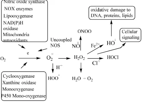
A variety of enzymatic and non-enzymatic sources of ROS exists in the biological system. Enzymes within the cell are primary sources of ROS/RNS. Superoxide is produced by one electron reduction of oxygen by several enzymes, such as NAD(P)H oxidase, xanthine oxidase and cytochrome P450 within the cell. In addition to these, a number of external mediators also contribute to the ROS generation, such as ionizing radiation. Heavy metals, like Hg, Pb, Cd, Cr, and Cu, metal complexes, nanoparticles, cigarette smoke, pollutants from automobiles, fossil burning furnaces, various drugs and certain types of other chemical compounds (Scheme 1) play a
role (Gilica et al., 2007; Halliwell, 2001; Krumova & Cosa,
2016).
Scheme 1. Generation of ROS
In mitochondria, superoxide ROS is produced as a natural by-product of electron transport chain activity. The superoxide leaks through the mitochondrial permeability transition pore in the outer membrane into the cytoplasm, where it is converted into hydrogen peroxide by MnSOD or in the cytosol by Cu/ZnSOD. The diffusible hydrogen peroxide also serves as a second messenger, and may cross cellular membranes through members of the aquaporin family (Halliwell, 2001; Krumova & Cosa, 2016; Turrens, 2003). The superoxide created by the mitochondrial metabolized oxygen is converted into hydrogen peroxide, hydroxyl radical, peroxy radical and other radicals capable of damaging proteins, lipids, mtDNA, neurons, brain tissue, cardiovascular tissue, endothelial tissue and skeletal tissue. Since mitochondria are the major source of ROS in mammalian cells, the closest attack also takes place at the
[image:3.595.308.557.421.599.2]mitochondrial DNA (mtDNA). Aging is a consequence of this mitochondrial dysfunction and other actions due to the ROS formation, leading to various age related diseases including cancer, Alzheimer’s, Parkinson’s and other neurological disorders, wherein is supported by various reports (Turrens,
2003; Sanz & Stefanatos, 2008; Dai et al., 2014; Bánhegyi &
Sümegi, 2014; Krzeszowiak & markiewicz-Górka, 2014;
Piotrowska & Bartnik, 2014; Indo et. al2015; Springo et al.,
2015; Li et al., 2014; Conti et al., 2015; Haddadi et al., 2014:
Patel et al., 2014; Ladiges et al., 2010; Liou & Storz, 2010; Sullivan & Chandel, 2014; Loeb et al., 2005; Balaban et al.,
2005; Barja, 2004; Junqueiraet al., 2004; Lin & Beal, 2006;
Hirai et al., 2001). Studies have shown that 8-oxo-dG, a
common oxidized product of DNA is found in higher levels in
the mtDNA than in nuclear DNA (Cui et al., 2012). The
accumulation of oxidative DNA damage products, e.g.,
8-oxo-dG from mtDNA, contributes to the aging process (Cui et al.,
2012; Maynard et al., 2009; Capel et al., 2005). Studies with
mice and fly models show that reduced free radical production
increased life span (Cui et al., 2012). C.elegans resistant to OS
have a longer life span, whereas mice lacking antioxidant
enzyme SOD exhibit a shorter life span (Cui et al., 2012).
Mice that lack DNA repair enzymes, such as 8-oxaguanineglycosylase and Muty homolog 1, have shorter life
spans (Cui et al., 2012). Mitochondrial ROS were shown to
regulate the NF-kB pathway, together with age–related inflammatory activation of endothelium, which leads to vascular dysfunction and oxidative stress-related diseases (Li
et al., 2015; Zinovkin et al., 2014). Clinical studies showed mitochondrial ROS-induced cataract in aging eye (Babizhayev and Yegorov, 2016). A study provides epidemiological evidence supporting free radical/oxidative stress using derivatives of reactive oxygen species metabolites and total
thiols as biomarkers (Schȍttker et al., 2015). Dietary
restrictions decrease mitochondrial ROS production, leading to animal longevity (Pamplona and Barja, 2007). Lowering methionine levels in tissue proteins controls mitochondrial oxidative stress and increases the longevity of mammals and birds (Pamplona and Barja, 2006). Superoxide (CuZn) dismutase deficiency causes accelerated vascular aging process (Chen and Chen, 2006). ROS have historically been viewed as toxic metabolic byproducts and have been identified in many
physiological dysfunctions and pathologies, such as
atherosclerosis, diabetes, cancer, neurodegeneration, and aging. More recent work, however, indicates ROS are important intermediates in cellular signaling pathways, initiating signaling in a broad variety of cellular processes, such as proliferation and survival (MAP kinases, P13 kinase, PTEN, and protein tyrosine phosphatases, ROS homeostasis, antioxidant gene regulation, mitochondrial oxidative stress, apoptosis and aging (Thannickal and Fanburg, 2000;
Hamanaka and Chandel, 2010; Marchi et al., 2012; Yan, 2014;
Ludovico and Burhans, 2014; Liochev, 2013; Labunskyy and
Gladyshev, 2013; Ray et al., 2012; Collins et al., 2012; Back
et al., 2012).
Stem cell and reactive oxygen species
Stem cells, which are capable of self-renewal and differentiation, are essential for the normal homeostatic maintenance and repair of tissue throughout the life span of an organism. Studies indicate self-renewal ability of adult stem cells declines with advancing age, suggesting that stem cell function plays a central role in aging. Hypoxia and low ROS play an important role in regulating stem and progenitor cell
function in various physiological and pathological responses
(Maraldi et al., 2015; Bigarella et al., 2014; Shyh-Chang et al.,
2013). Studies deal with ROS mechanism, metabolism and
aging in stem cells (Bigarella et al., 2014; Sharpless &
DePinho, 2007). Aging increases the susceptibility of mesenchymal stem cells to ROS and impairs their therapeutic
transplantation for myocardial infarction (Atashi et al., 2014).
A similar report deals with ROS in mesenchymal stem cell
aging and its implication in lung diseases (Yang et al., 2015).
Genetic studies of mice deficient in gene implicated in ROS regulation have demonstrated that elevated levels of ROS within the stem cell compartments lead to a rapid decline in stem cell self-renewal (Ito, 2004; Mantel, 2012; Rossi, 2007).
Accelerating neuronal aging in in vitro model brain stem cell
disorders was shown to involve ROS (Campos et al., 2014).
Protein oxidation
It is known for a long time that aging is associated with the accumulation of altered forms of a number of enzymes (Rohtstein, 1984). Modified or oxidized proteins are dysfunctional as enzymes or as structural proteins. Oxidative attack of the polypeptide backbone is initiated by the ROS and RNS dependent abstraction of the α-hydrogen atom of the amino acid residue to form a carbon-centered radical. All amino acid residues of proteins are susceptible to oxidation by ROS and RNS radicals (Berlett and Stadtman, 2013). Protein oxidative modifications can be classified into two types, irreversible oxidation and reversible oxidation (Cai and Yan, 2013). Studies have shown the detrimental effects of protein oxidation in aging (Stadtman, 2001; Stadtman, 2006). An investigation showed the effects of both irreversible and reversible protein oxidation products in health and disease (Cai and Yan, 2013). Several other investigations relate to protein
oxidation and aging (Baraibar et al., 2012; Shringapure and
Davis, 2002; Höhn et al., 2013; Sitte et al., 2000; Sitte et al.,
2000; Friguet, 2006).
Telomere shortening, senescence, ROS and aging theory
Telomeres, nucleoprotein structures located at the ends of chromosomes, are subjected to shortening at each cycle of cell division. Telomeres prevent chromosome ends from being recognized as double-strand breaks and protect them from end to end fusion and degradation. Unlike stem cells, telomeres in somatic cells shorten with each division leading to cellular senescence (aging). The enzyme telomerase is involved in telomere stability by synthesizing a new copy of the repeat by using its RNA template. Oxidative DNA damage can lead to dysfunctional telomere. DNA damaged senescence cells were found to contain 30% more oxidative modified guanine in their DNA. Evidence points to oxidative modifications and shortening by ROS leading to aging of the somatic cells
(Kawanishi and Oikawa, 2004; Stte et al., 1998; Kamsler.
Kamsler et al., 2001; Tchirkov and Lansdorp, 2003; Duan et
al., 2005; Blasco, 2005; Xin and Broccoli, 2004; Hausmann et
al., 2003; Camisi, 2005; Jeyapalan & Sedivy, 2008; Collado et
al., 2007). Fig. 2 summarizes the role of ROS/RNS in aging.
Cardiovascular System
ROS/RNS
Beneficial effects
Cell signaling Lipid peroxidation
Aging
Damage to mitochondraial DNA
Accumulation of mtDNA mutations Decreased DNA repair
Aging
Oxiduzed mitochondria protein and enzymes
Aging Telomere shortening
senescence
chromosomal aberration
Aging Stem renewal and differentiation
Fig.2. ROS/RNS in aging
The review discusses involvement of ET-ROS-OS-AO. The liporfuscin test showed enhanced OS with aging after 20 years. The review deals with radical species in cardiac tissue injury. ROS formation is associated with cardiovascular toxins. The heart is characterized by a number of adverse factors, including high oxygen consumption, abundance of mitochondria and deficiency of AO defenses. There are large number of substances harmful to the heart that fit the oxidative stress theory. Among these are medicines, such as, anthracyclines, quinolones, gentamycin, methyldopa, and amphotericin B. Harmful abused drugs include alcohol, cocaine, tobacco, nicotine, N-nitrosamines, polynuclear aromatic hydrocarbons, amphetamines and MPTP. The cardiovascular system is adversely affected by a significant number of metal compounds, mainly those of Cd, Co, Pb, Mn, Ni, V, As, Cr, Cu, Fe and Hg. The metals generally exhibit reduction potentials favorable for ET leading to ROS. The adverse effects are alleviated by AOs of various types. Included in the pesticides and herbicides categories are lindane, paraquat, organophophates and carbon disulfide. Toxic industrial chemicals include haloalkanes, alkenes, acrolein and allylamines. In addition, there are large numbers of miscellaneous compounds, e.g., catecholamines, endotoxin, alloxan, nitric oxide, PCBs,dioxin, phenylhydrazines, sulfur dioxide and dinitrotoluene. AOs that have exhibited effectiveness in decreasing risk of mortality from heart disease include flavonoids, vitamin E, selenium, vitamin C, probucole, ubiquinol, cysteine, SOD, catalase and GSH.
Alzheimer’s Disease
This illness (AD) is characterized by three conditions, namely, senile plaque (SP), neurofibulary tangles and synapse loss (Butterfield, 2003). Clinical manifestations include loss of memory, speech, cognition and normal behavior. AD is one of the main causes of aging and death. Research indicates that the main component of SP, namely amyloid βpeptide (Aβ) is central to the pathological condition, including OS. The AD brain is under intense OS as shown by protein oxidation. Lipid peroxidation, ROS formation, advanced glycation end products and DNA oxidation. The generation of superoxide and NO leads to peroxynitrite, a neurotoxic species, lipid peroxidation, resulting from radicals associated with Aβ, can damage membrane proteins. Another product of lipid peroxidation is
the toxic 4-hydroxy-2-nonenal (HNE). Protein oxidation is caused by Aβ. The three major ways of introducing carbonyl groups into protein by OS are peptide breakdown scission by radicals, selective oxidation on amino acid side chains and protein modification by alkenals. Lipid peroxidation is caused by an Aβ-induced lipid peroxidation in the AD brain. Studies indicate that a therapeutic strategy is to block OS associated with Aβ using brain accessible AOs.
Dementia
Dementia is a complex brain disorder composed mainly of AD, Lewy bodies (Mao, 2013), and vascular types (Bennett, 2009) involving OS. Other factors are endothelial dysfunction, in addition to neuronal cell death and damage. The condition is characterized by increase in oxidative DNA damage, including
formation of 8-oxyguanine (Gackowski et al., 2008).
Resveratrol, an AO, anti-inflammatory and anti-carcinogen,
exhibits beneficial effects in dementia (Ma et al., 2013). A
decrease in AO levels and increase in oxidative damage are apparently involved in the pathophysiology associated with
diabetes-related dementia (Hatanka et al., 2015). Oxidative
damage is an early indicator of frontotemporal dementia (Gerst
et al., 1999). The AO S-alkyl cystiene alleviates OS related to cognitive impairment and neurodegeneration in mice with
Alzheimer’s dementia (Khan et al., 2011). Selenium
compound protected against free radical deterioration of cognitive functions and neurobehavioral, in addition to
memory loss (Chiapinottoet al., 2015). SOD activity was
protected. OS may be a feature of cognitive impairment and
AD (Cervellati et al., 2014). There is discussion of OS with
involvement in dementia.
Parkinson’s Disease
Parkinsonson’s disease (PD) usually occurs after 65 and slowly
progresses until death (Adams et al., 2001). The disease is
caused by the death of dopaminergic neurons in the brain. OS is involved in the process of killing these cells, entailing ROS. Mitochondrial dysfunction, which generates superoxide, has been implicated in PD. Dopaminergic neurons die by apoptosis in a process involving OS. ROS abstract hydrogen from DNA forming radicals that fragment, leading to apoptosis. Tyrosine hydroxlase can give rise to ROS in a redox mechanism.
Dopamine may be oxidized to form ROS.
3,4-Dihydroxyphenylacetaldehyde is also involved in the generation of ROS. There are many mechanisms for ROS generation in dopaminergic neurons, none of which should be ignored. Monoamine oxidase, may also be important in PD.
Prostate
Prostate cancer is the most common type and is the second main cause of death from cancer involving USA males (Sikha, 2003). Various models of action play a role, such as genetics and OS, the latter being supported by extensive evidence. The ROS involved are produced by carcinogens, illness, infection, inflammation, aging, nutrients and pollutants. The ET-ROS-OS mechanism is believed to be involved. Also participating are RNS, such as NO and peroxynitrite.
Lung
The lung is another organ which is involved in aging and death. A recent review addresses pulmonary toxicity based on the unifying theme of ET-ROS-OS (Kovacic and Somanathan,
2009). The pulmonary system is a main target for toxicity. In the industrial age, there has been a substantial increase in atmospheric pollutants. In lung tissues, many adverse effects result from exposure to lung pollutants that fit into the unifying theme of ET-ROS-OS. More familiar examples include the
following: ozone, SO2, chlorine, benzene, chloroform, carbon
tetrachloride, pentachlorophenol, anesthetics, metals and metal compounds, particulates, asbestos, silica, tobacco (N-nitrosoamines), cocaine, nitroaromatics (Kovacic and Cooksy. 2010) and diacetyls (Kovacic and Somanathan, 2014). Exposure to pollutants results in various illnesses related to aging, including asthma, COPD and cancers (Kovacic and Somanathan, 2009).
Mitochondria
There exists considerable literature linking mitochondria to aging, much of which relates to the unifying theory of ETR-ROS-OS. Some of the reports are present in other sections. This portion is mostly concerned with more recent materials. ROS, generated by mitochondria or other sites, damage various components including the mitochondria, and induce harmful
degradation of body components (Bonomini et al., 2015). Such
toxic manifestations make up a significant portion of aging. The review addresses metabolic syndrome in connection with accelerated senescence. Two items are closely related to species longevity, namely rate of ROS formation by mitochondria and degrees of unsaturated fatty acids in tissue (Barja, 2014). Both are low in longevity. Other factors involved are also treated. Mitochondrial ROS importantly participate in the health span of many essential body organs, as
discussed in other sections of the present review (Dai et al.,
2014). There is a related article (Kong, 2014). ROS at a low, non-toxic concentration can operate as cell signaling agents
that protect against damaging events (Liu et al., 2014). Also,
peroxiamine, closely related to mitochondria, appear to play a role in longevity. Events following oxidative damage induce
inflammation, followed by apoptosis (Venkatarama et al.,
2013). OS related to the cardiovascular and central nervous systems is discussed with emphasis on aging-related diseases. A review discusses the free radical theory from various perspectives, including mitochondrial pathways involving apoptosis that causes subsequent functional tissue alterations (Ivanova and Yankova, 2013). Included is discussion of delay in aging by diet or drug therapy. A 2013 review presents an updated view of the mitochondrial free radical theory (Barja, 2013). Key aspects are emphasized. The two general characteristics responsible for animal longevity appear to be low rate of endogenous damage and macromolecular tissue makeup that is very resistant to oxidative damage.
Cancer
Cancer is one of the main factors involved in shortening the length of life. After a continuing rise in cancer death rates, there has been a steady decline since 1990 (Newcott, 1916). The illness is multifaceted making for difficulty in prevention and treatment with a multitude of targets involved. In relation to mode of action, various factors have been discussed,
including genes, DNA, mutagenesis, estrogens and
inflammation. This review deals with the unifying mechanism involving ET-ROS-OS-AO (Kovacic and Jacintho, 2001a). The Introduction provides more detailed information. Carcinogenic ET quinones are represented by adriamycin, daunomycin, pyrene quinones and estrogen quinone. The
requisite quinone is frequently generated from metabolic
precursors, such as benzene, phenols (catechols),
hydroquinones, biphenyls and polynuclear aromatic
hydrocarbons. Other examples of ET carcinogens are aromatic benzenoid and heterocyclic nitro compounds, such as nitropyrene and 4-nitroquinoline-N-oxide, aromatic pri-amines
(benzenoid and heterocyclic), such as benzidine,
imidazolquinoxalines and imidazopyridines. Another
carcinogenic category includes agents, which alkylate macromolecules, primarily DNA. These substances generate ROS, followed by oxidation of DNA. There is scarcity of mechanistic details. There are other N-containing compounds that are carcinogens, such as hydrazines and N-nitroso compounds. Several studies indicate involvement of ROS. In relation to the various carcinogens, the supporting evidence has been characterized as overwhelming. However, AO research with humans revealed little or no protective effect which proves to be difficult to rationalize.
Other Literature
Two types of hereditary diseases are discussed, namely those involving chromosome instability, e.g., Fanconi’s, and genotypis illnesses, such as Dounis syndrome and cystic
fibrosis (Korkina et al., 1998). All are associated with cancers
and premature aging due to OS. Chromosome instability in Fanconi’s anemia is related to DNA repair defect caused by ROS. The relation of high expectancy has been demonstrated for various factors, such as ROS generation in mitochondria, modifications of mitochondrial DNA and involvement of polyunsaturated fatty acids (Dubinina and Pustygina, 2007). OS plays a role, e.g., in various neural disorders, An important aspect is oxidized proteins, as in Alzheimer’s, Parkinson’s and Lou Gherig’s diseases. A report deals with free radicals and mitochondrial aging (Jendryczko, M. Drózdz, 1989). The factors discussed include metabolic rate, AO addition and limited caloric intake. There is a related article (Bobyrev, 1989). A review treats mitochondrial and other sources of radical entities (Arutiunian and Kozina, 2009). Relation of AO enzyme activity and life expectancy isdiscussed. Data deal with AO compounds and protection against aging. Later a “vicious cycle” theory was proposed in which ROS from
respiration impair mitochondrial DNA (Szarka et al., 2014).
Generation of the mutations is accelerated by the “vicious cycle” which is involved in accelerated aging. An article presents OS as a universal cause for aging in humans, yeast and bacteria (Ksiazek, 2010). SOD and superoxide are the center of attention in the free radical theory of aging (Gusev and Panchenko, 1982). There is discussion of cell division and differentiation. Reduced efficiency of the repair process in
damaged cells and OS are discussed in an article (Michalak et
al., 2014). Factors involved are oxidative damage to
molecules, such as proteins, lipids and nucleic acids. DNA damage is an important focus with emphasis on mutation. Lipid peroxidation and repair systems are treated. Evidence indicates that ROS play a role in pathogenesis of the skin
(Kozina et al., 2012). The role of ROS and AOs is addressed.
A focus is on damage by exposure to UV light.
Conclusion
support for the deleterious role of oxidative stress. Certain organs are importantly involved, such as heart, brain (Alzheimer’s disease, dementia and Parkinson’s disease), mitochondria, lung and prostate, oxidative stress in these organs contribute to aging. Cancer is a significant contributing factor. Other aspects addressed are stem cells, protein oxidation, and telomeres and they all contribute to aging.
Acknowledgement
Editorial assistance by Thelma Chavez is acknowledged.
REFERENCES
Adams Jr JO, Chang ML, Klaidman L. 2001. Parkinson’s
disease-redox mechanisms. Curr. Med. Chem. 8, 809-814.
Arutiunian AV, Kozina LS. 2009. Mechanisms of free radical
oxidation and its role in aging. Adv. Gerontol., 22,
104-116.
Atashi F, Modarressi A, Pepper MS. 2015. The role of reactive oxygen species in mesenchymal stem cell adipogenic and
osteogenic differentiation: a review. Stem Cells Dev., 24,
doi:10.1089/scd.2014.0484.
Babizhayev MA, Yegorov YE. 2016. Reactive oxygen species and the aging eye: specific role of metabolically active mitochondria in maintaining lens function in the initiation of the oxidation-induced maturity onset cataract-a novel platform of mitochondria-targeted antioxidants with broad therapeutic potential for redox regulation and detoxification
of oxidants in eye diseases. Am. J. Therap., 23, e98-e117.
Back P, Braeckman BP, Matthijssens F. 2012. ROS in aging
Caenorhabditis elegans: damage or signaling. Oxid. Med. Cell. Long. , doi:10.1155/2012/608478.
Balaban RS, Nemoto S, Finkel T. 2005. Mitochondria,
oxidants, and aging. Cell, 120, 483-495.
Bánhegyi G, Sümegi, B. 2014. Mitochondria, oxidative stress
and aging. Orv. Hetil., 155, 447-452.
Baraibar MA, Liu L, Ahamed EK, Friguet B. 2012. Protein oxidative damage at the crossroads of cellular senescence,
aging, and age-related diseases., Oxid. Med. Cell. Longev.,
doi:10.1155/2012/919832.
Barja G. 2004. Free radicals and aging. Trends Neurosci., 27,
595-600.
Barja G. 2013. Updating the mitochondrial free radical theory of aging: an integrated view, key aspects, and confounding
concepts. Antioxid. Redox. Sign., 19, 1420-1445.
Barja G. 2014. The mitochondrial free radical theory of aging.
Prog. Mol. Biol. Transl. Sci., 127. doi: 10.1016/B978-0-12-394625-6-00001-5.
Bennett S. 2009. Oxidative stress in vascular dementia and
Alzheimer’s disease: a common pathology. J. Alzheimers
Dis., 17, 245-257.
Berlett BS, Stadtman ER. 2013. Protein oxidation in aging,
disease, and oxidative stress. J. Biol. Chem., 272,
20313-20316.
Bigarella CL, Liang R, Ghaffari S. 2014. Stem cells and the
impact of ROS signaling. Development,141, 4206-4218.
doi: 10.1242/dev.107086.
Blasco MA. 2005. Mice with bad ends: mouse models for the study of telomere and telomerase in cancer and aging.
EMBO, 24, 1095-1103.
Bobyrev VN. 1989. Free-radical oxidation in the pathogenesis
of the diseases associated with aging. Patol. Fiziol. Eksp.,
Ter. 90-94.
Bokova A, Chaudhurib A, Richason A. 2004. The role of
oxidative damage and stress in ageing. Mech. Ageing Dev.,
125, 811-826.
Bonomini, F., L. F. Rodella, R. Rezzani. 2015. Metabolic syndrome, aging and involvement of oxidative stress.
Aging Dis., 6, 109-120.
Cadenas E, Davies KJA. 2000. Mitochondrial free radical
generation, oxidative stress and ageing. Free Rad. Biol.
Med., 29, 222-230.
Cai Z, Yan L-J. 2013. Protein oxidative modifications:
beneficial roles in disease and health. J. Biochem.
Pharamacol. Res., 1, 15-26.
Campisi J. 2005. Senescent cells, tumor suppression, and
organismal aging: good citizens, bad neighbors. Cell,120,
513-522.
Campos PB, Paulsen BS, Rehen SK. 2014. Accelerating neuronal aging in in vitro model brain disorders: a focus on
reactive oxygen species. Front. Aging Neurosci., 6, doi:
10.3389/fnagi.2014.00292.
Capel F, Rimbert V, Lioger D. 2005. Due to reverse electron
transfer, mitochondrial H2O2 release increases with age in
human vastus lateralis muscle although oxidative capacity
is preserved. Mech. Aging. Develop., 126, 505-511.
Cervellati C, Romani A, Seripa D, Cremonini E, Bosi C, Magon S, Bergamini CM, Valacchi G, Pilotto A, Zuliani G. 2014. Systemic oxidative stress and conversion to dementia of elderly patients with mild cognitive impairment.
Biomed.Res.Int., pp.309507.doi.org/10.1155/2014/309507
Chen D-D, Chen AF. 2006. CuZn superoxide dismutase deficiency culprit of accelerated vascular aging process.
Hypertension, 48, 1026-1028.
Chiapinotto SC, Bucco Soares M, Pinto Izaguirry A, Musacchio Vargas L, Zanchi MM, Frasson Pavin N, Ferreira Affeidt R, Seibert Lüdtke D, M. Prigol M, Santos FW. 2015. Selenofuranoside ameliorates memory loss in
Alzheimer-like sporadic dementia: AChE activity,
oxidative stress, and inflammation involvement. Oxid.
Med. Cell Longev., 2015 pp.
976908.doi.org/10.1155/2015/976908
Collado M, Blasco MA, Serrano M. 2007. Cellular senescence
in cancer and aging. Cell, 130, 223. doi:
10.1016/j.cell.2007.07.003.
Collins Y, Chouchani ET, James AM, Menger KE, Cochemé HM, Murphy MP. 2012. Mitochondrial redox signaling at
a glance. J. Cell Sci., 125, 801-106.
Conti V, Corbi G, Russomanno G, Manzo V, Ferrara N, Filippelli A. 2015. Aging-related changes in oxidative
stress response of human endothelial cells. Aging Clin.
Exp. Res., 27, 547-553.
Cui H, Kong Y, Zhang H. 2012. Oxidative stress,
mitochondrial dysfunction, and aging. J. Signal. Transduc.,
doi: 10.1155/2012/646354.
Butterfield, D. A. 2003. Amyloid β-peptide [1-42]-associated free radical-induced oxidative stress and neurodegeneration
in Alzheimer’s disease brain: mechanisms and
consequences. Curr. Med.Chem., 10, 265102659.
Dai D-F, Chiao YA, Marcinek DJ, Szero HH, Rabinovitch PS.2014. Mitochondrial oxidative stress in aging and
healthspan. Longe. Healthspan., 3:6.
doi:10.1186/2046-2395-3-6.
Davis KJ. 1995. Oxidative stress: the paradox of aerobic life.
Biochem. Soc. Symp., 61, 1-31.
de Magalhães JP, Church GM. 2006. Cells discover fire: employing reactive oxygen species in development and
consequences for aging. Exp. Gerontol., 41, 1-10.
Duan J, Zhang Z, Tong T. 2005. Irreversible cellular senescence by prolonged exposure to H2O2 involves DNA-damage-and repair genes and telomere shortening.
Int. J. Biochem. Cell Biol., 37, 1407-1420.
Dubinina EE, Pustygina AV. 2007. Free radical processes in aging, neurodegenerative diseases and other pathological
states. Biomed. Khim., 53, 351-372.
Fontana L, Klein S. 2007. Ageing, adiposity and calorie
restrictions. JAMA, 297, 986-994.
Friguet B. 2006. Oxidized protein degradation and repair in
aging and oxidative stress. FEBS Letters, 580, 2910-2916.
Gackowski D, Rozalski R, Siomek A, Dziaman T, Nicpon K, Klimarczyk M, Araszkiewicz A, Olinski R. 2008. Oxidative stress and oxidative DNA damage is characterestic for mixed Alzheimer’s disease/vascular
dementia. J. Neurol. Sci., 266, 57-62.
Germs D, de la Gurdia Y. 2012. Alternative perspectives on aging in Caenorhabditis elegans: reactive oxygen species or
hyperfunction? Antioxid. Redox Signal., doi:
10.1089/ars.2012.4840.
Gerst JL, Siedlak SL, Nunomura A, Casrellani R, Perry G, Smith MA. 1999. Role of oxidative stress in frontotemporal
dementia. Dement. Geriat. Cogn. Disord., 10, 85-87.
Gilca M, Stoian I, Atanasiu V, Virgolic B. 2007. The
oxidative hypothesis of senescence. J. Postgrad. Med., 53,
207-213.
Goto S, Radák Z. 2010. Hormetic effects of reactive oxygen species by exercise: a view from animal studies for
successful aging human. Dose-Response: An Int. J., 8,
68-72.doi:10.2203/dose-response.09-044.Goto.
Guerra-Araiza C, Álvarez-Mejía AL, Sánchez-Torres S, Farfan-García E, Mondragon-Lozano R, Pinto-Almazan R, Salgado-Ceballos H. 2013. Effect of natural exogenous
antioxidants on aging and neurodegenerative diseases. Free
Rad. Res., 47, 451-462.
Gusev VA, Panchenko LF. 1982. Superoxide radical and superoxide dismutase in the free-radical theory of aging.
Vopr. Med. Khim. 28, 8-25.
Haddadi M, Jahromi SR, Sagar BK, Shivanandappa T, Ramesh SR. 2014. Brain aging, memory impairment and oxidative
stress; a study in Drosophila melanogaster. Behav. Brain
Res., 259, 60-69.
Halliwell B, Gutteridge JMC. 1999. Free Radicals in Biology
and Medicine; Oxford University Press, New York, 1-897. Halliwell B. 2001. Free radical and other reactive species in
disease. Encyclopedia of Life Science, pp. 1-7
Hamanaka RB, Chandel NS. 2010. Mitochondrial reactive oxygen species regulate cellular signaling and dictate
biological outcomes. Trends Biochem. Sci., 35, 505-513.
Harman D. 2000. Antioxidant supplements: effects on disease
and aging in the United States population. J. Amer. Aging
Assoc., 23, 25-31.
Harman D. 2009. Origin and evolution of the free radical theory of aging: a brief personal history, 1954-2009.
Biogerontology,10,773-781.
Hatanka H, Hanyu H, Fukasawa R, Sato T, Shimizu S, Sakurai H. 2016. Peripheral oxidative stress markers in
diabetes-related dementia. Geriatr. Gerontol. Int., 16, 1312-1318.
Hausmann MF, Winkler DW, O’Reilly KM, Huntington CE, Nisbet LC, Vleck CM. 2003. Telomeres shorten more slowly in long-lived birds and mammals than in short lived
ones. Proc.Biol. Biol. Sci., 270, 1387-1392.
Hirai K, Aliev G, Nunomura A. 2001. Mitochondrial
abnormalities in Alzheimer’s disease. J. Neurosci., 21,
3017-1023.
Höhn A, König J, Grune T. 2013. Protein oxidation in aging
and the removal of oxidized proteins. J. Proteomics, 92,
132-159.
Indo HP, Yen HC, Nakanishi I, Matsumoto K, Tamura M, Nagano Y, Matsui H, Gusev O, Okuda T, Minamiyama Y, Ichikawa H, Oki M, Sato T, Ozawa T, Clair DK, Majima HJ. 2015. A mitochondrial superoxide theory for oxidative
stress diseases and aging. J. Clin. Biochem. Nutr., 56, 1-7.
Ito K. 2004. Regulation of oxidative stress by ATM is required
for self-renewal of haematopoietic stem cells. Nature,431,
997-1002.
Ivanova DG, Yankova TM. 2013. The free radical theory of
aging in search of a strategy for increasing life span. Folia
Med. (Plovdiv) 55, 33-41.PMID:23905485
Jendryczko A, Drózdz M. 1989. Free radical-related processes of mitochondrial aging. Wiad. Lek. 42, 110-113.
Jeyapalan JC, Sedivy JM. 2008. Cellular senescence and
organismal aging. Mech. Aging Dev., 129, 467-474.
Junqueira VBC, Barros SBM, Chan SS, Rodrigues L, Givarotti L, Abud RL, Deucher GP. 2004. Aging and oxidative
stress. Mol. Aspects Med., 25, 5-16.
Kamsler A, Daily D, Hochman A, Stern N, Shiloh Y, Rotman G, Brazilai A. 2001. Increased oxidative stress in ataxia telangiectasia evidence by alterations in redox state of
brains from atm-deficient mice. Cancer Res., 61,
1849-1854.
Kawanishi S, Oikawa S. 2004. Mechanism of telomere
shortening by oxidative stress. Ann. N. Y. Acad. Sci., 1019,
278-284.
Khan MM, Khan A, Vaibhav K, AhmedKhuwaja AG, Ahmed ME, Raza SS, Ashafaq M, Tabassum R, Siddiqui MS, El-Agnaf CM, Sathi MM, Islam F. 2011. S-allyl cysteine attenuates oxidative stress associated cognitive impairment and neurodegeneration in mouse model of
streptotocin-induced experimental dementia of Alzheimer’s type. Brain
Res., 1389, 133-142.
Kong Y, Trabucco SE, Zhang H. 2014. Oxidative stress, mitochondrial dysfunction and the mitochondria theory of
aging. Interdis. Top Gerantol., 39, 86-107.
Korkina LG, Trakhtman PE, Pagano D. 1998. Comparative characterization of oxidative stress in some hereditary diseases differing in predisposition of neoplasma and early
aging. Vestn. Ross Akad. Med. Nauk., 51-55.
Kovacic P, Becvar LE. 2000. Mode of action of anti-infective agents: focus on oxidative stress and electron transfer.
Curr. Pharmaceut. Des., 6, 143-167.
Kovacic P, Cooksy AL. 2005. Unifying mechanism for toxicity and addiction of abused drug, electron transfer and
reactive oxygen species. Med. Hypotheses, 64, 357-367.
Kovacic P, Cooksy AL. 2010. Electron transfer as a possible
cause of diacetyl toxicity in popcorn lung disease. Rev.
Environ. Contam. Toxicol., 204, 133-148.
Kovacic P, Jacintho JD. 2001a. Mechanism of carcinogenesis.
Focus on oxidative stress and electron transfer. Curr. Med.
Chem., 8, 773-796.
Kovacic P, Jacintho JD. 2001b. Reproductive toxins. Pervasive
theme of oxidative stress and electron transfer. Curr. Med.
Chem., 8, 863-892.
Kovacic P, Osuna JA. 2000. Mechanisms of anti-cancer agnets: emphasis on oxidative stress and electron transfer.
Curr. Pharmaceut. Des., 6, 277-309.
structure-activity relationships. Curr. Med. Chem. 5, 22601-2623.
Kovacic P, Sacman A, Wu-Weis M. 2002. Nephrotoxins: Widespread role of oxidative stress and electron transfer.
Curr. Med. Chem., 9, 823-847.
Kovacic P, Somanathan R, Abadjian M-CZ. 2015. Natural monophenols as therapeutics, antioxidants and toxins:
electron transfer, radicals and oxidative stress. Nat. Prod. J.
5, 142-151.
Kovacic P, Somanathan R. 2011. Cell signaling and receptors in toxicity of advanced glycation end products (AGEs):
α-dicarbonyls, radicals, oxidative stress and antioxidants. J.
Recept. Sig, Transd., 31, 332-339.
Kovacic P, Somanathan R. 2014. Nitroaromatic compounds: environmental toxicity, carcinogenicity, mutagenicity,
therapy and mechanism. J. Appl. Toxicol., 34, 810-824.
doi:10.1002/jat.2950.
Kovacic P, Somanathan R. 2005. Neurotoxicity: The broad framework of electron transfer, oxidative stress and
protection by antioxidants. Curr. Med. Chem-CNS Agents.,
5, 249-258. Doi:10.2174/092986705774370646.
Kovacic P, Somanathan R. 2008. Ototoxicity and noise trauma: Electron transfer, reactive oxygen species, cell signaling, electrical effects, and protection by antioxidants:
Practical medical aspects. Med. Hypotheses, 70, 914-923.
Kovacic P, Somanathan R. 2009. In: Rev. Environ. Contam.
Toxicol. Whitacre, D. E. (Ed); Springer, New York, 201, 41-69.
Kovacic P, Somanathan R. 2009. Pulmonary toxicity and environmental contamination: radicals, electron transfer,
and protection by antioxidants. Review Environ. Contam.
Toxicol., 201, 41-69.
Kovacic P, Somanathan R. 2010. Mechanism of conjugated imine and iminium species, including marine alkaloids: electron transfer, reactive oxygen species, therapeutics and
toxicity. Curr. Bioact. Compds., 6, 46-59.
Kovacic P, Thurn LA. 2005. Cardiovascular toxicity from the perspective of oxidative stress, electron transfer, and
prevention by antioxidants. Curr. Vasc. Pharmacol. 3,
107-117.
Kovacic P., P, Somanathan R. 2006. Beneficial effects of
antioxidants in relation to carcinogens, toxins and various illnesses, In Antioxidants:New Research. Ed. V. Panglossi. Nova Science Publishers, NY. Ch. 1. 1-37. Kozina LS, Borzova IV, Arutinuov VA, Ryzhak GA. 2012.
The role of oxidative stress in skin aging. Adv. Gerontol.
25, 217-222.
Krumova K, Cosa G. 2016. Overview of reactive oxygen
species. Singlet Oxygen: Application in Bioscience and Neurosciences. Ed. S. Nonell and C. Flores. Chap. 1. Royal Society of Chemistry.doi:10.1039/9781782622208-00001. Krzeszowiak J, Markiewicz-Górka I. 2014. The correlation
between aging of the human body, oxidative stress and
reduced efficiency of repair. Postepy Hig. Med. Dosw., 68,
1483-1491.
Ksiazek A. 2010. Oxidative stress as an universal cause of aging-from human somatic cells to the unicellular yeast and
bacteria. Postepy Biochem., 56, 260-268.
Labunskyy VM, Gladyshev VN. 2013. Role of reactive oxygen
species-mediated signaling in aging. Antioxid. Redox. Sig.,
19, 1362-1372.
Ladiges W, Wanagat J, Preston B, Loeb L, Rabinovitch P. 2010. A mitochondrial view of aging, reactive oxygen
species and metastatic cancer. Aging Cell., 9, 462-465.
Li S, Xue K, Zhan J. 2015. Endothelium aging and oxidative
stress. Progress Physiol., 46, 23-27.
Li, Y. Guo, H. Zhai, Y. Yin, J. Zhang, H. Chen, L. Wang, N. Li, R. Liu, Y. Xia. 2014. Aging increases the susceptibility of MSCs to reactive oxygen species and impairs their
therapeutic potency for myocardial infarction. PLos One, 9,
pp.e111850.
Lin MT. Beal MF. 2006. Mitochondrial dysfunction and
oxidative stress in neurodegenerative diseases. Nature,
443, 787-795.
Liochev SI. 2013. Reactive oxygen species and the free radical
theory of aging. Free Rad. Biol. Med. 60, 1-4.
Liou, G-Y.; Storz, P. 2010. Raective oxygen species in cancer.
Free Rad. Res. 44, 479-499. Doi:10.3109/107157610036 67554.
Liu Y, Long J, Liu J. 2014. Mitochondrial free radical theory
of aging: who moved my premise? Geriatr. Gerontol. Int.,
14, 740-749.
Loeb LA, Wallace DC, Martin GM. 2005. The mitochondrial theory of aging and its relationship to reactive oxygen species damage and somatic mtDNA mutation. PNAS 102, 18769-18770.
Ludovico P, Burhans WC. 2014. Reactive oxygen species,
aging and hormesis police. FEMS Yeast Res., 14, 33-39.
Ma X, Sun Z, Liu Y, Jia Y, Zhang B, Zhang J. 2013. Resveratrol improves cognition and reduces oxidative
stress in rats with vascular dementia. Neural Regen. Res.,
8:2. 2050-9. doi: 10.3969/j.issn.1673-5374.2013.22.004. Mantel C. 2012. Mouse hematopoietic cell-targeted STAT3
deletion: stem/progenitor cell defects, mitochondrial dysfunction, ROS overproduction, and a rapid aging-like
phenotype. Blood, 120, 2589-2599.
Mao P. 2013. Oxidative stress and its clinical applications in
dementia. J. Neurodegener. Dis., pp. 319898.
Maraldi T, Angeloni C, Giannoni E, Sell C. 2015. Reactive
oxygen species in stem cells. Oxid. Med. Cell. Longev.
doi: 10.1155/2015/159080.
Marchi S, Giorgi C, Agnoletto C, Bononi A, Bonora M, De Marchi E, Missiroli S, Patergnani S, Poletti F, Rimessi A, Dusznki J, Wieckowski MR, Pinton P.2012.
Mitochondria-ROS cross talk in the control of cell death and aging. J. Sig.
Transd., doi:10.1155/2012/329635.
Martin GM, Austad SN, Johnson TE. 1996. Genetic analysis of ageing: role of oxidative damage and environmental stress.
Nat. Genet., 13, 25-34.
Martin GM. 1987. Interaction of ageing and environmental
agents: the gerontological perspective. Prog. Clin. Bio.
Res., 228, 25-80.
Maynard S,Schurman SH, Harboe C, de Souza-Pinto NC, Bohr VA. 2009. Base excision repair of oxidative DNA damage
and association with cancer and aging.Carcinogenesis, 30,
2-10.
Michalak A, Krzeszowiak J, Markiewicz-Gorka I. 2014. The correlation between aging of the human body, oxidative
stress and reduced efficiency of repair systems. Postepy.
Hig. Med. Dosw., 68, 1483-11491.
Newcott B. 2016.The new war on cancer. AARP Bulletin, 57,
16-24.
Nordberg J, Arnér ESJ. 2001. Reavtive oxygen species,
antioxidants and the mammaliuan thioredoxin system. Free
Rad. Biol. Med., 31, 1287-1312.
Olinski R, Siomek A, Rozalski R, Gackowski D, Foksinski M, Guz J, Dziaman T, Szpila A, Tudek B. 2007. Oxidative damage to DNA and antioxidant status in aging and
age-related diseases. Acta Biochim. Polonia, 54, 11-26.
Pamplona R, Barja G. 2006. Mitochondrial stress, aging and caloric restriction: the protein and methionine connection.
Biochim. Biophys. Acta. 1757, 496-508.
Pamplona R, Barja G. 2007. Highly resistant macromolecular components and low rate of generation of endogenous
damage: two key traits of longevity. Aging Res. Rev., 6,
189-210.
Pamplona R. 2008. Membrane phospholipids, lipoxidative damage and molecular integrity: a casual role in aging and
longevity. Biochim. Biophys. Acta., 1777, 1249-1262.
Patel MK, Riley MA, Hobbs S, Cortez-Cooper M, Robinson VJ. 2014. Can α-lipoic acid mitigate progression of
aging-related decline caused by oxidative stress? South Med. J.,
107, 780-787.
Piotrowska A, Bartnik E. 2014. The role of reactive oxygen
species and mitochondria in aging. Postepy Biochem., 60,
240-247.
Poli G, Cheeseman KH, Dianzani MU, Slater TF.1989. Free
Radicals in the Pathogenesis of Liver Injury, Pergamon, New York, pp.1-330.
Poljsak B, Milisav I. 2013. Aging, oxidative stress and
antioxidants, In Oxidative stress and chronic degenerative disease-A role for antioxidants, Ed., J. A. Morales-González, Chap., 14, doi: 10.5772/51609.
Poljsak B, Šuput D, Milisav I. 2013. Achieving the balance between ROS and antioxidants: when to use the synthetic
antioxidants. Oxid. Med. Cell. Longev., Article ID
956792.doi: org/10.1155/2013/956792.
Rahman T, Hosen I, Islam MMT, Shekhar HU. 2012.
Oxidative stress and human health. Adv. Biosci.
Biotechnol., 3, 997-1019.
Ray PD, Huang B-W, Tsuji Y. 2012. Reactive oxygen species (ROS) homeostasis and redox regulation in cellular
signaling.Cell Signal., 24, 981-990.
Roberts L, T. Fulop (Ed.,) 2014. Aging facts and theories, 39,
pp. 86-107. Karger, Basel. Gambhir IS, Indian J. Med.
Res., 2016 Mar; 143(3): 385–386l.
Rohtstein M. 1984. Changes in enzymatic proteins during
aging: In R. A. K. Chatterjee, Ed. Molecular Basis of Aging, New York, Academic Press, Pp. 209-232.
Rossi DJ.2007. 2007. Deficiencies in DNA damage repair limit the function of hematopoietic stem cells with age.
Nature, 447, 725-729.
Sanz A, Stefanatos RK. 2008. The mitochondria free radical
theory of aging: a critical view. Curr. Aging Sci., 1, 10-21.
Schȍttker B, Brenner H, Jansen EHJ, Gardiner J, Peasey A, Kubinova R, Pajak A, Topor-Madry R, Tamosiunas A, Saum A-U, Holleczek B. 2015. Evidence for the free radical/oxidative stress theory of aging from the CHANCES consortium: a meta-analysis of individual
participant data. BMC Med., 13: 300. doi:
10.1186/s12916-0156-0537-7.
D. Stojilković, D. Pavlović, I. Arsić. Oxidative stress, skin aging and antioxidant therapy.Sci. J. Faculty Med. In Niś, 2014, 31, 207-217. doi: 10.2478/afmnai-2014-0026. Seifried HE, Anderson DE, Fisher EI, Milner JA. 2007. A
review of the interaction among dietary antioxidants and
reactive oxygen species. J. Nutr. Biochem., 18, 567-579.
Sharpless NE, DePinho RA. 2007. How stem cells age and
why this makes us grow old. Mol. Cell Biol., 8, 703-713.
Shringapure R, Davis KJA. 2002. Protein turnover by the
proteasome in aging and disease. Free Rad. Biol. Med., 32,
1084-1089.
Shyh-Chang N, Daley GQ, Cantley LC. 2013. Stem cell
metabolism in tissue development and aging. Development,
140, 2535-2547. doi: 10.1242/dev.091777.
Sikha SC. 2003. Role of oxidative stress response elements and antioxidants in prostate cancer pathology and
chemoprevention-a mechanistic approach. Curr. Med.
Chem., 10, 2679-2692.
Sitte N, Merker K, von Zglinicki T, Grune T, Davis KJA. 2000. Protein oxidation and degradation during cellular senescence of human BJ fibroblasts. Part I-effects of
proliferative senescence. FASEB J., 14, 2495-2502.
Sitte N, Merker K, von Zglinicki T, Grune T. 2000. Protein oxidation and degradation during proliferative senescence
of human MRC-5 fibroblasts. Free Rad. Biol. Med., 28,
701-708.
Sitte N, Saretzi G, Von Zglinicki T. 1998. Accelerated telomere shortening in fibroblasts after extended period of
confluency. Free Rad. Biol. Med., 24, 885-893.
Springo Z, Toth P, Tucsek Z, Koller A, Sonntag WE. 2015.
Aging exacerbates pressure-induced mitochondrial
oxidative stress in mouse cerebral arteries. J. Gerontol. A
Biol. Sci. Med. Sci., 70, 1355-1359.
Stadtman ER. 2001. Protein oxidation in aging and age-related
diseases. Am. NY Acad. Sci., 928, 22-38.
Stadtman ER. 2004. Role of oxidant species in aging. Curr.
Med. Chem., 11, 1105-1112.
Stadtman ER. 2006. Protein oxidation and aging. Free Rad.
Res., 40, 1250-1258.
Stojilković D, Pavlović D, Arsić I. 2014. Oxidative stress, skin aging and antioxidant therapy.
Sullivan, L. B.; Chandel, N. S. 2014. Mitochondrial reactive
oxygen species and cancer. Cancer Metab., 2:17. Doi:
10.1186/2049-3002-2-17.
Szarka A, Bánhegyi G, Sümegi B. 2014. Mitochondria,
oxidative stress and aging. Orv. Hetil., 155, 447-452.
Tchirkov A, Lansdorp PM. 2003. Role of oxidative stress in telomere shortening in cultured fibroblasts from normal
individuals and patients with ataxic-telangiectasia. Hum.
Mol. Genet., 12, 227-232.
Thannickal VJ, Fanburg AL. 2000. Reactive oxygen species in
cell signaling. Am. J. Physiol. Cell Mol. Physiol., 279,
L1005-L1028.
Turrens JF. 2003. Mitochondrial formation of reactive oxygen
species. J. Physiol., 552, 335-344.
Uttara B, Singh AV, Zamboni P, Mahajan RT. 2009. Oxidative stress and neurodegenerative diseases: a review of upstream and downstream antioxidant therapeutic options.
Curr. Neuropharmacol., 7, 65-74.
Valko M, Liebfritz D, Moncol J, Cronin MTD, Mazur M, Telser J. 2007. Free radical antioxidants in normal
physiological function and human disease. Int. J. Biochem.
Cell Biol., 39, 44-84.
Venkataraman K, Khurana S, Tai TC. 2013. Oxidative stress in
agingmatters of the heart and mind. Int. J. Mol. Sci., 14,
17897-17925.
Wei YH, Lee HC. 2002. Oxidative stress, mitochondrial DNA mutation, and impairment of antioxidant enzymes in aging.
Exp. Biol. Med. (Maywood)., 227, 671-682.
Xin Z, Broccoli D. 2004. Manipulating mouse telomeres:
models of tumorogenesis and aging. Cytogenet Genome
Res., 105, 471-478.
Yadev R, Chanu SI, Raj K, Sarkar S. 2013. Rise and fall of reactive oxygen species (ROS): implications in aging and
neurodegenerative disorders. Cell Dev. Biol., 2,4. doi:
Yan LJ, E. Christians S, Kiu L, Xiao X, Sohal RS, Benjamin IJ. 2002. Mouse heat shock transcription factor 1 deficiency alters cardiac redox homeostasis and increases
mitochondrial oxidative damage. EMBO J., 21, 5164-5172.
Yan L-J. 2014. Positive oxidative stress in aging and
aging-related disease tolerance. Redox Biol., 2, 165-169.
Yang S-R, Park J-R, Kang K-S. 2015. Reactive oxygen species in mesenchymal stem cell aging: implication to lung
diseases. Oxid. Med. Cell. Long. doi: 10.1155/2015/
486263.
Zinovkin RA, Romaschenko VP, Galkin II, Zakharova VV, Pletjushkina OY, B. V. Chernyak BV, Popova EN. 2014. Role of mitochondrial reactive oxygen species in
age-related inflammatory activation of endothelium. Aging, 6,
661-674.
*******