CHEMOTHERAPEUTIC POTENTIAL OF GRAPE SEED EXTRACT
(VITIS VINIFERA)
AGAINST
CYCLOPHOSPHAMIDE
INDUCED
OXIDATIVE STRESS IN MICE
*Naima Z. Mohamed1 and Hanan F. Aly2
1
Assistant professor, Therapeutic Chemistry Department, Pharmaceutical and Drug Industries, Division, National Research Centre, Dokki, Giza, Egypt.
2
Professor, Therapeutic Chemistry Department, Pharmaceutical and Drug Industries Division, National Research Centre, Dokki, Giza, Egypt.
ABSTRACT
Cyclophosphamide (CP) is one of the most potent chemotherapeutic antitumor drugs. Oxidative stress has been proven to be involved in cyclophosphamide-induced toxicity. Damage to normal tissues due to toxic metabolites limits the usage of CP efficiently for treating various cancers. Therefore, the present study was undertaken to examine the antioxidant potential of low and high dose (100 and 300 mg/kg body weight) of grape seed extract (GSE) against the toxicity of cyclophosphamide have been evaluated in adult Swiss albino mice. Cyclophosphamide treated animals revealed significant elevation of liver markere enzymes ; alanine aminotransferase (ALT) aspartate aminotransferase (AST) and alkaline phosphatase (ALP), while the activities of antioxidant enzymes glutathione reductase (GR) and thioredoxin reductase (TrxR) were decreased in liver, colon and testis of mice. Furthermore, mice administered cyclophosphamide showed a marked increase in DNA tail length, percentage of DNA in tail as well as DNA tail moment (comet assay parameters) and GSE significantly attenuated them.
Key Words: Cyclophosphamide, Grape seed extract, Antioxidants, Liver, Colon, Testis.
INTRODUCTION
Cyclophosphamide (CP) is one of the important therapeutic chemotherapy drug used worldwide. CP is an alkylating cytotoxic cancer drug that depending on its dose and timing of
Volume 3, Issue 4, 231-249. Research Article ISSN 2277 – 7105
Article Received on 15 April 2014,
Revised on 10 May 2014, Accepted on 05 June 2014
*Author for Correspondence
Dr. Naima Z. Mohamed
Assistant professor,
Therapeutic Chemistry
Department, Pharmaceutical
and Drug Industries, Division,
National Research Centre,
administration has been used as a chemotherapeutic and disease-modifying agent or to enhance immune responses [1, 2, 3, 4, 5, 6]. Reports dating from the 1980s have shown that under some conditions low-dose of CP can potentiate antitumor immunity in mouse models [4].
In fact CP is commonly used chemotherapeutic and immunosuppressive agent for the treatment of a wide range of neoplastic as well as some autoimmune diseases [7]. With increased success rate of cancer treatment, due in part to the aggressive use of high combination drug therapies, there has been growing concern about the long term side effects (carcinogenic) of these alkylating agents and other neoplastic drugs. There are several reports indicating the carcinogenic effects of CP in humans and animals [8,9]. An increased interest has been shown around the globe in rediscovering natural sources and food materials that could be helpful as therapeutic agents for the prevention of acute chemotherapeutic injuries.
It's well known that aspartate transaminase is similar to alanine transaminase in that it is another enzyme associated with liver parenchmal cells. It rose in acute liver damage, but is also present in red blood cells, cardiac and skeletal muscle and is therefore, not specific to liver. The ratio of AST to ALT is sometimes useful in differentiating between causes of liver damage. Elevated AST levels are not specific for liver damage and AST has also been used as a cardiac marker [10,11]. While, ALT is an enzyme present in hepatocytes (liver cells).When a cell is damage, it releases this enzyme into the blood, where it is measured. ALT rises dramatically in acute liver damage, such as viral hepatitis or paracetamol overdose. Elevations are often measured in multiple of the upper limit of normal (ULM) [10,12]. However, alkaline phosphatase (ALP) is an enzyme in the cells lining the billary ducts of the liver. ALP levels in plasma will rise with large bile duct obstruction, intrahepatic cholestasis or infiltrative diseases of the liver. ALP is also present in bone and placental tissue [12].
In addition, thioredoxin reductase (TrxR), is the enzyme catalyze the NADPH-dependent reduction of thioredoxin and is noticed to play an important role in multiple cellular events related to carcinogenesis including cell proliferation, apoptosis, and cell signaling. This enzyme represents a promising target for the development of cytostatic agents [13].
maintaining cellular redox homeostasis [14]. Concerning, alpha- fetoprotein (AFP) is a glycoprotein normally produced in large quantities during embryonic life in the foetal yolk sac and liver [15]. Elevation of AFP level up to pathological range in adults correlates with the appearance of several malignancies such as hepatocellular carcinoma (HCC) and chronic liver disease [16].
Also, tumor necrosis factor alpha (TNF-α) is produced by macrophages and it plays an important role in tumor conditions [17, 18]. It has been reported that, TNF-α is an essential factor in tumor promotion [19]. It was found that, CP significantly increased TNF-α suggesting, CP preferentially affects macrophages functions [20, 21, 22]. Indeed, TNF-α plays a causal role in the development of liver injury [23]. Furthermore, TNF-α has been proven to play an important role in inflammation by mediating the proliferation and differentiation of immune cells and development of immune response [24]. TNF-α is one of the major inflammatory mediators secreted by activated macrophage and involved in many crucial events for the initiation of both acute and chronic inflammation, such as regulating the production of several cytokines, up regulation of adhesion molecule expression and activation of leukocyte – specific chemotactic cytokines [25].
On the other hand, natural products and herbal medicines have been used traditionally for various ailments to avoid any side-effects [26]. Phytomedicines become more popular due to its cultural, historical reasons and to meet primary health care requirements [27]. Natural compounds and indigenous plant based compounds could also have protective effect againt CP induced hepatotoxicities [28]. Grape seed extract (GSE) is a natural extract from the seeds
of Vitis vinifera. It contains the most beneficial groups of plant flavonoids,
proanthocyanidins oligomers. These flavonoids are potent antioxidants and exert many health-promoting effects [29]. Their effects include the ability to increase intracellular vitamin C levels, decrease capillary permeability, fragility, scavenge oxidants and free radicals. There is great evidence that GSE prevents oxidative injury by modulating the expression of antioxidant enzyme systems [30]. The oxidative DNA damage in the brain regions of aged rats was also modulated by GSE administration [31]. In addition, GSE has been shown to be protective against nitrosative/oxidative stress [32], and has exhibited superior antioxidant performance over vitamins C, E and beta-carotene in both in vivo and in
vitro models [33]. It has been demonstrated that the activity of proanthocyanidins oligomers
antioxidant action [34]. Moreover, GSE has a significant cytotoxicity towards human breast, lung, gastric and colon adenocarcinoma cells, while enhancing the growth and viability of normal cells [35,36]. Moreover, GSE enhances anti-tumor effects of doxorubicin both in vitro
and in vivo [37]. Furthermore , Kaur et al .[38] reported that GSE inhibit colorectal cell
growth .These studies demonstrated that GSE is a potent scavenger of free radicals, bio-available and provide significant protection towards multiple target organs against structurally diverse drug- and chemical induced toxic manifestation [33]. In view of the above findings, the present study is designed to evaluate the potential protective effects of orally administered GSE against cyclophosphamide-induced oxidative stress in mice. Thus, the current study was initiated to determine whether CP could target TrxR in vivo as well as ifit caused a preferential TrxR inhibition over other antioxidant enzymes, such as glutathione peroxidase, catalase and superoxide dismutase. Besides liver function enzymes, inflammatory markers were evaluated. In addition, the chemo-preventive effects of GSE to attenuate CP induced oxidative stress in different mice organs were determined.
MATERIALS AND METHODS 1. Drugs
Grape seed extract (GSE) was obtained from the Division of Research, Development and Quality Control, Pharco Pharmaceuticals, Alexandria, Egypt. GSE was dissolved in distilled water just before use and was administrated by an oral gavage at two different dose levels: 100 and 300 mg/kg body weight (Low and High GSE therapeutic dose respectively) calculated according to Koga et al. [39] for 7 and 14 days.
2. Cyclophosphamide
Cyclophosphamide was injected intraperitoneally (IP) in a dose of 50 mg/kg body weight daily for 5 consecutive days [40]. Intraperitoneal injection of CP was performed due to it's rapidly clearance after 3-12 hours by urine [41].
Experiment Animals
were designed and conducted according to the Ethical Committee of National Research Center.
Experimental Protocol
Animals were divided into six separated groups. Each group contains 10 mice
Group Ι: Control mice treated with normal saline. Group ΙΙ: Mice intoxicated by IP injection of CP . Group ΙΙΙ: Intoxicated mice treated with GSE (100 mg kg-1b.wt. orally for 7 days consequently).Group ΙV: Intoxicated mice treated with GSE (300 mg kg-1
b.wt. orally for 7 days consequently). Groups V and VI: Intoxicated mice treated with GSE (100 and 300 mg kg-1b.wt. respectively orally for 14 days consequently). At the end of experimental period the animals were fating and blood was obtained by cutting , the sublingual vein centrifuge the blood at 3000 rpm for 15 min. then serum was separated and storage at -80 oC, for biochemical analysis. Then mice sacrificed by cervical decapitation after overnight fasting. Liver, colon and testis tissues were immediately washed with ice-cold physiological saline and homogenized in 0.1M Tris-HCL buffer (рH 7.4) and aliquots were used for the assays.
Biochemical estimation
1-Serum biomarkers for liver function tests and total protein content
AST, ALT and ALP were measured by the method of Gella et al. [42], where the transfer of amino group from aspartate or alanine formed oxalacetate or pyruvate, respectively and the developed color was measured at 520 nm. Total protein content was assayed by the method of Bradford [43], where Coomassie Brilliant Blue dye reacted with Bradford reagent and gave a blue complex at 595 nm. Alkaline phosphatase, catalyzed in alkaline medium the transfer of phosphate group from 4 nitrophosphatase to 2-amino-2-methyl-1-propanol (AMP) and librated 4-nitrophenol. The developed color was measured at 510 nm [44].
2- Determination of antioxidant enzymes
Glutathione reductase activity was assayed in the liver, colon and testis tissues according to the method of Erden and Bor [45]. The oxidation of NADPH was followed at 340 nm and one unit of activity is defined as the oxidation of 1 nmole NADPH/min/ mg protein.
3- Inflammatory markers
Tumor necrosis factor-α and α-fetoprotein, were quantified according to the manufacturer’s instructions and guidelines using enzyme-linked immunosorbent assay (ELISA) kits. These particular assay kits were selected because of their high degree of sensitivity, specificity, inter- and intra assay precision and small amount of serum sample required to conduct the assay.
Comet assay (single cell gel electrophoresis, SCGE)
Cell suspension (100 μl) of liver and colon was mixed with 600 μl of low-melting agarose (0.8% in PBS). 100 μl of this mixture was spread on pre-coated slides. The coated slides were immersed in lyses buffer (0.045 M TBE, pH 8.4, containing 2.5% SDS) for 15 min. The slides were placed in electrophoresis chamber containing the same TBE buffer, but devoid of SDS. The electrophoresis conditions were 2 V/cm for 2 min and 100 mA. Staining with ethidium bromide 20 μg/ml at 4°C. The observation was with the samples still humid, the DNA fragment migration patterns of 100 cells for each dose level were evaluated with a fluorescence microscope (With excitation filter 420-490nm [issue 510nm]).
The comets tails lengths were measured from the middle of the nucleus to the end of the tail with 40x increase for the count and measure the size of the comet. For visualization of DNA damage, observations are made of EtBrstained DNA using a 40X objective on a fluorescent microscope. Although any image analysis system may be suitable for the quantitation of SCGE data, we use a Komet 5 image analysis software developed by Kinetic Imaging, Ltd. (Liverpool, UK) linked to a CCD camera to assess the quantitative and qualitative extent of DNA damage in the cells by measuring the length of DNA migration and the percentage of migrated DNA. Finally, the program calculates tail moment. Generally, 50 to 100 randomly selected cells are analyzed per sample [47].
Statistical analysis
Data were analyzed by comparing values for different treatment groups with the values for individual controls. Results are expressed as means ± SD. The significant differences among values were analyzed using analysis of variance (one way Anova) coupled with post-hoc and least significance difference (LSD). Anova at p ≤ 0.05 using Co-stat program.
RESULTS
Effect of GSE on serum transaminases and alkaline phosphatase
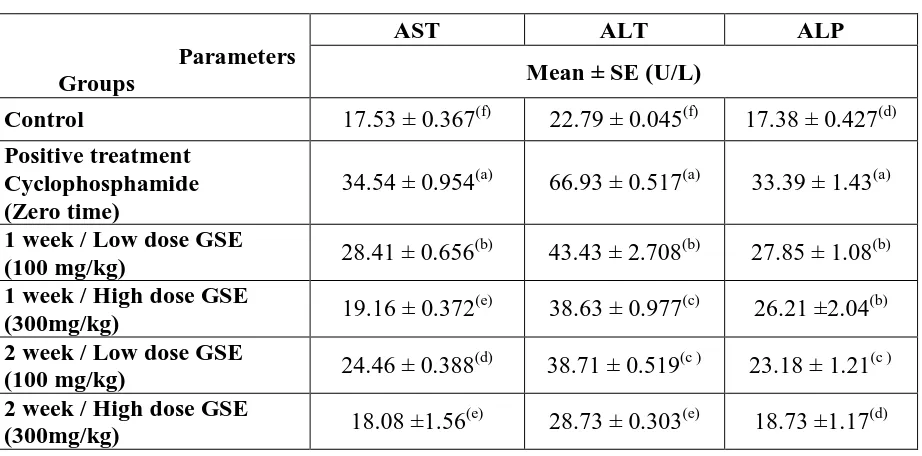
CP was metabolized in the liver by its microsomal enzymes. CP intoxication could leads to abnormal biochemical changes which were reflected in the serum. Table 1 represents the activities of serum marker enzymes; AST, ALT, and ALP in control and experimental mice that reflects the tissue damage. In CP challenged mice, as compared to control, almost two and three fold increase in the activities of AST and ALT were observed in the CP intoxicated mice. Administration of GSE resulted in markedly decrease in the activities of marker enzymes. Figure 1 depicts the levels of AST and ALT in the liver of experimental animals. Decrease in the activities of these enzymes in CP intoxicated mice and significant normalization during treatment with GSE was observed.
Effect of GSE on antioxidant levels and total protein content
CP group recorded significant decrease in hepatic GR (53.33%) and TrxR (56.82%) while colon GR exhibited percentage reached to 18.95%, TrxR 3.91%. Also, testis GR recorded 24.0% and TrxR 69.23%. In addition, hepatic total protein content showed significant decrease in CP intoxicated mice by percentage reached to 5.13%. While, the percentage of reduction in total protein content reached to 29.76% in colon. However, the percentage decrease in total protein content recorded 35.45% in testis. Treatment with low and high doses of GSE ameliorated hepatic GR level by 22.86 and 44.76% respectively and TrxR level by 40.91 and 86.36%, respectively. While, GR of colon improved by 35.95, 63.40%, respectively and TrxR by 34.78 and 82.61%, respectively.
Effect of GSE on DNA damage
Table (1): Levels of AST, ALT and ALP in sera of different experimental groups.
Parameters Groups
AST ALT ALP
Mean ± SE (U/L)
Control 17.53 ± 0.367(f) 22.79 ± 0.045(f) 17.38 ± 0.427(d) Positive treatment
Cyclophosphamide (Zero time)
34.54 ± 0.954(a) 66.93 ± 0.517(a) 33.39 ± 1.43(a)
1 week / Low dose GSE
(100 mg/kg) 28.41 ± 0.656
(b)
43.43 ± 2.708(b) 27.85 ± 1.08(b) 1 week / High dose GSE
(300mg/kg) 19.16 ± 0.372
(e)
38.63 ± 0.977(c) 26.21 ±2.04(b) 2 week / Low dose GSE
(100 mg/kg) 24.46 ± 0.388
(d)
38.71 ± 0.519(c ) 23.18 ± 1.21(c ) 2 week / High dose GSE
(300mg/kg) 18.08 ±1.56
(e)
28.73 ± 0.303(e) 18.73 ±1.17(d)
Data are means ± SD of ten mice in each group. Statistical analysis is carried out using one way analysis of variance (ANOVA), and Co-state computer program. Unshared superscript letters between groups are significant values at p ≤ 0.05.
Table (2): Levels of hepatic antioxidants enzymes; GR and TrxR in different experimental groups.
Parameters Groups
GR n mole
NADPH/min./mg protein
TrxR n mole NADPH/min/mg protein
Control
Mean ± SE
0.105 ± 0.013(c) 0.044 ± 0.004(c) Positive treatment
Cyclophosphamide (Zero time)
0.049 ± 0.002(d) 0.019 ± 0.0002(e)
1 week / Low dose (100mg/kg)
0.062 ± 0.002(d) 0.025 ± 0.001(e)
1 week / High dose
(300mg/kg) 0.079± 0.015
(d)
0.033 ± 0.002(d) 2 week / Low dose
(100mg/kg) 0.073 ± 0.020
(b)
0.037 ± 0.001(d) 2 week / High dose
(300mg/kg) 0.096 ± 0.020
(c)
0.057± 0.009(a)
[image:8.595.70.531.86.314.2]Table (3): Levels of colon antioxidants enzymes; GR and TrxR in different experimental groups
Parameters Groups
GR n mole
NADPH/min./mg protein
TrxR n mole NADPH/min/mg protein
Control
Mean ±SE
0.153±0.065(bc) 0.023 ± 0.005(e) Positive treatment
Cyclophosphamide (Zero time)
0.124 ± 0.012(c) 0.006 ± 0.002(a)
1 week / Low dose
(100 mg/kg) 0.118 ± 0.085
(c)
0.015 ± 0.003(b) 1 week / High dose
(300 mg/kg) 0.241 ± 0.037
(a)
0.022 ± 0.003(b) 2 week / Low dose
(100 mg/kg) 0.179 ± 0.003
(ab) 0.014 ± 0.001(e)
2 week / High dose
(300 mg/kg) 0.221 ± 0.104
(d)
0.025 ± 0.002(d)
*Values are presented as means ± SD (n= 10 mice /group). Unshared superscript letters between groups are significant values at p ≤ 0.05.
Table (4): Levels of testis antioxidants enzymes; GR and TrxR in different experimental groups.
Parameters Groups
GR n mole
NADPH/min./mg protein
TrxR n mole NADPH/min/mg protein
Control
Mean ±SE
0.025 ± 0.0001(b 0.039 ± 0.000(e) Positive treatment
Cyclophosphamide (Zero time)
0.011 ± 0.001(c) 0.012 ± 0.000(a)
1 week / Low dose
(100 mg/kg) 0.019 ± 0.001
(a)
0.031 ± 0.001(b) 1 week / High dose
(300 mg/kg) 0.022± 0.001
(b)
0.032 ± 0.000(b) 2 week / Low dose
(100 mg/kg) 0.023 ±0.002
(b)
0.035 ±0.001(e) 2 week / High dose
(300 mg/kg) 0.028 ± 0.000
(b)
0.037 ± 0.006(d)
[image:9.595.75.520.83.331.2]Table (5): Levels of TNF-α and AFP in liver tissues of different experimental groups. Parameters Groups TNF-α Pg/100mg AFP ng/100mg Control Mean ±SE
15.67± 0.001(a) 0.389± 0.000(a) Positive treatment
Cyclophosphamide (ZERO)
25.41 ± 0.001(b) 1.38± 0.002(b)
Low dose
(100 mg/kg) 17.37 ± 0.002
(a)
0.68± 0.003(c) High dose
(300 mg/kg) 14.85 ± 0.001
(b)
0.41± 0.004(b)
*Values are presented as means ± SD (n= 10 mice /group). Unshared superscript letters between groups are significant values at p ≤ 0.05.
Table (6):Levels of TNF-α and AFP in colon tissues of different experimental groups. Parameters Groups TNF-α Pg/100mg AFP ng/100mg Control Mean ±SE
20.88± 0.07(a) 0.447± 0.004(a) Positive treatment
Cyclophosphamide (ZERO)
28.57 ± 0.07 (b) 2.33± 0.001(b)
Low dose
(100 mg/kg) 25.07± 0.06
(a)
1.88± 0.002 (c) High dose
(300 mg/kg) 22.18± 0.05
(b)
0.75± 0.006 (b)
*Values are presented as means ± SD (n= 10 mice /group). Unshared superscript letters between groups are significant values at p ≤ 0.05.
0 10 20 30 40 50 60 70 80
Control CP (zero time)
1W/LD 1W/HD 2W/LD 2W/HD
[image:10.595.94.495.87.250.2]um ol e/ m in ./m g.p ro te in
Fig.1: Effect of GSE on AST and ALT in the liver of control and experimental mice. Results are expressed as Mean SD(n=10). Comparison were made between CP and control, CP+GSE (low and high dose) with CP. Statistically significant at P ≤
0.05
AST
[image:10.595.64.532.318.483.2] [image:10.595.105.469.521.730.2]
Figure 2: Comet assay parameters in the hepatic tissues of mice of different experimental
groups. Results are given as mean ± S.D. for 5 mice.
0 1 2 3 4 5 6 7
T
a
il
leng
th µ
m
D Groups
0 2 4 6 8
%
D
N
A
in
t
ai
l
E Groups
0 2 4 6 8
Tai
l m
o
m
en
t
F Groups
Figure 3: Comet assay parameters in the colon tissues of mice of different experimental groups. Results are given as mean ± S.D. for 5 mice.
[image:11.595.75.527.65.203.2] [image:11.595.78.520.285.417.2] [image:11.595.86.510.499.644.2]Figure 5: Photomicrographs of comets in the colon cells stained with ethidium bromide in different experimental groups. Control (5), CP (6), low dose of GSE (7), high dose (8).
DISCUSSION
Chemotherapy with CP can cause secondary tumors in humans by activating hepatic mixed function oxidases. Phosphoramide mustard, the major antineoplastic metabolite of CP, is an alkylating agent that induces a variety of changes in DNA [48,49], through its ability to form labile covalent DNA adducts and cross linkages [50].
Administration of chemotherapeutic drugs could leads to single nucleotide polymorphisms (SNPs) in chemotherapeutic drug metabolizing enzymes that are responsible for adverse drug
reactions (ADR) like alopecia, nausea, vomiting etc. with abnormal liver functions [51]. Cytochrome P450 group of enzymes have extensive functions in liver that includes the
detoxification of xenobiotics [52]. In the present study, elevation of serum marker enzymes in CP intoxication reflected the liver damage. Hepatopathy could leads to the leakage of marker enzymes; AST, ALT and ALP into the circulation confirming the extent of liver damage [53]. Also decreased levels in liver tissues and increased serum levels of both AST and ALT could be due to toxic compounds affecting the integrity of liver cells [54].
Elevation of AFP level up to pathological range in adults correlates with the appearance of several malignancies such as HCC and chronic liver disease [58]. Increased AFP and TNF-α levels observed in our study were due to the consequences of CP intoxication. On the other hand, GSE down–regulated the concentration of these cytokines and reduced the severity of injury. This may be explained on the basis of GSE interfering with cancer cell growth and proliferation, as well as inducing cell death appears to be one of its greatest highlights, which may be contributing to some of the clinical benefits demonstrated by the extract Kundu et al [59].
The amelioration effect by the post-administration of GSE is probably due to its renowned anti-inflammatory and antioxidant potency. GSE is also reported to possess significant multi organ histological protection against various toxic insults [60, 61, 62, 63, 64]. In addition, Veluri et al. [65] reported that gallic acid which is considered as major active constituents of GSE, showed a very strong dose- and time-dependent growth inhibition as well as apoptotic death of human prostate cancer DU145 cells. The antioxidant activity of GSE may be explained on the basis of it trapping free radicals (hydroxyl, lipid free radicals, free iron molecules and lipid peroxides), delaying fat oxidation, inhibiting the major substance responsible for generating oxygen derived free radicals (xanthin oxidase) and reducing the concentration of H2O2 [66] that produced by the oxidative stress resulted from O
-Nitrotoluene treatment.
In accordance with the present study Balu et al. [67] demonstrated that the antioxidant activity of grape extract was increased when the extract concentration increased. Besides, antioxidant/antiradical activity of grape seed extract, it was shown to possess many biological properties including the inhibition of DNA damage [68] and COX-2 gene expression [69]. Thus, it could be concluded that, GSE treatment showed ameliorating effects of precancerous stage in liver, colon and testis tissues induced by CP administration in dose- and time-dependent. While, these preliminary results appear promising and need further studies to elucidate the modulatory effects of GSE on early and late stages of liver, colon and testis cancer. Moreover, the present study confirmed the toxicity of CP as chemotherapeutic drug.
REFERENCES
2. Lutsiak ME, Semnani RT, De Pascalis R, Kashmiri SV, Schlom J, and Sabzevari H. Inhibition of CD4+25+ T regulatory cell function implicated in enhanced immune response by low-dose cyclophosphamide. Blood, 2005; 105(7): 2862–2868.
3. Ghiringhelli F, Larmonier N, Schmitt E, et al. CD4+CD25+ regulatory T cells suppress tumor immunity but are sensitive to cyclophosphamide which allows immunotherapy of established tumors to be curative. European Journal of Immunology, 2004; 34(2): 336– 344.
4. Awwad M and North RJ. Cyclophosphamide-induced immunologically mediated regression of a cyclophosphamide resistant murine tumor: a consequence of eliminating precursor L3T4+ suppressor T-cells. Cancer Research, 1989; 49(7): 1649–1654.
5. Ercolini AM, Ladle BH, Manning EA, et al. Recruitment of latent pools of high-avidity CD8+ T cells to the antitumor immune response. Journal of Experimental Medicine, 2005; 201(10): 1591–1602.
6. Xu L, Xu W, Jiang Z, Zhang F, Chu Y, and Xiong S. Depletion of CD4+CD25 high regulatory T cells from tumor infiltrating lymphocytes predominantly induces Th1 type immune response in vivo which inhibits tumor growth in adoptive immunotherapy. Cancer Biology and Therapy, 2009; 8(1): 66–72.
7. Popov B, Georgieva Sv and Gadjeva V. Modulatory effects of total extract of haberlea rhodopensis against the cyclophosphamide induced genotoxicity in rabbit lymphocytes in vivo. Trakia Journal of Sciences, 2011; 9(1): 51-57.
8. Ember I, Raposa T, Vargo C, Herceg L and Kiss I. Carcinogenic effects of cytostatic protocols in CBA/Ca mice. In Vivo, 1995; 9: 65-69.
9. Ridder D, de Poppel HV and Demonty L. Bladder cancer in patients with multiple sclerosis treated with cyclophosphamide. J. Uro, 1998; 159: 1881-1884.
10. Nyblom H, Berggren U, Balldin J, Olsson R. High AST/ALT ratio may indicate advanced alcoholic liver disease rather than heavy drinking. Alcohol, 2002; 39 (4): 336–9.
11. Ostapowicz G, Fontana RJ, Schiodt FV. Results of a prospective study of acute liver failure at 17 tertiary care centers in the United States. Ann.Intern.Med, 2002; 137(12): 947-54.
12. Nyblom H, Björnsson E, Simrén M, Aldenborg F, AlmermS and Olsson R."The AST/ ALT ratio as an indicator of cirrhosis in patients with PBC". Liver Int, 2006; 26 (7): 840– 5.
requires both transcriptional and translational modulation. Carcinogenesis, 2003; 24(3): 497–503.
14. Kim IS, Shin SY, Kim YS, Kim HY, Yoon HS. Expression of a glutathione reductase from Brassica rapa subsp. pekinensis enhanced cellular redox homeostasis by modulating antioxidant proteins in Escherichia coli. Mol Cells, 2009; 28(5): 479-87.
15. Yap SF, Peh SC. Alpha-Fetoprotein in Hepatocellular Carcinoma: A serologlcal and Histochemical Study in Malaysian Patients Malaysian. J Pathol, 1991; 13(2): 115-118. 16. Jawed AB, Junaid MA, Syed RM, Muhammad B, Rabia S, Ishrat S, Abdul Waheed.
Hepptocellular Carcinoma (HCC) and Diagnostic Significance of alpha-Fetoprotein (AFP). J Ayub Med Coll Abbottabad, 2009; 21(1): 72-75.
17. Abdel-Wahhab MA, Ahmed HH and Hagazi MM. Prevention of aflatoxin B1 initiated
hepatotoxicity in rat by marine algae extracts. J. Appl. Toxicol, 2006; 26: 229-238.
18. Moon EY, Rhee DK and Pyo S 1999. Involvement of NO, H2O2 and TNF-[alpha] in the
reduced antitumor activity of murine peritoneal macrophages by aflatoxin B1. Cancer
Lett, 1999; 136: 167-176.
19. Suganuma M, Sueoka E, Sueoka N, Okabe S and Fujiki H. Mechanisms of cancer prevention by tea polyphenols based on inhibition of TNF-alpha expression. Biofactors, 2000; 13: 67-72.
20. Marcinkiewicz J, Bryniarski K and Ptak W. Cyclophosphamide uncovers two separate macrophage subpopulations with opposite immunogenic potential and different patterns of monokine production. Cytokine, 1994; 6 (5): 472-477.
21. Bryniarski K, Ptak M and Ptak W. The in vivo and in vitro effects of an alkylating agent, mechlormethamine, on IL-6 production in mice and the role of macrophages. Immuno pharmacology,1996; 34: 73-78.
22. Bryniarski K, Szczepanik M, Ptak M, Zemelka M and Ptak W. Influence of cyclophosphamide and its metabolic products on the activity of peritoneal macrophages in mice. Pharmacological Reports, 2009; 61: 550-557.
23. Barton CC, Barton EX, Ganey PE, Kunkel SL, and Roth RA. Bacterial lipopolysaccharide enhances aflatoxin B1 hepatotoxicity in rats by a mechanism that
depends on tumor necrosis factor. Hepatol, 2001; 33: 66-73.
25. Zhou HP, Lutterodt H, Cheng ZH and Yu LL. Anti- inflammatory and anti- proliferative activities of trifolirhizin, a flavonoid from sophora flavescens roots. J. Agric. Food Chem, 2009; 57: 4580-4585.
26. Abdel-Hamid NM, Nazmy MH, Mahmoud AW, Fawzy MA and Youssof M. A survey on herbal management of hepatocellular carcinoma. World J. Hepatol, 2011; 3: 175-183. 27. Alakilli SYM. Evaluation of camphor mutagenicity in somatic cells of pregnant rats.
Asian J. Biotechnol, 2009; 1: 111-117.
28.Senthikumar S, Ebenezar KK, Sathish V,Yogeeta S and Devaki T. Modulation of the tissue defense system by squalene in cyclophosphamide induced toxicity in rats. Arch. Med. Sci, 2006a; 2: 94-100.
29. Yilmaz Y, Toledo RT. Health aspects of functional grape seed constituents. Trends food Sci. Technol., 2004; 15: 422-33.
30. Puiggros F, Llopiz N, Ardevol A, Blade C, Arola L, Salvado MJ. Grape seed procyanidins prevent oxidative injury by modulating the expression of antioxidative enzyme systems. J. Agric Food Chem., 2005; 53: 6080-6.
31. Balu M, Sangeetha P, Murali G, Panneerselvam C. Modulatory role of grape seed extract on age-related oxidative DNA damage in central nervous system of rats. Brain Res. Bull, 2006; 68: 469-73.
32. Roychowdhury S, Wolf G, Keilhoff G, Bagchi D and Horn T. Protection of primary glial cells by grape seed proanthocyanidin extract against nitrosative/oxidative stress. Nitric Oxide, 2001; 5: 137-49.
33. Bagchi D, Ray SD, Patel D, Bagchi M. Protection against drug- and chemical induced multiorgan toxicity by a novel IH636 grape seed proanthocyanidin extract. Drugs Exp. Clin. Res, 2001; 27: 3-15.
34. Shi J, Yu J, Pohorly E, Kakuda Y. Polyphenolic in grape seeds-biochemistry and functionality. J. Med. Food, 2003; 6: 291-99.
35. Ye X, Krohn RL, Liu W, Joshi SS, Kuszynski CA, McGinn TR, Bagchi M, Preuss HG, Stohs SJ and Bagchi D. The cytotoxic effects of a novel IH636 grape seed proanthocyanidin extract on cultured human cancer cells. Mol. Cell Biochem, 1999; 196: 99-108.
37. Zhang XY, Li WG, Wu YJ, Bai DC, Liu NF. Proanthocyanidin from grape seeds enhances doxorubicin-induced antitumor effect and reverses drug resistance in doxorubicin-resistant K562/DOX cells. Can J. Physiol. Pharmacol, 2005; 83: 309-18. 38. Kaur M, Singh RP, Gu M, Agarwal R, Agarwal C. Grape seed extract inhibits in vitro and
in vivo growth of human colorectal carcinoma cells. Clin Cancer Res, 2006; 12: 6194-6202.
39. Koga T, Moro K, Nakamori K, et al. Increase of antioxidative potential of rat plasma by oral administration of proanthocyanidin-rich extract from grape seeds. J Agric Food Chem, 1999; 47: 1892-1897.
40. Meirow D, Lewis H, Nugent D and Epstein M. Subclinical depletion of primordial follicular reserve in mice treated with cyclophosphamide: Clinical importance and proposed accurate investigative tool. Hum. Reprod, 1999; 14: 1903-1907.
41.Takimoto CH, Calvo E, Pazdur R, Wagman LD, Camphausen KA, Hoskins WJ. Principles of Oncologic Pharmacotherapy in (Eds) Cancer Management: A Multidisciplinary Approach, 2008; 11 ed.
42. Gella FJ, Olivella T, Cruz PM, Arenas J, Moreno R, Durban R and Gomez JA. A simple procedure for routine determination of aspartate aminotransferase and alanine aminotransferase with pyridoxal phosphate. Clin Chem Acta, 1985; 153: 241-247.
43. Bradford MM. A rapid and sensitive method for the quantitation of microgram quantities of protein utilizing the principle of protein-dye binding. Anal Biochem, 1976; 72: 248-254.
44. Rosalki SB, Foo AY, Burlina A. Multicenter evaluation of iso-ALP test kit for measurement of bone alkaline phosphatase activity in serum and plasma. Clin Chem, 1993; 39: 648-652.
45. Erden M, Bor NM. Changes in reduced glutathione, glutathione reductase and glutathione peroxidase after radiation in guinea pigs. Biochem Med, 1984; 31: 217-227.
46. ARNÉR E, HOLMGREN A. Physiological functions of thioredoxin and thioredoxin reductase. Eur J Biochem, 2000; 267: 6102-6109.
47. Singh NP, McCoy MT, Tice RR and Schneider EL: A simple technique for quantitation of low levels of DNA damage in individual cells. Exp Cell Res, 1988; 175: 184-191. 48. IARC. Some antineoplastic and immunosuppressive agents. In: Monographs on the
49. Krishna G, Nath J, Peterson M and Ong T., Cyclophosphamide- induced cytogenetic effects in mouse bone marrow and spleen cells in vivo and in vivo/in vitro assays. Teratog Carcinog Mutagen, 1987; 7: 183-195.
50. Anderson D, Bishop JB, Garner RC, Ostrosky-Wegman P and Selby PB. Cyclophosphamide: review of its mutagenicity for an assessment of potential germ cell risks. Mutat Res, 1995; 330(1-2): 115-81.
51. Khan S, Jamil K, Das GP, Vamsy CM and Murthy S. Polymorphic sites (1236 and 3435) in multi drug resistance gene 1 influencing drug response in breast cancer patients. Int. J. Pharmacol, 2007; 3: 453-460.
52. Tirona RG, Lee W, Leake BF, Lan LB and Cline CB et al. The orphan nuclear receptor HNF4- alpha determines PXR-and CAR-mediated xenobiotic induction of CYP3A4. Nature Med, 2003; 9: 220-224.
53. Nkosi CZ. Opoku AR and Terblanche SE. Effect of pumpkin seed (Cucurbita pepo) protein isolate on the activity levels of certain plasma enzymes in CCL4–induced liver
injury in low-protein fed rats. Phytother. Res, 2005; 19: 341-345.
54. Senthilkumar S, Ebenezar KK, Sathish V, Yogeeta S and Devaki T. Modulation of the tissue defense system by squalene in cyclophosphamide induced toxicity in rats. Arch. Med. Sci, 2006a; 2: 94-100.
55. Schmidt E. Strategy and Evaluation of Enzyme Determinations in Serum in Disease of the Liver and the Biliary System. In: Evaluation of Liver Function: A Multifaceted Approach to Clinical Diagnosis, Demers LM. and Shaw LM (Eds.). Urban and Schwarzenberg, Baltimore, MD., USA, 1978; 79-101.
56. Puiggros FN, Liopiz A, Ardevol C, Blade LA and Salvado MJ. Grape seed procyanidins prevent oxidative injury by modulating the expression of antioxidative enzyme systems. J. Agric. Food Chem,2005; 53: 6080–6086.
57. Özer T, Yahya O, Ender D, Umit T, Feriha E and Goksel T. Grape Seed Extract Treatment Reduces Hepatic Ischemia-Reperfusion Injury in Rats. Phytother. Res, 2008; 22: 43–48.
58. Jawed AB, Junaid MA, Syed RM, Muhammad B, Rabia S, Ishrat S, Abdul Waheed. Hepptocellular Carcinoma (HCC) and Diagnostic Significance of alpha-Fetoprotein (AFP). J Ayub Med Coll Abbottabad, 2009; 21(1): 72-75.
60. El-Beshbishy HA, Mohamadin A, Nagy A and Abdel-Naim A. Amelioration of tamoxifen-induced liver injury in rats by grape seed extract, black seed extract and curcumin. Indian J. Exp. Biol, 2010; 48: 280-8.
61. El-Ashmawy IM, Gad SB and Salama OM. Grape seed extract prevents azathioprine toxicity in rats. Phytother. Res, 2010; 24: 1710-5.
62. Safa J, Argani H, Bastani B, Nezami N, Rahimi Ardebili B, Ghorbanihaghjo A, Kalagheichi H, Amirfirouzi A, Mesgari M and Soleimany Rad J. Protective effect of grape seed extract on gentamicin induced acute kidney injury. Iran. J. KidneyDis, 2010; 4: 285-91.
63. Yalçin E, Oruç E, Cavuşoğlu K and Yapar K. Protective role of grape seed extract against doxorubicin-induced cardiotoxicity and genotoxicity in albino mice. J. Med. Food, 2010; 13: 917-25.
64. Khalifa FK, Khalil FA, Barakat HA and Hassan MM. Protective role of wheat germ and grape seed oils in chlorpyrifos-induced oxidative stress, biochemical and histological alterations in liver of rats. Aust. J. Basic App. Sci, 2011; 5: 54-66
65. Veluri R, Singh RP, Liu Z, Thompson JA, Agarwal R and Agarwal C. Fractionation of grape seed extract and identification of gallic acid as one of the major active constituents causing growth inhibition and apoptotic death of DU145 human prostate carcinoma cells. Carcinogenesis, 2006; 27(7): 1445-53.
66. Sugisawa A, Inoue S and Umegaki K. Grape seed extract prevents H2O2 induced
chromosomal damage in human lymphoblastoid cells. Biol. Pharm. Bull, 2004; 27: 1459-1461.
67. Baydar NG, Ozkan G and Yasar S. Evaluation of the antiradical and antioxidant potential of grape extracts. Food Control, 2007; 18: 1131-1136.
68. Balu M, Sangeetha P, Murali G and Panneerselvam C. Modulatory role of grape seed extract on agerelated oxidative DNA damage in central nervous system of rats. Brain Res. Bull, 2006; 68: 469-473.