EVALUATION OF THE EXPIRATORY FLOW RATE CHANGES IN
PREGNANCY (HOSPITAL-BASED STUDY)
Dr. Saif Abdulhussein Hassan*
*M.B.Ch.B, High Diploma in Respiratory Medicine. Ministry of Health, Baghdad –Iraq.
ABSTRACT
Background: Pregnancy is associated with physiological changes in the control of breathing, in lung volumes, in the mechanics of
respiration and in acid base balance. Objective: To compare the FEF25-75 and PEFR of non-pregnant & pregnant women in different
trimesters of pregnancy and to determine the effect of gestational age
on the PEFR and FEF 25-75 measurements. Patients and Methods: This cross sectional analytical study undertaken in the unit of
pulmonary function in association with the Department of Gynecology
and Obstetrics, in Baghdad teaching hospital in Baghdad. The study
started from June 2016 to June 2017. Results: The study did not follow the same female in her different stages of pregnancy; we took
random samples in different trimesters of the pregnancy (some of them were in the 1st, some
in the 2nd and some in the third trimester). FEF25percentage was the only small airway
parameter that is affected between the pregnant and the non- pregnant (P value was less than
0.05) BUT not in different trimesters of same pregnancy. In addition, there was change in the
PEFR between the pregnant and the non- pregnant (P value was less than 0.05), But no
change between the different trimesters. Conclusion: We conclude that, there is significant difference in FEF25percentage between pregnant and non- pregnant women but no difference
among different trimesters of pregnancy. In addition, there are no difference in
FEF50percentage, FEF75percentage between pregnant and non- pregnant women, and no
difference among different trimesters of pregnancy. Recommendation: We recommend that
every pregnant woman should have the FEF25percentage as an important parameter for small
airway diseases severity assessment in pregnancy.
KEYWORD: Evaluation, expiratory, flow, rate, pregnancy.
Volume 7, Issue 17, 98-106. Research Article ISSN 2277– 7105
Article Received on 26 July 2018,
Revised on 16 August 2018, Accepted on 06 Sept. 2018
DOI: 10.20959/wjpr201817-13247
*Corresponding Author
Dr. Saif Abdulhussein
Hassan
M.B.Ch.B, High Diploma
in Respiratory Medicine.
INTRODUCTION
Studies had shown that there is a good evidence mirroring relationships between pregnancy
period and respiratory system.[1] The different adaptive changes in the respiratory system in
pregnant women and thus preventing any unnecessary or unneeded treatment for the
physiologically changed respiratory functions misinterpreted as abnormal the pregnancy is
associated with physiological changes in the control of breathing, in lung volumes, in the
mechanics of respiration and in acid base balance.[1]
Maternal respiratory changes in turn affect the metabolism and the well-being of the fetus by
their effect on placental gas exchange. Three cardinal changes in the thorax which happen
during pregnancy were: an increase in the circumference of the lower chest wall (with
increases in anterio posterior and the transverse diameters)[2,3,4]; a 50% widening of the costal
angle (1 -3) and elevation of the diaphragm (a cephalic displacement of approximately 4 cm
to 5 cm).[5] These changes becoming more prominent around the 37th week of pregnancy and
returns back to normal within 6 months post-delivery.[6] Pulmonary function during
pregnancy is influence by changes of the airway, thoracic cage, and respiratory drive.
Additionally, there is capillary engorgement and congestion throughout the respiratory tract,
which results in mucosal edema and hyperemia.[7] Different biochemical changes like
increase in cyclic nucleotide, prostaglandins, progesterone, corticosteroid and estrogen levels
happens concomitantly during the course of pregnancy.[8]
The thoracic circumference increases about 6cm but not sufficiently to present a marked
reduction in the Residual Volume of air in the lungs controlled by the elevated diaphragm.
The most important change in lung function is the increase in Minute Ventilation[9], which
increases by nearly 36% by the eighth week of pregnancy reaching levels, which are about
50% above the non- pregnant need. This adaptation is required to satisfy the increase in
oxygen consumption of 30-35% by the growing fetus. This increase in Minute Ventilation
results in a slight decrease in alveolar PCO2 and lower PaCO2 from 38 mm Hg in non-
pregnant females to approximately 32-34 mm HG at term.[10] This study aimed to compare
the FEF25-75 and PEFR of non-pregnant & pregnant women in different trimesters of
pregnancy and to determine the effect of gestational age on the PEFR and FEF 25-75
Patients and methods: This cross sectional analytical study undertaken in the unit of pulmonary function in association with the Department of Gynecology and Obstetrics, in
Baghdad teaching hospital in Baghdad. The study started from June 2016 to June 2017. The
study consists of recording the Pulmonary Function Tests for two major groups of Iraqi
female of childbearing age. Including 120 pregnant women of various phases of gestational
period subdivided into 10 weeks (I trimester), around 24 weeks (II trimester), 37 weeks (III
trimester) and control group of 40 non pregnant women of age and sex compatible. The
different lung function parameters measured in this study were peaked expiratory flow rate
(PEFR), forced mid expiratory flow 25-75%(FEF25-75percentage). All the participants were
included in the study detailed history, and a complete clinical examination. The study
includes two-group (pregnant and non-pregnant) aged from 16-44 years, of different weight;
height (which were recorder) and different conception from 1st, 2nd and 3rd trimesters were
included. The nature and the purpose of the study explained to each participant. Physical
examination done to each participant especially regarding the cardiovascular and the
respiratory system. The Statistical Package for Social Sciences (SPSS) version 22 program
for Windows used to analyze this data. The mean +standard deviation (SD) used to analyze
for each group.
Parameters involved
PEFR: is the maximum flow rate during the forced vital capacity in the initial 0.1 second. Normal values in young adult equals 500L/MIN. It reflects the airway status in general and it
is more relate for larger airways rather than small airways.
Forced mid expiratory flow 25%-50%-75percentage: Is the maximum flow rate during the mid-expiratory part of Forced vital capacity maneuver. Normal values equals 300L/MIN. It
Figure 1: The PEFR, FEF25%, FEF50%, FEF75%.
Inclusion criteria: Healthy pregnant Iraqi women in the childbearing age who accepted doing the study were included in the study. The visible health status of the women
determined by history taking and clinical examination. Also weight, height, oxygen saturation
using pulse Oximeter (which was normal for all) and blood pressure also is recorded.
Exclusion criteria: Women suffering from acute respiratory infection in the last three months. Chronic respiratory infection including asthma. Also the clinical or history of
cardiovascular diseases. Diabetes mellitus (controlled or uncontrolled), hypertension
(controlled or uncontrolled). In addition, any smoking history (even small number of
cigarettes for any period), alcohol consumption., endocrine disease, morbid obesity, anemia
(moderate to severe).
Recording the PFT
The test were performing in the early morning hours (9 am to 12am). The test done in calm
conditions with subjects in sitting position according to ATS guidelines. Using master lab
spirometer Jaegersn.(105052-175111:model 1998.germany). The pregnant and non- pregnant
women did the procedure by their own agreement. The aim of the study was explained to
them. Their written agreement was taken from each of the participant. Before recording the
PFT, the procedure also, explained and demonstrated in detail until proper understanding. If
any questions, it has answered to their satisfaction and instructions about the importance of
the tests are given. Each subject was ask it to sit comfortably in a chair facing the
computerized flexi flow machine. Subject’s age; height, weight, and specific room
temperature were entered in the computer.
RESULTS
This table shows that the descriptive statistics for FEF25%, the mean and the S.D. for the
[image:5.595.115.484.243.333.2]study and the control groups.
Table 1: Mean and SD for FEF25.
State Mean Std. Deviation N
non pregnant 80.6525 15.25671 40
first trimester pregnancy 69.7975 23.35779 40 second trimester pregnancy 74.2550 21.44466 40 third trimester pregnancy 72.1500 14.77524 40
Total 74.2137 19.33058 160
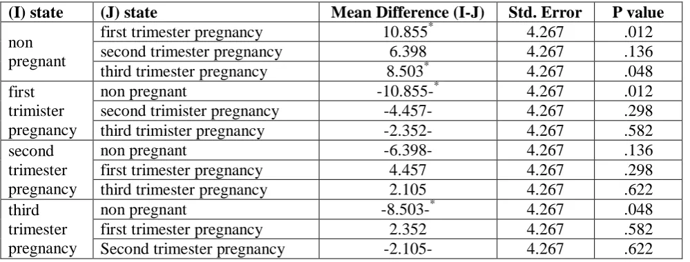
This table shows the statistical results for FEF25percentage, there was significant change
between the pregnant and the non-pregnant but no change in the different trimesters of the
different pregnancies.
Table (2): Mean differences and SE for FEF25percentage.
(I) state (J) state Mean Difference (I-J) Std. Error P value non
pregnant
first trimester pregnancy 10.855* 4.267 .012 second trimester pregnancy 6.398 4.267 .136 third trimester pregnancy 8.503* 4.267 .048 first
trimister pregnancy
non pregnant -10.855-* 4.267 .012
second trimister pregnancy -4.457- 4.267 .298 third trimister pregnancy -2.352- 4.267 .582 second
trimester pregnancy
non pregnant -6.398- 4.267 .136
first trimester pregnancy 4.457 4.267 .298 third trimester pregnancy 2.105 4.267 .622 third
trimester pregnancy
non pregnant -8.503-* 4.267 .048
first trimester pregnancy 2.352 4.267 .582 Second trimester pregnancy -2.105- 4.267 .622
Table 3 Abbreviates the descriptive statistics for FEF50percentage. The mean and the S.D.
[image:5.595.66.566.440.630.2]Table (3): Mean and SD for FEF50percentage.
STATE Mean S.D. NO.
non pregnant 83.2800 19.11358 40
first trimester pregnancy 79.0750 25.33031 40 second trimester pregnancy 81.4300 28.51465 40 third trimester pregnancy 83.2250 18.15027 40
Total 81.7525 23.02587 160
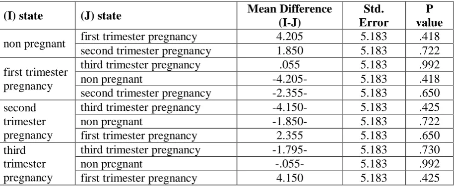
FEF50 in non-pregnant and pregnant female; in this table shows the statics for the FEF50
percentage, which shows that there was no statistically difference between the pregnant and
the non -pregnant and also no change between different trimesters of the different
pregnancies.
Table (4): Mean differences and SE for FEF50percentage.
(I) state (J) state Mean Difference
(I-J)
Std. Error
P value non pregnant first trimester pregnancy 4.205 5.183 .418
second trimester pregnancy 1.850 5.183 .722
first trimester pregnancy
third trimester pregnancy .055 5.183 .992
non pregnant -4.205- 5.183 .418
second trimester pregnancy -2.355- 5.183 .650 second
trimester pregnancy
third trimester pregnancy -4.150- 5.183 .425
non pregnant -1.850- 5.183 .722
first trimester pregnancy 2.355 5.183 .650 third
trimester pregnancy
third trimester pregnancy -1.795- 5.183 .730
non pregnant -.055- 5.183 .992
first trimester pregnancy 4.150 5.183 .425
Descriptive statistics for FEF75percentage. In this table shows that the mean and S.D. For
each group.
Table 5: Mean and SD for FEF75%.
State Mean Std. Deviation N
non pregnant 80.8825 19.93008 40
first trimester pregnancy 73.9525 26.95131 40 second trimester pregnancy 76.2600 49.93274 40 third trimester pregnancy 82.8475 28.44124 40
Total 78.4856 33.13929 160
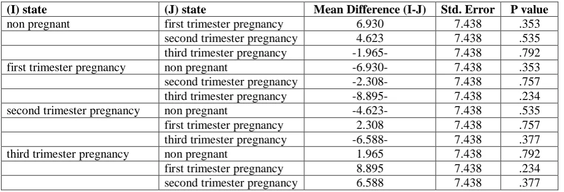
FEF75 in non-pregnant and pregnant female:- This table includes the statistics for the variable FEF75. There was no different between the pregnant and the non-pregnant and no
[image:6.595.70.528.309.498.2]Table 6: Mean differences and SE for FEF75.
(I) state (J) state Mean Difference (I-J) Std. Error P value non pregnant first trimester pregnancy 6.930 7.438 .353
second trimester pregnancy 4.623 7.438 .535 third trimester pregnancy -1.965- 7.438 .792 first trimester pregnancy non pregnant -6.930- 7.438 .353 second trimester pregnancy -2.308- 7.438 .757 third trimester pregnancy -8.895- 7.438 .234 second trimester pregnancy non pregnant -4.623- 7.438 .535 first trimester pregnancy 2.308 7.438 .757 third trimester pregnancy -6.588- 7.438 .377 third trimester pregnancy non pregnant 1.965 7.438 .792 first trimester pregnancy 8.895 7.438 .234 second trimester pregnancy 6.588 7.438 .377
DISCUSSION
According to the aims of this study, our result found that there was statistically significant
changes in FEF25percentage between pregnant and non-pregnant but not in the different
trimesters of the different pregnancies. The possible cause could be due to smooth muscle
relaxing effects of progesterone, Relax in and Corticosteroids during pregnancy. In addition,
there was no change between pregnant and non-pregnant regarding FEF 50%-75%.
Although some workers have already studied the effect of pregnancy on pulmonary Function,
but in my study I focused mainly on the difference of expiratory flow rates at different stages
of pregnancy particularly the changes at the small airways level. Previous studies have
concluded that forced spirometry values largely remain unchanged in normal pregnancy,
compared with a non-pregnant control group.[11,12]
According to other study[13], they found that there is no significance change in FEF25-FEF
75% between the pregnant and the non-pregnant in different stages of the different
pregnancy. Spiro metric values despite being lower than those of the controls are remained
within normal physiological ranges throughout pregnancy. These changes in the maternal
pulmonary function during pregnancy are actually adaptive in nature. In spite of the
mechanical disadvantage to the respiratory apparatus, pregnant women are able to achieve
adequate ventilation, which facilitates feto maternal gas exchange. Other study done by
Emilia kolarzyk et al showed that there is no statistical significant change in
In a study conducted by NEERAJ et al, they found that there was decrease in the
FEF25-75percentage in the third trimester of pregnancy. This decrease was because of decrease in
alveolar Pco2 caused by hyperventilation, which acts as Broncho constrictor. Hormonal
changes also play a role in altering & compromising the FEF25-75percentage.[15]
A study by SAVITA SINGH et al have reported that there was decrease in FEF25%, FEF
50% & FEF 75% in second trimester as compared with third trimester. The cause assumed
that, the fetal bulk does a greater restriction on the breathing of pregnant women of Indian
race who are generally diminutive compared to their western counterparts.[16] In a study by
RUPA. M et al, they found that the values of MMF (maximal mid expiratory flow) were
significantly lower in first trimester compared to control.[17]
To establish the cause of decrease in respiratory parameters more in FEF25percentage more
than FEF50percentage and FEF75percentage, further longitudinal studies are be done on
acid-base balance, hormonal assay in different trimesters to know the possible compensatory
mechanism.
CONCLUSION
We conclude that, there is significant difference in FEF25percentage between pregnant and
non- pregnant women but no difference among different trimesters of pregnancy. In addition,
there are no difference in FEF50percentage, FEF75percentage between pregnant and non-
pregnant women, and no difference among different trimesters of pregnancy.
Recommendation: We recommend that every pregnant woman should have the FEF25percentage as an important parameter for small airway diseases severity assessment in
pregnancy.
Limitations of the study: Because of time limit, the research conducted only on a small size of population who were attending the hospital. As well, the bias and hesitation of the
volunteers affect the research and the busy schedule of our hospital makes collection data a
difficult job. In addition, Studies on larger population for longer periods are require to set a
standard reference range of the PFT values in the different trimesters of pregnancy. Such
norms would help in accurate evaluation of the changes in maternal respiratory function by
REFERENCES
1. Gold smith LT, weiss G, relaxin and its role in pregancny. Endocrinology metabolic clinic
north AM, 1995; 24(1): 171-86.
2. Pandya KD, chand wani S, Desai CA. Study of the vital capacity and timed vital capacity in
normal and pregnant women. J obstet gynecol Ind, 1984; 36: 1053-57.
3. LR Brancazlo, SA laifer & TS Chartz. Pregnancy and advancing gestation on peak expiratory
rate. Obstetrics & gynecology, 1997; 89: 383.
4. A merican college of obstetrician and gynecologist. Pulmonary diseases in preganancy.
ACOG bulletin NO.224 Washington DC: 1996.
5. Weinberger SE, Weiss ST, Gohen WR. Pregnancy and the lungs. AM Rev Respiartory, 1980;
121(3): 559-81.
6. Ellegard EK, pregnancy rhinitis, 2006; 26(1): 119-35.
7. Toppozada H, Michales L. The human respiratory nasal mucosa on pregnancy. An electron
microscopic and histochemical study, 1999; (7): 613-26.
8. P Bhatia, K Bhatia. Pregnancy and the lungs. Post graduate medical journal, 2000; 76:
683-93.
9. Rees GB, Pipkin FB. Longitudinal study of respiratory in normal human pregnancy. AMJ
obstet gynecol, 1990; 162: 826-30.
10.Nelson piercy. respiratory disease. Hand book of obstetric medicine, oxford, 1997; 15-65.
11.Kolarzyk E, Szot WM, Lyszczarz J. Lung function and breathing regulation parameters
during pregnancy. Arch Gynecol Obstet, 2005; 272: 53.
12.McAuliffe F, Kametas N, Costello J, Rafferty GF, Greenough A, Nicolaides K. Respiratory
function in singleton and twin pregnancy. BJOG, 2002; 109: 765–9.
13.Das TK, Jana H. Maternal airways function during normal pregnancy. Indian J Med Sci.,
1991; 45: 265–8.
14.Kolarzyk E, Szot WM, Lyszczarz J. Lung function and breathing regulation parameters
during pregnancy. Arch Gynecol Obstet, 2005; 272(1): 53-8.
15.Neeraj, Sodhi C, John P, Singh J & Kaur V. Effect of advanced uncomplicated pregnancy on
pulmonary function parameters of North Indian subjects. Indian J Physiol Pharmacol, 2010;
54(1): 69-72.
16.Singh S, Singh KC, Sircar SS, Sharma KN. Airway functions in pregnant Indian women,
Indian J Physiol Pharmacol, 1995; 39(2): 160-62.
17. Mokkapati R, Prasad EC, Venkatraman, Fatima K. Ventilatory functions in pregnancy Indian