APOPTOTIC EFFECT OF GOLD NANOPARTICLES ON HUMAN
COLON CANCER CELL LINES CACO-2 BUT NOT ON THE NORMAL
EPITHELIAL CELL LINES WISH
Marwa Alkayaty1, Nahla.G.Mohamed2, Rehab. E. El-Hennamy1 and Soad Nady*1
1
Zoology and Entomology Department, Faculty of Science, Helwan University.
2
G.M. of Applied Research Department, VACSERA.
ABSTRACT
Colorectal cancer (CRC) is the third most common cancer worldwide. Consequently, the discovery of new strategies for therapeutic intervention remains a priority. The gold nanoparticles (AuNPs) have been discovered to exert cytotoxicity and induce apoptosis in some cancer cells. The aim of the present study was to investigate the effect of AuNPs on human colon cancer cell lines (Caco-2) and normal human epithelial cell lines (WISH) in vitro. The two cell lines were treated with different concentrations of AuNPs for 5 days and cytotoxicity of AuNPs was assessed. RT-PCR was used to analyze the expression profiles of pro- and anti-apoptotic genes. Tumor necrosis factor alpha (TNF-α) and interlukin-6 (IL-6) levels were measured in the culture supernatant using ELISA. Results revealed that AuNPs has a cytotoxic effect on Caco-2 cell lines at high and low concentrations, but has no cytotoxic effect on WISH cell lines. Upregulation of the pro-apoptotic gene (bax) was induced in AuNPs treated Caco-2 cell lines but not WISH cells. The levels of IL-6 secreted by Caco-2 cell line treated with AuNPs increased gradually up to 96 h then began to decline thereafter. On the other hand, the levels of TNF-α reached its peak after 24h then started to decrease gradually. In conclusion, AuNPs did not cause acute cytotoxicity for normal human epithelial cell lines (WISH), but they have the ability to induce apoptosis in human colon cancer cell lines (Caco-2).
KEYWORDS: Colon cancer, Gold nanoparticles, Apoptosis, TNF-α, Interlukin-6.
Volume 6, Issue 7, 127-148. Research Article ISSN 2277–7105
*Corresponding Author
Soad Nady
Zoology and Entomology
Department, Faculty of
Science, Helwan University. Article Received on
26 April 2017,
Revised on 17 May 2017, Accepted on 06 June 2017
INTRODUCTION
Colorectal cancer is the abnormal growth of epithelial cells which form the lining of the colon or rectum.[1] These small growths (known as polyps) are often benign, although some have the potential to develop and become cancerous. Up to two thirds of colorectal polyps are pre-malignant and associated with a risk of colorectal cancer.[2]
The most common kind of polyp is called an adenomatous polyp or adenoma. Adenomas arise from glandular cells, which produce mucus to lubricate the colorectum.[1,3] Although all adenomas have the capacity to become cancerous, fewer than 10% are estimated to progress to invasive cancer.[1] An adenoma will evolve into cancer as much as it becomes larger.[4] Treatment of colon cancer depends on tumor type, the disease stage, age of patient, patient's level of health, and his attitude towards life.[3] Currently, most anti-cancer drugs in use are selected on their ability to induce apoptotic cell death so the use of nanoparticles for cancer therapy has been gaining popularity in recent years.[5]
Nanoparticles are of great scientific interest as they are considered a bridge between bulk materials and atomic or molecular structures.[6] Nanoparticles are characterized by the large surface area of the material, compared to that made by the small bulk of the material.[7] They have been increasingly used in biological and medical applications including clinical diagnostics, therapeutics development and drug delivery, as well as advanced imaging.[8]
Nanoparticles can target tumor cells by an accumulation band entrapment process, known as permeation and retention effect imposed by angiogenic vessels and improper lymphatic flow.[9] They can accumulate selectively inside the tumor cells, showing bright scattering at higher concentrations than the normal cells.[10,11]
Gold nanoparticles were recently investigated with regard to cytotoxicity and biocompatibility according to their interaction with cells.[12,13] Gold possess unique physical and chemical properties through its conjugation with a variety of drugs to serve as carrier for drug delivery. The potential to use AuNPs as a targeted drug delivery agent stems from the absence of any report on AuNP-induced cytotoxicity.[14] AuNPs used also as contrast agents for imaging enhancement, and for topical thermal therapy.[15]
progression. Among these, interleukin-6 (IL-6) seems to take a center stage in human cancer development.[16] IL-6 is produced by various cell types, mainly by monocytes and macrophages during acute and T cells during chronic inflammation.[17] An increased expression of IL-6 has been detected and associated with an unfavorable prognosis in patients with various types of cancer including both sporadic and colitis-associated CRC.[18] Experimental studies found an activation of important oncogenic pathways in cancer cells through IL-6.[16] As well, Tumor necrosis factor alpha (TNF-α) is one of the cytokines that exhibit dual roles in tumorigenesis. TNF-α is cytotoxic to tumor cells under certain conditions; however, it also fuels tumor-promoting inflammation and angiogenesis.[19,20]
So, the aim of the present study was to examine the cytotoxic effects of gold nanoparticles on the viability of human colon cancer cell line (Caco-2) and normal human epithelial cell line (WISH). Also the effect of AuNPs on the expression of pro-apoptotic (bax and p53) and anti-apoptotic (bcl-2) genes as well as the induction of TNF-α and IL-6 cytokines were investigated.
MATERIALS AND METHODS
Cell line and cell culture
Human colon cancer cell line (Caco-2) and normal human epithelial cell line (WISH) were purchased from tissue culture department of the holding company for biological products and vaccines (VACSERA, 51 Wezaret EL Zeraa Street, Agouza, Giza). Caco-2 cell lines were cultured in RPMI 1640 media (Lonza, Co.) while WISH cell lines were cultured in DMEM media (Lonza, Co.) supplemented with 10% heat inactivated fetal bovine serum (FBS) (Lonza, Co.), 2mM L-Glutamine (Biochrome, Co.), antibiotics (Penicillin 100IU/ml, Streptomycin 100μg/ml) (Biochrome, Co.) and 1mM Na.Pyruvate (Lonza, Co.) in T-75 culture flask (Falcon, Becton Dickinson Labware, Franklin Lakes, NJ, USA) and kept under standard culture conditions (95% humidified air incubator at 37C° and 5% CO2.
Gold nanoparticles and Cisplatin
The Citrate capped-Gold Nanoparticles (AuNPs) were purchased from NanoTech (Egypt for
(0.5mg/ml). Cisplatin (positive control, concentration of1.0 mg/ml) was purchased from a commercial pharmacy.
Human colon cancer cell lines (Caco-2) and normal human epithelial cell lines (WISH)
cytotoxicity assay using Sulphorhodamine B
SRB assay measure drug-induced cytotoxicity even at large-scale application.[21] Sulphorhodamine B (SRB) is a bright pink Aminoxanthine dye with two sulfonic groups. Under mild acidic conditions, SRB binds dye to basic amino acid residues in Trichloro acetic acid (TCA) fixed cells to provide a sensitive index of cellular protein content. The monolayer cell cultures of Caco-2 and WISH cell lines were separately trypsinized (0.25% trypsin with 0.025% EDTA), then suspended in fresh RPMI-1640 and DMEM media respectively, supplemented with 10% FBS at a final cell density of 10*104 cell/ml. Cells were seeded in a tissue culture 96-well flat bottom plate and kept under standard culture conditions (37oC, 95% humidified air and 5% CO2. After 24 hours the growth medium was discarded and a 100μl of serum free media were added directly to the cells containing serial dilutions of the AuNPs and Cisplatin (12.5μg/ml-0.024μg/ml) each one in quadruplicate and the plates were then incubated for 72 hours with microscopic examination was carried out and observations recorded every 24 hour After an incubation period, media was discarded and cell monolayers were fixed with 10% trichloroacetic acid(TCA) (wt/vol) for one hour at +4ºc then washed by distilled water for three times to remove TCA completely from cells. Then the plates were stained with 0.4% SRB dissolved in 1.0% acetic acid for 10 minutes at room temperature (70μl/well). After which the excess dye is removed by washing repeatedly with 1% (vol/vol) acetic acid. The plates were then air - dried. The protein-bound dye is dissolved in 10mM Tris base solution for optical density (O.D) determination at 570nm using a microplate reader.
Treatment of human colon cancer cell lines (Caco-2) and normal human epithelial cell
lines (WISH) with gold nanoparticles (AuNPs)
A total of twenty 75cm² flasks of Caco-2 cells were treated with 0.048 and 0.39µg/100µl AuNPs and Cisplatin in serum free RPMI-1640 media for 24, 48, 72, 96 and 120 hours. Also
a total of ten 75cm² flasks of WISH cells were treated with 0.048µg/100µl AuNPs and
extraction while supernatant was kept in -80 for measuring tumor necrosis factor alpha (TNF-α) and interlukin-6 (IL-6) cytokines by ELISA.
Measurement of tumor necrosis factor alpha (TNF-α) and interlukin-6 (IL-6) cytokines
using ELISA
Human IL-6 and TNF-α ELISA Kits (Boster immunoleader, USA) were used for quantitative detection of human IL-6 and TNF-α in cell culture supernatants according to the manufacturer’s protocol.[18,19]
Color reaction developed was measured as OD units at 450 nm. The concentration of each cytokines was determined by using standard curve constructed with kit’s standards and was expressed in pg/ml. The minimum detectable concentration of the IL-6 and TNF-α ELISA kit was less than 2 pg/ml.
RNA extraction
RNA extracted using RNA-spin™ Total RNA Extraction Kit which is designed for rapid isolation of total RNA from cells and the procedure was performed according to pamphlet.[22]
Preparation of cDNA
DNA was synthesized complementary to expressed pro-apoptotic genes (p53 and bax) and anti-apoptotic genes (bcl-2).
Maxime RT PreMix Kit was the product in which every component for cDNA synthesis was mixed in each tube for 1 rxn PCR. The cDNA synthesis reaction was performed according to the manufacturer’s protocol and using PCR machine (Table 1).
Table 1: cDNA synthesis reaction.
Reaction step Temp. Time
cDNA synthesis 45 ◦C 60 min RTase inactivation step 95 ◦C 5 min
Polymerase chain reaction (PCR)
Polymerase chain reaction (PCR) was performed using iNtRON's Maxime PCR PreMix Kit, according to manufacturer’s protocol.[22]
Gel-Electrophoresis
after electrophoresis and letting it to cool to approximately 45 C and poured into a casting tray which serves as a mold. Finally photographed the gel in U.V apparatus to examine bands.
Table 2: primer sequences of genes.
Gene Primer sequence Primer
length
RT/PCR product
GAPDH forward 5’- GAAGGTGAAGGTCGGAGTCA - 3’ 20 226bp
reverse 5’-GAAGATGGTGATGGGATTTC-3’ 20
BAX forward 5’- CCGCCGTGGACACAGAC-3’ 17 133bp reverse 5’-CAGAAAACATGTCAGCTGCCA-3 21
BCL-2 forward 5’-TCCGATCAGGAAGGCTAGAGT- 3’ 21 108bp
reverse 5’-TCGGTCTCCTAAAAGCAGGC-3’ 20
P53 forward 5’- CAGCCAAGTCTGTGACTTGCACGTAC-3’ 26 226bp reverse 5’- CTATGTCGAAAAGTGTTTCTGTCATC-3’ 26
Statistical analysis
Statistical analysis was performed using Graph Pad Prism software. Paired Student’s t test was used to compare between different treatments. ANOVA followed by Tukey-Kramer test as post ANOVA to compare different activation time points. The data obtained were represented as mean ±S.D. Results with a P value <0.05 were considered significant.
RESULTS
In this study the effect of gold nanoparticles (AuNPs) was investigated on Caco-2 and WISH cell lines as models of human colon cancer cell line and normal human epithelial cell line respectively. Cisplatin was used as positive control.
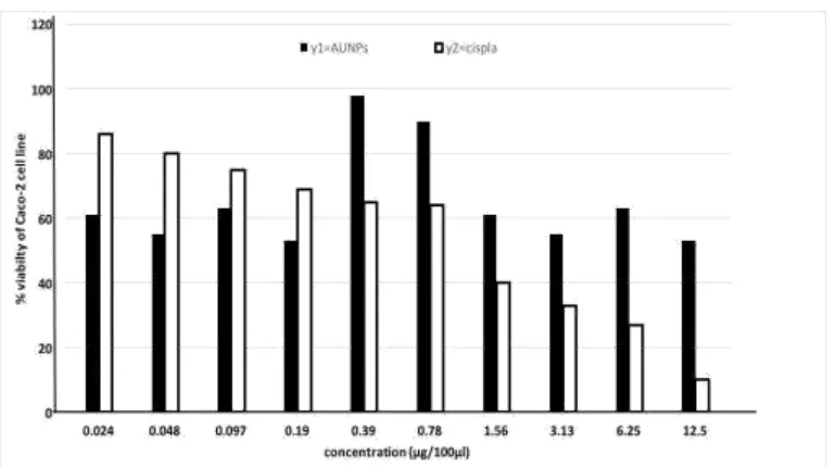
Cytotoxicity of gold nanoparticles (AuNPs) on human colon cancer cell line (Caco-2)
Figure 1a shows the cell viability variation of Caco-2 cell line exposed to 2 fold dilutions of AuNPs and Cisplatin for 72h. Caco-2 cell line treated with AuNPs showed high viability 98%
Fig. 1(a): Cytotoxicity of AUNPs and Cisplatin on Caco-2 cell line after 72hr of
treatment.
Cytotoxicity of gold nanoparticles (AuNPs) and cisplatin on normal human epithelial
cell lines (WISH) cell lines
Figure 1b shows the cell viability variation of WISH cell lines exposed to 2 folds dilutions of AuNPs and Cisplatin for 72h. WISH cell lines treated with AuNPs showed high viability at all
concentrations (0.024, 0.048, 0.079, 0.195, 0.39, 0.78, 1.56, 3.12, 6.25 and 12.5μg/100µl) by percent changes of 132%, 103%, 111%, 117%, 117%, 119%, 118%, 127%, 114% and 106.% respectively.
[image:7.595.107.492.67.282.2]Fig. 1(b): Cytotoxicity of AuNPs and Cisplatin on WISH cell lines after 72h of
treatment.
Effect of gold nanoparticles (AuNPs) on expression level of pro- and anti-apoptotic
genes
A single band with predicted size 226bp was GAPDH, bcl-2 band with size 108bp, bax band with size 133bp and p53 band with size 292bp, the molecular-weight size marker was in the form of a 100bp RNA ladder. Caco-2 and WISH cell lines treated with AuNPs or Cisplatin
were positive for Bcl-2 mRNA expression at all time intervals and at all concentrations (Fig. 2, 3 and 4). p53 mRNA was expressed in Caco2 and WISH cell lines treated with 0.048µg/100µl AuNPs (Fig. 2a and 4a). After treatment with 0.048 and 0.39µg/100µl of
AuNPs for 72, 96 and 120h Caco-2 cell lines expressed bax (Fig. 2a and 3a), while in case of
treatment with 0.048µg/100µl of Cisplatin the bax gene expressed after 48h (Fig. 2b) while at the higher concentration of Cisplatin (0.39µg/100µl) it was expressed after 24h (Fig. 3b). WISH.
[image:8.595.122.483.65.233.2]Fig [2 (a, b)] Fig [3 (a, b)]
Fig. 2 and 3: Expression of anti-apoptotic gene (bcl-2 at 108bp) and pro-apoptotic genes
(bax at 133bp and p53 at 292) in Caco-2 cell lines treated with 0.048µg/100µl and 0.39
[image:8.595.121.477.544.704.2](1, 2): Expression of bcl-2/GAPDH and bax/p53 after 24h. (3, 4): Expression of bcl-2/GAPDH and bax/p53 after 48h. (5, 6): Expression of bcl-2/GAPDH and bax/p53 after 72h. (7, 8): Expression of bcl-2/GAPDH and bax/p53 after 96h. (9, 10): Expression of bcl-2/GAPDH and bax/p53 after 120h. M: marker.
C1: Expression of bcl-2/GAPDH in untreated cells. C2: Expression of bax/p53 in untreated cells.
GAPDH represent positive control which predicted at 226bp.
Cell lines treated with 0.048µg/100µl of AuNPs bax mRNA was detected after 48, 72, 96 and
[image:9.595.153.443.335.526.2]120h of treatment (Fig. 4a) but it was only expressed after 48h of treatment with 0.048µg/100µl of Cisplatin (Fig. 4b).
Fig. 4: Expression of anti-apoptotic genes (bcl-2 at 108bp) and pro-apoptotic genes (bax
at 133bp and p53 at 292) in WISH cell lines treated with 0.048µg/100µl of AuNPs (a)
and Cisplatin (b).
(1, 2): Expression of bcl-2/GAPDH and bax/p53 after 24h. (3, 4): Expression of bcl-2/GAPDH and bax/p53 after 48h. (5, 6): Expression of bcl-2/GAPDH and bax/p53 after 72h. (7, 8): Expression of bcl-2/GAPDH and bax/p53 after 96h. (9, 10): Expression of bcl-2/GAPDH and bax/p53 after 120h. M: marker.
C2: Expression of bax/p53 in untreated cells.
GAPDH represent positive control which predicted at 226bp.
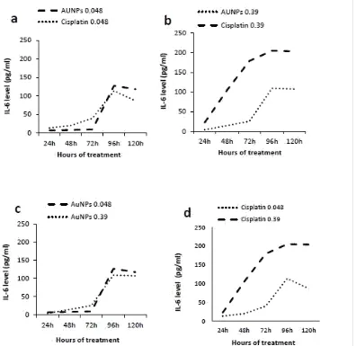
Estimation of IL-6 levels secreted by human colon cancer cell lines (Caco-2) in response
to gold nanoparticles (AuNPs) treatment
The level of IL-6 released from Caco-2 cell lines stimulated with AuNPs (0.048µg/100µl)
showed significant changes (P< 0.05) after 24h, 48h, 72h, 96h and 120h as compared to untreated cells. However, Caco-2 cell lines stimulated with Cisplatin (0.048µg/100µl) released significant level of IL-6 (P< 0.05) after 24h, 48h, 72h, 96h and 120h as compared to untreated cells. Comparing IL-6 levels released from Caco-2 cell lines stimulated with AuNPs
(0.048µg/100µl) to the corresponding concentration of Cisplatin there were significant changes (P< 0.05) after 24h, 48h, 72h and 120h while it showed non-significant change (P=0.065) at 96h (Fig. 5.a).
Similarly, Caco-2 cell lines stimulated with AuNPs (0.39µg/100µl) produced significant level
of IL-6 (P< 0.05) after 24h, 48h, 72h, 96h and 120h as compared to untreated cells. As well, Caco-2 cell lines stimulated with Cisplatin (0.39µg/100µl) produced significant level of IL-6 (P< 0.05) after 24h, 48h, 72h, 96h and 120h as compared to untreated cells.
Comparing IL-6 levels released from Caco-2 cell lines stimulated with AuNPs
(0.39µg/100µl) to the corresponding concentration of Cisplatin, significant changes (P< 0.05) were observed after 24h, 48h, 72h, 96h and 120h (Fig. 5.b).
Comparing IL-6 levels released from Caco-2 cell lines stimulated with AuNPs
(0.048µg/100µl) to the other concentration (0.39µg/100µl) showed significant changes (P< 0.05) after 24h, 48h, 72h, 96h and 120h (Fig. 5.c).
Fig. 5: Levels of IL-6 secreted by Caco-2 cell lines in response to AuNPs and Cisplatin
treatment. (a) Concentration of (0.048μg/100μl) AuNPs and Cisplatin, (b)
Concentration of (0.39μg/μl) AuNPs and Cisplatin, (c) Concentration of AuNPs (0.048
and 0.39μg/100μl) and (d) Concentration of Cisplatin (0.048 and 0.39μg/100μl).
Data represented as % to untreated cells
Estimation of IL-6 levels secreted by normal human epithelial cell lines (WISH) in
response to gold nanoparticles (AuNPs) treatment
(P<0.05) after 24h and but after 72h, 96h and 120h there was a non-significant level of IL-6 (Fig. 6). The differences among all time intervals was significant as analyzed by ANOVA. (P=1.00).
Fig. 6: Levels of IL-6 secreted by WISH cell lines in response to AuNPs and Cisplatin
treatment.
Data represented as % to untreated cells
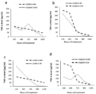
Estimation of TNF-α levels secreted by human colon cancer cell lines (Caco-2) in
response to gold nanoparticles (AuNPs) treatment
The level of TNF-α released from Caco-2 cell lines stimulated with AuNPs (0.048µg/100µl)
showed significant changes (P<0.05) after 24h, 48h, 72h, 96h and 120h as compared to untreated cells. However, Caco-2 cell lines stimulated with cisplatin (0.048µg/100µl) released significant level of TNF-α after 24h, 48h, 72h, 96h and 120h compared to untreated cells. Comparing TNF-α levels released from Caco-2 cell lines stimulated with AuNPs
(0.048µg/100µl) to the corresponding concentration of Cisplatin showed significant changes (P<0.05) after 24h, 48h, 72h, 96h and 120h (Fig. 7a). Similarly, Caco-2 cell lines stimulated with AuNPs (0.39µg/100µl) produced significant level of TNF-α (P<0.05) after 24h, 48h, 72h
and 96h as compared to untreated cells but after 120h there was a non-significant level of TNF-α as compared to untreated cells.
[image:12.595.144.453.147.358.2]Comparing TNF-α levels released from Caco-2 cell lines stimulated with AuNPs
(0.39µg/100µl) to the corresponding concentration of Cisplatin showed significant changes (P<0.05) after 24h, 48h, 72h, 96h and 120h (Fig. 7b).
Comparing TNF-α levels released from Caco-2 cell lines stimulated with AuNPs
[image:13.595.151.485.302.629.2](0.048µg/100µl) to the other concentration (0.39µg/100µl) showed significant changes (P<0.05) after 24h, 48h, 72h, 96h and 120h (Fig. 7c). Comparing TNF-α levels released from Caco-2 cell lines stimulated with cisplatin (0.048µg/100µl) to the other concentration (0.39µg/100µl) showed significant changes (P<0.05) after 24h, 48h, 72h and 96h while it showed non-significant change (P=0.288) at 120h (Fig. 7d). The differences among all time intervals was significant as analyzed by ANOVA.
Fig. 7: levels of TNF-α secreted by Caco-2 cell lines in response to AuNPs and Cisplatin
treatment.(a) Concentration of (0.048μg/100μl) AuNPs and Cisplatin, (b) Concentration
of (0.39μg/100μl) AuNPs and Cisplatin, (c) Concentration of AuNPs (0.048 and
0.39μg/100μl) and(d) Concentration of Cisplatin (0.048 and 0.39μg/100μl).
Estimation of TNF-α levels secreted by normal human epithelial cell lines (WISH) in
response to gold nanoparticles (AuNPs) treatment
In response to the treatment by 0.048µg/100µl AuNPs, the WISH cell lines produced
significant level of TNF-α (P<0.05) after 24h, 48h, 72h, 96h and 120h as compared to untreated cells. Similarly, WISH cell lines stimulated with 0.048µg/100µl Cisplatin produced significant level of TNF-α (P<0.05) after 24h, 48h, 72h, 96h and 120h as compared to untreated cells.
Comparing TNF-α levels released from WISH cell lines stimulated with 0.048µg/100µl AuNPs to the corresponding concentration of 0.048µg/100µl Cisplatin showed significant
[image:14.595.120.479.336.538.2]changes (P<0.05) after 24h, 48h, 72h, 96h and 120h (Fig. 8). The differences among all time intervals was significant as analyzed by ANOVA.
Fig. 8: levels of TNF-α secreted by WISH cell lines in response to AuNPs and Cisplatin
treatment.
Data represented as % to untreated cells.
Comparing the levels of TNF-α and IL-6 secreted by human colon cancer cell lines
(Caco-2) and normal human epithelial cell lines (WISH) in response to gold
nanoparticles (AuNPs) treatment
diminish by time until 120h recorded its lowest level. In the contrast, IL-6 had its lowest level after 24h then gradually increased to reach its peak after 120h (Fig. 9a).
In case of WISH cell lines, the levels of TNF-α also reached its peak after 24h then decreased, while IL-6 showed no detectable levels through treatment (Fig. 9b).
Fig. 9(a): Comparison between TNF-α and IL-6 levels secreted by Caco-2 cell lines
inresponse to AuNPs treatment.
Data represented as % to untreated cells.
Fig. 9(b): Comparison between TNF-α and IL-6 levels secreted by WISH cell lines in
response to AuNPs treatment.
Data represented as % to untreated cell.
DISCUSSION
Cytotoxic effects of AuNPs at the cellular level appeared as inhibition of Caco-2 cell growth
[image:15.595.97.514.192.332.2] [image:15.595.92.509.446.595.2]exposure (200 μM and 300 μM), with the most evident inhibitory effect detected at concentration of 300μM at 72 h[22]
which complies with this study results.
According to cytotoxicity assay, at the moderate concentrations (0.39 and 0.78µg/100µl) AuNPs showed less toxic effects on Caco-2 cell line, while, it became more toxic at lower
and higher concentrations. And cisplatin (as a positive control) showed less toxic effects on Caco-2 cell line at lower concentrations while it became more toxic at higher concentrations. Meanwhile, in case of normal epithelial cell line (WISH) the AuNPs was non-toxic at all
concentrations used in the current study while cisplatin was toxic at high concentrations only. There was no significant dead cells were observed in cells treated with AuNPs. This may
indicate that AuNPs had no effect or very slight cell cytotoxic effect compared to cisplatin on
normal cells.
Selim and Hendi[23] reported that gold nanoparticles produce significant cytotoxicity to breast cancer cells (MCF-7) in a dose-dependent manner with the concentration range of 25- 200 μg/mL. Furthermore, quantitative real-time PCR analysis displayed that mRNA levels involved in the apoptosis was altered by gold nanoparticles. These data reported that gold nanoparticles may induce apoptosis in MCF-7 cells via p53, bax/bcl-2 and casapase pathways. This in vitro study showed the induction of apoptosis by gold nanoparticles recommended further investigation to determine if in vivo exposure consequences may exist for gold nanoparticles application.
In agreement with the result, Lan, et al.[24] found that the high dose of AuNPs was cytotoxic
to NPC cells as it reduces cell viability in a concentration dependent manner. More than half of nasopharyngeal cancer cells have been killed after treatment with 100mM AuNPs for three
days.
Arnida, et al.[25] observed that gold nanoparticles were taken up by human prostate cancer cells but do not cause severe toxicity. The uptake of gold nanoparticles was size dependent.
Particles with core diameters of 30 and 50nm were taken up by the cells to a higher extent compared with larger particles.
or cisplatin, while p53 mRNA was only expressed in Caco-2 and WISH cell line treated with 0.048µg/100µl AuNPs.
Complement to recent results, Selim and Hendi,[23] analyzed the mRNA expression levels of five apoptosis associated genes; p53, bax, bcl-2, caspase-3 and caspase-9 in response to gold nanoparticles exposure in MCF-7 cells. Quantitative real-time PCR results showed that gold nanoparticles up-regulated mRNA level of cell cycle checkpoint protein p53 and pro-apoptotic bax while expression of anti-pro-apoptotic bcl-2 was down-regulated in cells exposed to gold nanoparticles.
p53 is a transcriptional modulator of bax gene which promotes apoptosis[26] and a p53 negative response element has been identified in the Bcl-2 gene.[27] Based on these findings, Iimura, et al.[28] postulated that bax induced apoptosis could be operative in inflamed colonic mucosa in ulcerative colitis (UC).
Molecular biomarkers related to AuNPs exposures were identified by changes in RNA
expression levels. Broad range of responses which potentially mediate inhibition of cellular growth and other functions of Caco-2 cells in response to nano gold, were identified. These cellular processes affected by exposure to 5 nm AuNPs include, for the early time point
examined (24 h), down regulation of genes involved in regulation of transcription (transcriptional factors), biogenesis of RNA and GTP binding among others. At the later time point studied (72 h), more dramatic changes in differentially expressed gene expression patterns evoked by 5 nm AuNPs were detected.[22]
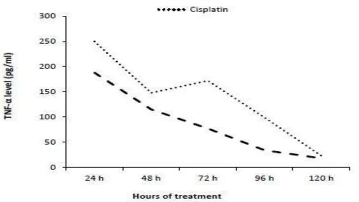
Tumor necrosis factor alpha (TNF-α) exhibits both tumor-promoting and tumor-inhibitory properties, depending on the experimental context within which the conclusions are made.
The results showed that TNF-α reached its peak after 24h and started to decrease gradually reaching its lowest level after 120h in response to Caco-2 cell line treated with AuNPs, so as
for the WISH cell line treated with AuNPs levels of TNF-α recorded its peak after 24h and
lowest level after 120h.
collapse.[29] It is now accepted that chronic inflammation is a major risk factor for carcinogenesis, and emerging evidence shows that TNF-α has key roles in this process.[30]
IL-6 is a multifunctional cytokine that was originally characterized as a regulator of immune and inflammatory responses; however, elevated expression of IL-6 has been detected in multiple epithelial tumors[31] Given the reported involvement of IL-6 and its downstream targets in the regulation of cell proliferation, survival, and metabolism, it is not surprising that IL-6 signaling has also been implicated in tumorigenesis.[32] However, the nature of IL-6’s involvement in cancer has been quite controversial, as dichotomous roles for IL-6 in both tumor-promoting and -suppressive activities have been reported. For example, IL-6 signaling has been linked to both pro- and anti-apoptotic activity in breast cancer cells.[33,34]
In this study, the levels of IL-6 secreted by Caco-2 cell line was increased gradually up to its highest level after 120h of treatment with AuNPs, while in WISH cell line treated with
AuNPs, there was no detected levels of IL-6 after all times of treatment.
Multiple studies have documented high IL-6 levels in the serum of patients with certain carcinomas (i.e., breast, lung and lymphoma) and have correlated high IL-6 levels with a poor clinical prognosis. These data imply an oncogenic role for IL-6; however, lacking is an understanding of the mechanisms governing IL-6 production in tumors and the biological role of this cytokine in tumorigenesis.
The results of another in vitro study on murine macrophages has suggested that 60nm AuNPs
are not cytotoxic or do not elicit pro-inflammatory IL-6 or TNF-α responses[35] that back up our results. On the other hand, Yen, et al.[36] found that both silver and gold nanoparticles enter the cells but that only the AuNPs (especially those with smaller diameter) upregulate the
expression of pro-inflammatory genes (IL-1, IL-6, TNF-α). The authors speculated that part of the negatively charged AuNPs may adsorb serum protein and enter cells via the more
complicated endocytotic pathway, resulting in higher cytotoxicity and immunological response of AuNPs than silver nanoparticles.
and increases blood levels of IL-1b and TNF-α; mRNA expressions of inflammation-related genes were also elevated in the cultured peritoneal macrophages harvested from the treated mice. A single intra-tracheal instillation of platinum and silver nanoparticles causes progressive increase in pro-inflammatory cytokines (IL-1, IL-6, TNF-α) by day 28 post-instillation.[39,40] Intra-gastric administration of titanium dioxide nanoparticles has been shown to increase significantly the mRNA expression of several inflammatory cytokines in mouse hepatocytes.[41] The above literature clearly indicates that AuNPs are comparatively
safer and more biocompatible than other nanomaterials.
In conclusion, although it was confirmed that AuNPs do not cause acute cytotoxicity for
normal epithelial cell lines (WISH), it had shown that in colon cancer cell lines (Caco-2) the AuNPs have been able to induce apoptosis. These results predict a new era of targeting
therapies and avoid the negative side effects of other therapies in the market that affects both normal and tumor cells.
REFERENCES
1. Risio, M. The natural history of adenomas. Best Pract Res Clin Gastroenterol, 2010; 24: 271-280.
2. Levin, B.; Lieberman, D.A.; McFarland, B.; Andrews, K.S.; Brooks, D.; Bond, J.; Dash, C.; Giardiello, F.M.; Glick, S.; Johnson, D.; Johnson, C.D.; Levin, T.R.; Pickhardt, P.J.; Rex, D.K.; Smith, R.A.; Thorson, A. and Winawer, S.J. Screening and surveillance for the early detection of colorectal cancer and adenomatous polyps. Gastroenterology,
2008;134(5): 1570-1595.
3. Mishra, J.; Dromund, J.; Quazi, S.H.; Karanki, S.S.; Shaw, J.J.; Chen, B. and Kumar, N. Prospective of colon cancer treatments and scope for combinatorial approach to enhanced cancer cell apoptosis. Crit Rev Oncol Hematol, 2013; 86(3): 232–250.
4. Pickhardt, P.J.; Kim, D.H.; Pooler, B.D.; Hinshaw, J.L.; Barlow, D,; Jensen, D.; Reichelderfer, M. and Cash, B.D. Assessment of volumetric growth rates of small colorectal polyps with CT colonography: a longitudinal study of natural history. Lancet Oncol, 2013;14: 711-720.
6. Kim, Y.H.; Kwak, K.A.; Kim, T.S.; Seok, J.H.; Roh, H.S.; Lee, J.K.; Jeong, J.; Meang, E.H.; Hong, J.S.; Lee, Y.S. and Kang, J.S. Retinopathy induced by zinc oxide nanoparticles in rats assessed by micro-computed tomography and histopathology.
Toxicological research,2015;31(2): 157–63.
7. Heim, J.; Felder, E.; Tahir, M.N.; Kaltbeitzel, A.; Heinrich, U.R.; Brochhausen, C.; Mailänder, V.; Tremel, W. and Brieger, J. Genotoxic effects of zinc oxide nanoparticles.
Nanoscale,2015;7(19): 8931–8.
8. Guntaka, R.V.; Varma, B.R. and Weber, K.T. Triplex-forming oligonucleotides as modulators of gene expression. Int J Biochem Cell Biol, 2003; 35: 22-31.
9. Akerman, M.E;, Chan, W.C.; Laakkonen, P.; Bhatia, S.N.; Ruoslahti, E.; Chan; Laakkonen; Bhatia and Ruoslahti Nanocrystal targeting in vivo. proceedings of the national academy of sciences of the united states of America,2002;99(20): 12617–21. 10.El-Sayed, I.; Huang, X. and El-Sayed, M.A. Surface plasmon resonance scattering and
absorption of anti-EGFR antibody conjugated gold nanoparticles in cancer diagnostics; applications in oral cancer. Nano Lett, 2005; 4: 829-34.
11.El-Sayed, I.; Huang, X. and El-Sayed, M.A. Selective laser photo-thermal therapy of epithelial carcinoma using anti-EGFR antibody conjugated gold nanoparticles. Cancer Lett, 2006; 239: 129.
12.Hwu, J.R.; Lin, C.C.; Chuang, S.H.; King, K.Y.; Su, T.R. and Tsay, S.C. Aminyl and iminyl radicals from arylhydrazones in the photo-induced DNA cleavage. Bioorg Med Chem, 2004; 12: 2509-15.
13.Ceruti, J.M.; Scassa, M.E.; Flo, J.M.; Varone, C.L. and Cánepa, E.T. Induction of p19INK4d in response to ultraviolet light improves DNA repair and confers resistance to apoptosis in neuroblastoma cells. Oncogene, 2005; 24: 4065-80.
14.Connor, E.E.; Mwamuka, J.; Gole, A.; Murphy, C.J. and Wyatt, M.D. Gold nanoparticles are taken up by human cells but do not cause acute cytotoxicity. Small, 2005; 1: 325-7. 15.Coulter, J.A.; Jain, S.; Butterworth, K.T.; Taggart, L.E.; Dickson, G.R.; McMahon, S.J.;
Hyland, W.B.; Muir, M.F.; Trainor, C.; Hounsell, A.R.; O'Sullivan, J.M.; Schettino, G.; Currell, F.J.; Hirst, D.G. and Prise, K.M. Cell type-dependent uptake, localization, and cytotoxicity of 1.9 nm gold nanoparticles. Int J Nanomedicine, 2012; 7: 2673–2685. 16.Waldner, M.J.; Foersch, S. and Neurath, M.F. Interleukin-6 –a key regulator of colorectal
cancer development. Int. J. Biol. Sci, 2012; 8(9): 1248-1253.
18.Chung, H.W.; Seo, J.S.; Hue, S.E.; Kim, H.L.; Kim, J.Y.; Jung, J.H.; Kim, L.H.; Park, B.L. and Shin, H.D. Association of interlukin-6 romotor variant with bone mineral density in pre-menopausal women. J. Hum. Genet, 2003; 48: 243-248.
19.Ferrari, S.L.; Ahn-Luong, L.; Garnero, P.; Humphries, S.E. and Greenspan, S.L. Two promoter polymorphisms regulating interlukin-6 gene expression are associated with circulating levels of C-reactive protein and markers of bone resorption in postmenopausal women. J. Clin. Endocr. Metab, 2003; 88: 255-259.
20.Tse, B.W.; Scott, K. F. and Russell, P.J: Paradoxical roles of tumor necrosis factor-alpha in prostate cancer biology. Prostate Cancer, 2012; 10.1155/2012/1:8.
21. Skehan, P. Storeng, P. Scudiero, D. Colorimetric cytotoxicity assay for anticancer-drug screening. J. Natl Cancer Inst., 1990; 82: 1107-1112.
22.Bajak, E.; Fabbri, M.; Ponti, A.; Gizoria, S.; Ojea-Jiménez, I.; Collotta, A; Mariani, V.; Gilliland, D.; Rossi, F. and Gribaldo, L. Changes in Caco-2 cells transcriptome profiles upon exposure to gold nanoparticles. Toxicology Letters, 2015; 233: 187–199.
23.Selim, M.E. and Hendi, A.A. Gold nanoparticles induce apoptosis in mcf-7 human breast cancer cells. Asian Pacific J Cancer Prev., 2012; 13: 1617-1620.
24.Lan, M. Y.; Hsu, Y. B.; Hsu, C.H.; Ho, C. Y.; Lin, J. C. and Lee, S. W. Induction of apoptosis by high-dose gold nanoparticles in nasopharyngeal carcinoma cells. Auris Nasus Larynx, 2013; 40: 563–568.
25.Arnida; Malugin, A. and Ghandehari, H. Cellular uptake and toxicity of gold nanoparticles in prostate cancer cells: a comparative study of rods and spheres. J. Appl. Toxicol,2010;30: 212–217.
26.Miyashita, T.; Krajewski, S.; Krajewska, M.; Wang, H.G., Lin, H.K.; Liebermann, D.A.; Hoffman, B. and Reed, J.C. Tumor suppressor p53 is a regulator of bcl-2 and bax gene expression in vitro and in vivo. Oncogene, 1994; 9: 1799–805.
27.Miyashita, T.; Harigai, M.; Hanada, M. and Reed, J.C. Identification of a p53- dependent negative response element in the bcl-2 gene. Cancer Res, 1994; 54: 3131–5.
28.Iimura, M.; Nakamura, T.; Shinozaki, S.; Iizuka, B.; Inoue, Y.; Suzuki, S. and Hayashi, N. Bax is downregulated in inflamed colonic mucosa of ulcerative colitis. Gut, 2000; 47: 228–235.
29.Szlosarek, P.; Charles, K. and Balkwill, F. Tumor necrosis factor-α as a tumor promoter.
European Journal of Cancer, 2006; 42(6): 745–750.
31.Kishimoto, T. Interleukin-6: from basic science to medicine-40 years in immunology.
Annu. Rev. Immunol, 2005;23: 1–21.
32.Hodge, D.R.; Hurt, E.M. and Farrar, W.L. The role of IL-6 and STAT3 in inflammation and cancer. Eur. J. Cancer, 2005; 41: 2502–2512.
33.Chiu, J.J.; Sgagias, M.K. and Cowan, K.H. Interleukin 6 acts as a paracrine growth factor in human mammary carcinoma cell lines. Clin. Cancer Res, 1996; 2: 215–221.
34.Conze, D.; Weiss, L.; Regen, P.S.; Bhushan, A.; Weaver, D.; Johnson, P. and Rincón, M. Autocrine production of interleukin 6 causes multidrug resistance in breast cancer cells.
Cancer Res, 2001; 61: 8851–8858.
35.Zhang, Q.; Hitchins, V.M.; Schrand, A.M.; Hussain, S.M. and Goering, P.L. Uptake of gold nanoparticles in murine macrophage cells without cytotoxicity or production of pro-inflammatory mediators. Nanotoxicology, 2011; 5: 284-295.
36.Yen, H.J.; Hsu, S.H. and Tsai, C.L. Cytotoxicity and immunological response of gold and silver nanoparticles of different sizes. Small, 2009; 5: 1553-1561.
37.Carlson, C.; Hussain, S.M.; Schrand, A.M.; Braydich-Stolle, L.K.; Hess, K. L.; Jones, R. L. and Schlager, J. J. Unique cellular interaction of silver nanoparticles: size-dependent generation of reactive oxygen species. J. Phys. Chem. B, 2008; 112: 13608-13619.
38.Park, E.J. and Park, K. Oxidative stress and pro-inflammatory responses induced by silica nanoparticles in vivo and in vitro. Toxicol. Lett, 2009; 184: 18-25.
39.Park, E.J.; Kim, H.; Kim, Y. and Park, K. Intratracheal instillation of platinum nanoparticles may induce inflammatory responses in mice. Arch. Pharm. Res, 2010; 33: 727-735.
40.Park, E.J.; Choi, K. and Park, K. Induction of inflammatory responses and gene expression by intratracheal instillation of silver nanoparticles in mice. Arch. Pharm. Res,
2011; 34: 299-307.

![Fig [2 (a, b)] Fig [3 (a, b)]](https://thumb-us.123doks.com/thumbv2/123dok_us/836199.593475/8.595.121.477.544.704/fig-a-b-fig-a-b.webp)