Adhesion molecules such as selectins and in-tegrins expressed on the surface of leukocytes rep-resent essential components of immunity because they are involved in the interaction of the immu-nocompetent cells and in the communication of these cells with the intercellular matrix. Selectins are membrane-bound lectins that initiate the adhe-sion of leukocytes to endothelial cells, platelets, or other leukocytes on the vessel wall. An initial slow-ing of leukocytes mediated by selectins is followed by firm adhesion of leukocytes to the vascular en-dothelium and transmigration of leukocytes into
the site of inflammation. This process is mediated
by other adhesion molecules including β2-integrins
(Ebnet et al., 1996; Czarnik et al., 2007).
The mammary gland is protected by various de-fence mechanisms. There is some production of immunoglobulins secreted by plasma cells that are present in the tissue of the mammary gland (Lee et al., 1992). The migration of leukocytes from the blood into the mammary gland is an essential ele-ment of resistance to infections. Circulating leu-kocyte populations are a primary source of cells that transmigrate into the mammary gland of
Immunohistochemical localization of adhesion
molecules (cd62 and cd18) in the mammary
gland of dairy cows
M. Simon
1, S. Hluchý
2, Ľ. Horovská
1, J. Antalíková
1, J. Čuboň
21Institute of Animal Biochemistry and Genetics, Slovak Academy of Sciences, Ivanka pri Dunaji,
Slovak Republic
2Slovak University of Agriculture, Nitra, Slovak Republic
ABSTRAcT: Localization of the L-selectin (CD62L) and β2-integrin (CD18) bearing cells in different tissues of the bovine mammary gland was examined. Five dairy cows of Holstein-Friesian breed in the middle of their secondand third lactation cyclewere used in the study.Blood, milk and udder tissue samples were collected from each cow to estimate the milk somatic cell count (SCC) and bacteriological infection of the mammary gland. The expression of CD62L and CD18 on blood cells, milk cells and parenchymal tissues of udder, Fürstenberg’s rosette and the transverse section of the central part of the teat was tested. In the mammary gland quarters the value of SCC in milk secretion was also reflected in the presence of CD18+ and CD62L+ leukocytes in mammary tissues. In the quarters where SCC was higher than 105 a frequent incidenceof L-selectin and β2-integrincells was observed in the parenchyma and Fürstenberg’s rosette region, while in the quarters with low SCC, none or only a few reac-tive cells were found. In the mammary parenchymal tissue CD18 posireac-tive cells were present in both the epithelial and the connective tissue. In Fürstenberg’s rosette the cells were concentrated in the connective collagenous and loose tissue. None or only scattered L-selectin (CD62L) and β2-integrin (CD18) bearing cells were identified in the transverse section from the central part of the teat. When we compared the L-selectin and β2-integrin expression, the study revealed the down-regulation of L-selectin on the cells of mammary tissue.
Keywords: cattle; L-selectin; β2-integrin; mastitis
healthy and infected animals. The manner of the neutrophil traverse of the secretory epithelia is not quite clear yet. However, adhesion molecules are likely to be one group of molecules that regulate the entry of leukocytes into the mammary gland (Van Kampen et al., 1999). Variations in the ex-pression of integrins and selectins may indicate the important role of these molecules in regulating the movement of leukocytes into the mammary gland. The adhesion molecules on blood leukocytes are also present on somatic cells of milk. During the transendothelial migration of lymphocytes and neutrophils into the mammary gland the expres-sion of L-selectin is down-regulated in contrast to CD18 which is up-regulated (Persson-Waller and Colditz, 1998). Milk is not the only element of the udder defence as leukocytes are also present in mammary gland tissues. However, the distribution of the L-selectin (CD62L) or β2-integrin (CD18) bearing cells in bovine mammary gland tissues has not been studied yet. Therefore, the purpose of this study was to investigate the distribution of CD18 and CD62L positive cells in three different parts of the mammary gland of dairy cows (parenchyma, Fürstenberg’s rosette and teat).
MATERIAL ANd METHOdS
Animals, blood, milk and tissue samples
Five crossbred Slovak Pied × Holstein-Frisian cows aged 5–6 years and in the middle lactation stage were selected for this study. Blood and milk samples were collected from each animal. Post-mortem, the mammary gland parenchymal tis-sue was dissected from the central region of the upper body of the mammary gland and from the region surrounding the mammary gland cistern. Further tissue samples were taken from the region of Fürstenberg’s rosette and from the transverse section in the central part of teat immediately after slaughter. The NK-mastitis test was used to select the cows with infected and uninfected udder quar-ters. The samples were cut into cubes smaller than
1 cm3.
The tissues were quickly frozen in liquid nitrogen and stored at –70°C until a histochemical analysis. The blood from all animals and the milk from all selected quarters were examined. To determine “the incidence of bovine mastitis”, the milk sam-ples were cultured for bacteriology (cultivation on
GTK agar at the temperature of 30 ± 1°C during 72 ± 3 hours) and the estimation of SCC was per-formed with Fossomatic 90 (Foss Electric, Hillerod, Denmark).
Isolation of leukocytes from milk and blood
200 ml of milk was mixed with an equal volume of phosphate-buffered saline (PBS) (pH 7.2) contain-ing 20% of acid citrate dextrose and 20mM EDTA (Park et al., 1992). The mixture was filtered through a nylon net of 50 µm porosity and centrifuged at 400 × g for 30 min at 15°C. The supernatant was dis-carded and cell pellets were washed several times and used in an indirect immunofluorescence test to examine the CD18 and CD62L positive cells. Blood leukocytes were isolated after haemolysis of
erythrocytes with a lysing solution (0.15M NH4Cl).
Smears prepared from blood and mammary cells for a differential analysis were stained by Papenheim’s panoptic method. At least 100 cells were identified on the basis of morphological and standard staining characteristics.
Monoclonal antibodies
Hybridoma cell lines producing monoclonal antibodies IVA-35 (CD18) and IVA-94 (CD62L) were obtained after the immunisation of BALB/c mice with bovine peripheral blood leukocytes us-ing standard procedures for the fusion of SP2/0 with splenocytes of immunised mice, selection and cloning of hybridomas (Dušinský et al., 1988). The specificity of both antibodies was verified in
tests performed at the 3rd Workshop on Ruminant
Leukocyte Antigens (Naessens and Hopkins, 1996). The reactivity of IVA-35 and IVA-94 was analysed on various leukocyte populations and cell lines with a flow cytometer. Generally, both antibodies rec-ognised the target antigen on 95–100% of bovine blood granulocytes, monocytes and lymphocytes. However, the reaction of IVA-94 on lymphocytes was more restricted (70% positive cells).
Immunohistochemical staining and morphometric analysis of tissue sections
sample with Cryocut 1 800 (LEICA). The sections were fixed for 5 min in a cold ethanol-acetone
mixture (1:1), air-dried, treated with 0.6% H2O2
in PBS and then stained using an indirect immu-noperoxidase test. The sections were incubated with the first antibody (IVA-35 and IVA-94) for 45 min at 20°C. The slides were washed with PBS and then incubated for 45 min with peroxidase-conjugated porcine anti-mouse Ig (SEVAC Praha) diluted 1:50 in PBS containing 5% of normal bovine serum. After repeated washing in PBS, the sections were incubated in 0.06% (w/v) diaminobenzidine tetrachloride (Sigma Chemicals, St. Louis, MO) in
PBS containing 0.05% (v/v) H2O2 for 10 min at room
temperature. After washing, the slides were slightly contra-stained with Harris’s haematoxylin.
The preparations were evaluated subjectively and quantitatively by a morphometric method. The data were acquired by means of a digital camera using the NIKON and Image Analysis System. Tissue and cell count was carried out so as to evaluate 15 fields per section; one field depicts a rectangle of
353 600 µm2 at 350× magnification using a
moni-tor micrometer of 221 squares (Nikon) according to the point counting technique by the System for Image Processing and Analysis – Lucia 4.60. The relative volume of glandular parenchyma, connec-tive tissue, loose tissue and also the relaconnec-tive volume of epithelium and alveolar lumen and ducts were determined. The mean and standard deviation (SD) were calculated and the differences between the various tissues of udders were tested by Student’s
t-test.
Indirect immunofluorescence assay
An indirect immunofluorescence assay described by Boucheix et al. (1983) was used to test the bin-ding of IVA-35 and IVA-94 to blood or milk
leuko-cytes. Briefly, the separated cells were washed three times with PBS with 1% bovine serum albumin and 0.1% sodium azide. The cell suspensions were incubated with monoclonal antibodies in round-bottomed 96-well polystyrene plates for 45 mi- nutes at room temperature. Then the cells were washed 3 times and treated with diluted (1:20–30) fluorescein isothiocyanate (FITC)-conjugated por-cine anti-mouse immunoglobulin (SEVAC Praha), for 45 minutes at room temperature. After further washing the cell suspensions were placed on slides and examined under an epifluorescence micros-cope (Jenalumar).
RESULT ANd dIScUSSION
The analysis of milk secretions revealed two infection profiles of mammary glands (Table 1). Milk samples of cows number 2 and 4 showed the highest counts of microbes and further features of inflammation. These two cows had the highest values of SCC and an elevated number of PMN (polymorphonuclear) leukocytes in their milk. Cows number 1, 3 and 5 were quite free of infecti-on and mastitis. SCC per ml of milk were markedly
lower than 105 cells and the predominant type of
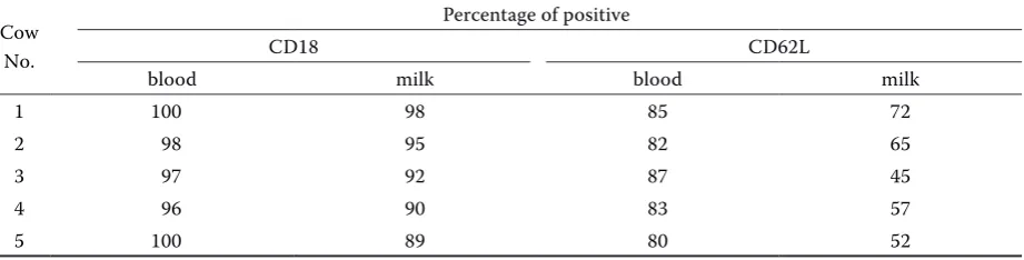
[image:3.595.64.531.644.755.2]cells were macrophages and lymphocytes. The labelling of blood and milk cells with mono-clonal antibodies IVA-35 and IVA-94 was tested on the whole leukocyte population. The informative values measured in blood and milk just before kil-ling the animals are given in Table 2. Considerable proportions (percentage) of immunofluorescence labelled leukocytes were found in the blood and milk secretion of all animals independently of the infection stage of the udder. Both the blood and milk leukocytes contained a lower percentage of L-selectin positive cells compared to β2-integrin bearing cells.
Table 1 Bacterial infection, NK mastitis test, SCC and leukogram of the milk of experimental cows
Cow
No. Mammary gland quarter bacterial CPMIncidence of NK values SCC × 103 PMN (%) Lymphocytes (%) Macrophages (%)
1 right fore 1 800 – 92 35 17 48
2 right fore 5 100 ++ 1 320 85 4 11
3 right fore 2 300 – 11 34 21 45
4 left fore 8 500 + 854 81 7 12
Table 2Expression of CD18 and CD62L on bovine blood and milk leukocytes tested by an indirect immunofluo-rescence test
Cow No.
Percentage of positive
CD18 CD62L
blood milk blood milk
1 100 98 85 72
2 98 95 82 65
3 97 92 87 45
4 96 90 83 57
5 100 89 80 52
There was a coincidence between SCC in milk and the presence of CD18 and CD62L positive leukocytes in the udder tissues. Integrin and sele-ctin bearing cells were rarely present (only a few scattered cells) in the mammary gland tissues of cows number 1, 3 and 5, showing low SCC in their milk. On the other hand, these cells were frequently observed in the udder tissues of cows number 2 and 4, whose milk contained elevated SCC. Similarly like Hodgkinson et al. (2007) we were not able to detect L-selectin positive cells in the udder tissue of healthy cows in the different physiological stages of mammary gland. In the mammary tissue, integrin- and selectin-bearing cells were concentrated in the region of parenchy-ma and Fürstenberg’s rosette. None or only few scattered cells were found in the sections from the central part of the teat (Table 3). In the mammary parenchymal tissue CD18 positive cells were pre-sent in both the epithelial and connective tissue region. In the epithelial region CD18 leukocytes were most frequently located in close proximi-ty to the mammary epithelium among epithelial cells (Figure 1a) and in the lumen of alveoli. Fewer leukocytes were reactive with CD62L monoclonal antibody in the same part of the mammary gland. Cells with lower intensity of staining were found mainly in the interalveolar connective loose tissue (Figure 1b). A high concentration of CD18 leu-kocytes was found in the region of Fürstenberg’s
rosette. In this region, the presence of β2 integrin
positive cells was restricted to the interalveolar and interlobular connective tissue (Figure 1c). Similarly to the parenchyma, a lower number of
L-selectin than β2-integrin positive cells with lower
intensity of staining was observed in the connecti-ve tissue of Fürstenberg’s rosette (Figure 1d).
Scattered β2-integrin positive cells (Figure 1e)
and L-selectin positive cells (Figure 1f ) were also
detected on the section from the central part of teat.
The relative frequency of the selectin and inte-grin positive cells was analysed morphometri-cally in three parts of the udder in the tissue of two cows (number 2 and 4) which showed an elevated value of somatic cells in their milk and mammary gland tissue (Table 4). A lower number of selectin positive cells was found in all three regions of the udder under study, although the difference between selectin and integrin positive cells in the parenchyma was not significant. A significantly lower percentage of both adhesi-on molecule positive cells was found in the teat compared with parenchyma and Fürstenberg’s
rosette (P < 0.001).
In this study the mammary tissue localisation
of the L-selectin and β2-integrin bearing
leuko-cytes was analysed in the cows with apparent signs of mastitis (elevated SCC value, bacterial infection) and in the cows free of inflammation. Remarkable differences were found in the
expres-sion of L-selectin and β2-integrin on tissue
leu-kocytes in the three mammary gland regions of the cows with mastitis. L-selectin was expressed at lower intensity on much fewer cells than was
β2-integrin. The down-regulation of L-selectin
could be explained as a result of the role of the CD62L antigen in the rolling of leukocytes along endothelial cells in the microvasculature. After L-selectin mediates the slow rolling of leukocytes on the vascular endothelium, proteases induce the shedding of L-selectin from the leukocyte surface (Radi et al., 2001).
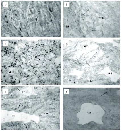
Figure 1.
(a) Immunoperoxidase reaction of monoclonal antibody IVA-35 (CD18) with leukocytes between epithelial cells in mammary parenchyma alveoli (arrows – reactions of IVA-35, A – alveolus, CT – collagenous connective tissue) (b) Lower intensity of staining of CD62L bearing cells in the interalveolar connective loose tissue of mammary gland parenchyma with monoclonal antibody IVA-94 (arrows – reactions of IVA-94, LT – loose connective tissue, CT – collagenous connective tissue)
(c) Intensive immunoperoxidase reaction of IVA-35 in the connective tissue of Fürstenberg’s rosette (arrows – reac-tions of IVA-35, A – alveolus, LT – loose connective tissue)
(d) Scattered L-selectin positive cells in the connective tissue of Fürstenberg’s rosette (arrows – reactions of IVA-94, A – alveolus, ILD – interlobular duct, LT – loose connective tissue, CT – collagenous connective tissue) (e) Scattered integrin positive cells in the central part of the teat stained with monoclonal antibody IVA35 (arrows – reactions of IVA-35, TC – part of teat cisternae, ILD – longitudinal section of interlobular duct, CT – collagenous connective tissue)
CD62L was found only in cow number 4. Nicker- son and Pankey (1984) observed a massive neutro-phil migration through the teat end tissues of the quarters experimentally infected with Staphylococcus aureus. Similarly, Persson et al. (1992) found an invasion of neutrophils to the teat epithelium af-ter endotoxin infusion into the teat cisaf-tern. A weak
invasion of β2-integrin and L-selectin positive cells
into the central part of the teat of the studied ani-mals might be due to the fact that their quarters were “naturally infected” while in the previous stu-dies a massive application of infectious agents was performed. Another reason for the lower number of the CD18 and CD62L bearing leukocytes in the teat tissue might be the strong “anatomical immune defence” present in the teat and a weaker level of cel-lular immunity. The teat canal is lined with keratin, which is crucial for the maintenance of the barrier function of the teat end. The removal of keratin was correlated with increased susceptibility to bacterial invasion and colonization (Capuco et al., 1992).
Acknowledgements
We are grateful to the Department of Microbio-logy of the Slovak University of Agriculture in Nitra for their microbiological analysis of milk samples and to the National Reference Laboratory for Raw Cow Milk in Nitra for their estimation of somatic cell counts in the milk samples. We
also thank to Zuzana Nádaždyová for very good technical assistance.
REFERENcES
Boucheix C., Perrot J.Y., Mirshahi M., Bernadou A., Rosenfeld C. (1983): A rapid method for detection of membrane antigens by immunofluorescence and its application to screening hybridoma antibodies. J. Im-munol. Methods, 57, 145–150.
Capuco A.V., Bright S.A., Pankey J.W., Wood D.L., Miller R.H., Bitman J. (1992): Increased susceptibility to the intramammary infection following removal of teat ca-nal keratin. J. Dairy Sci., 75, 2126–2131.
Czarnik U., Galiński M., Pareek Ch.S., Zabolewicz T., Wielgosz-Groth Z. (2007): Study of an association be-tween SNP 775C>T within the bovine ITBG2 gene and milk performance traits in Black and White cows. Czech J. Anim. Sci., 52, 1–6.
Dušinský R., Simon M., Nouzovská D. (1988): Production of monoclonal antibodies against cell surface antigens. Vet. Med., 33, 135–141. (In Czech)
Ebnet K., Kaldjian E.P., Anderson A.O., Shaw S. (1996): Orchestrated information transfer underlying leuko-cyte endothelial interactions. Annu. Rev. Immunol., 14, 155–177.
[image:7.595.68.533.102.273.2]Hodgkinson A.J., Carpenter E.A., Smith C.S., Molan P.C., Prosser C.G. (2007) : Adhesion molecule expression in bovine mammary gland. Vet. Immunol. Immunopathol., 115, 205–215.
Table 4.Percentage of CD18 and CD62L positive cells and tissues in different parts of the udder (mean ± SD)
Tissue Stained cells in whole tissue
Alveolar epithe-lium
Alveolar lumen
Collagenous con-nective tissue
Loose connective tissue Parenchyma
CD18 CD62L
9.95 ± 1.86 7.65 ± 1.31
30.32 ± 6.0 23.35 ± 4.27
28.51 ± 6.61 19.30 ± 3.91
17.65 ± 5.72 24.20 ± 6.82
13.57 ± 4.70 25.50 ± 7.41 Fürstenberg’s rosette
CD18 CD62L
8.35 ± 2.10 5.90 ± 1.93***
9.86 ± 1.53 14.57 ± 2.56
11.75 ± 2.45 19.00 ± 3.96
36.10 ± 4.72 30.67 ± 3.20
33.94 ± 2.88 29.86 ± 3.84 Central teat transverse section
CD18 CD62L
2.26 ± 1.28** 0.50 ± 0.26*
18.10 ± 4.80 11.31 ± 3.52
0.90 ± 0.45 7.69 ± 1.61
75.57 ± 26.24 45.53 ± 14.23
3.17 ± 24.96 34.97 ± 4.50
*P < 0.001 lower from CD18 and CD62L in parenchyma and Fürstenberg’s rosette; P < 0.001 lower from CD18 in the central teat transverse section
Lee C.S., Meeusen E., Brandom M.R. (1992): Local im-munity in mammary gland. Vet. Immunol. Immun-opathol., 32, 1–11.
Naessens J., Hopkins J. (1996): Introduction and summary of workshop findings. Vet. Immunol. Immunopathol., 52, 213–235.
Nickerson S.C., Pankey J.W. (1984): Neutrophil migration through teat end tissues of bovine mammary quarters experimentally challenged with Staphylococcus aureus. J. Dairy Sci., 67, 826–834.
Park Y.H., Fox L.K., Hamilton M.J., Davis W.C. (1992): Bovine mononuclear leukocyte subpopulations in peri-pheral blood and mammary gland secretions during lactation. J. Dairy Sci., 75, 998–1006.
Persson K., Sandgren CH.H., Rodriguez-Martinez H. (1992): Studies of endotoxin-induced neutrophil
mig-Corresponding Author
Ing. Michal Simon, DrSc., Institute of Animal Biochemistry and Genetics, Slovak Academy of Sciences, Moyzesova 61, 900 28 Ivanka pri Dunaji, Slovak Republic
Tel. +421 245 943 151, fax +421 245 943 932, e-mail: michal.simon@savba.sk
ration in bovine teat tissues, using indium-111-labeled neutrophils biopsies. Am. J. Vet. Res., 53, 2235–2240. Persson-Waller K., Colditz I.G. (1998): Expression of surface antigens on blood and mammary leukocytes in lactating and dry ewes. Vet. Immunol. Immuno-pathol., 62, 273–278.
Radi Z.A., Kehrli Jr. M.E., Ackermann M.R. (2001): Cell adhesion molecules, leukocyte trafficking and strategies to reduce leukocyte infiltration. J. Vet. Intern. Med., 15, 516–529.
Van Kampen C., Mallard B.A., Wilkie B.N. (1999): Adhe-sion molecules and lymphocyte subsets in milk and blood of periparturient Holstein cows. Vet. Immunol. Immunopathol., 69, 23–32.