Supported by the Ministry of Education, Youth and Sports of the Czech Republic, Project No. MSM 6198959215, Project No. ED0007/01/01 – Centre of the Region Haná for Biotechnological and Agricultural Research, and the Czech Science Foundation, Grant No. GACR 522/08/H003.
Reactive Oxygen and Nitrogen Species and Hormone
Signalling in Systemic Infection of Pea
by
Pea enation mosaic virus
Helena KYSELÁKOVÁ1, Michaela SEDLÁŘOVÁ2, Martin KUBALA3, Vladimíra NOŽKOVÁ1,
Jana PITERKOVÁ4, Lenka LUHOVÁ4, Ondřej NOVÁK 5 and Petr ILÍK 1
1Centre of the Region Haná for Biotechnological and Agricultural Research, 2Department
of Botany, 3Department of Biophysics and 4Department of Biochemistry, Faculty of Science,
Palacky University in Olomouc, Olomouc, Czech Republic; 5Laboratory of Growth Regulators,
Palacky University and Institute of Experimental Botany AS CR, Olomouc, Czech Republic
Abstract
Kyseláková H., Sedlářová M., Kubala M., Nožková V., Piterková J., Luhová L., Novák O., Ilík P. (2013): Reactive oxygen and nitrogen species and hormone signalling in systemic infection of pea by Pea enation mosaic virus. Plant Protect. Sci., 49: 105–119.
The physiological responses of pea plants during 40 days of compatible interaction with Pea enation mosaic virus
(PEMV) were evaluated. PEMV induces systemic changes in the concentration of phytohormones. At 5 days post inoculation (dpi), a simultaneous increase in abscisic acid (ABA) level and a decrease in salicylic acid (SA) level was observed, which is known to be involved in the suppression of hypersensitive reaction. In our pathosystem it preceded the virus presence in the systemic leaves. PEMV induces the accumulation of ABA, SA and Hsp70 and enhances peroxidase (POX) activity at 15 dpi, when it is already transmitted throughout the plant. The formation of enations was preceded by a local accumulation of nitric oxide, which was followed by the appearance of reactive oxygen spe-cies, mostly in the vicinity of leaf veins. Such heterogeneity suggests involvement of these molecules in the control of hyperplasia and tissue differentiation.
Keywords: PEMV; Pisum sativum;heat shock proteins; hydrogen peroxide; nitric oxide; phytohormones Abbreviations
ABA – abscisic acid; CLSM – confocal laser scanning microscopy; DAB – 3,3'-diaminobenzidine-4HCl; DAF-FM DA – 4-amino-5-methylamino-2’,7’-difluorofluorescein diacetate; dpi – days post inoculation; ΦPSII – yield of photosystem II photochemistry; FW – fresh weight; Hsp70 – heat shock protein family 70; H2DCF DA – 2',7'-dichlorofluorescein diacetate; JA – jasmonic acid; LC-MS/MS – liquid chromatography–tandem mass spectrometry; NBT – 2,2'-di-p -nitro-phenyl-5,5'-diphenyl-3,3',3,3'-dimethoxy-4,4'-diphenylene]-ditetrazolium chloride; POX – peroxidase; ROS – reactive oxygen species; RONS – reactive oxygen and nitrogen species; SA – salicylic acid
It is well known that viruses, in order to facilitate their own replication and transmission, are able to trigger numerous local and systemic responses in susceptible plants (Whitham & Wang 2004).
passes through plasmodesmata attached to mi-crotubules. In the absence of an active resistance response, the virus progressively spreads to most tissues, which leads to the appearance of disease symptoms (Maule et al. 2002). Based on the ex-periments comparing the inoculation by isolated nucleic acids and integral viral particles, virion structural proteins were proposed to act as elici-tors of plant resistance. Additionally, the fact that the systemic signal is not viral was confirmed by a rapid systemic response preceding virus trans-mission (Love et al. 2005).
Strong attention has been paid to the quest for the sources and markers of plant resistance to viruses. Genetic engineering strategies have been based on pathogen-derived concept that involves “coat-protein-mediated” protection in transgenic plants or subsequently found RNA-silencing, triggered by a non-coding viral RNA. Alternative strategies such as virus-specific antibodies have also been successfully applied, for review see Prins et al.
(2008). Surprisingly, the physiology of compatible plant-virus interactions is less understood despite the fact that the damage of crops by disease results in a significant economic loss in agriculture world-wide (Grünwald et al. 2004; Love et al. 2005).
Pea is susceptible to a large group of aphid-transmitted viruses that cause diseases either in-dividually, or in combinations. Pea enation mosaic is incited by obligatory associated enamovirus Pea
enation mosaic virus-1 (PEMV-1) and umbravirus
Pea enation mosaic virus-2 (PEMV-2), both
encap-sidated in separate icosahedral particles (Demler
et al. 1996).Being spread by green or pea aphids,
PEMV infects legumes (pea, broadbean, sweet pea, alfalfa, etc.) mainly in temperate regions. Viral particles are transmitted in a circulatory manner within a host thus causing severe symptoms in juvenile plants, or attenuated ones in adult plants infected after onset of bloom (Grünwald et al.
2004). Commercially grown pea cultivars still lack the resistance to PEMV, because breeding is complicated by the fact that pea enation mosaic is caused by two mutualistic RNA viruses.
PEMV symptoms include chlorotic, translucent or necrotic lesions, malformation of leaves and stipules, and plant distortion. However, the most characteristic symptom is the formation of ena-tions on the abaxial, i.e. downy, leaf side. Enaena-tions are derived from the cells of vascular bundles undergoing hyperplasia (Grünwald et al. 2004). Only a few publications discuss the metabolic
processes underlying this interesting plant-virus interaction. Similarly to the initiation of galls or cancer, the formation of enations is presumably linked to the alteration of phytohormone levels, especially cytokinins and auxins, which leads to the loss of cell cycle control (Choi et al. 2011). Our previous work has been focused on the changes in pea photosynthesis during 40 days of PEMV pathogenesis (Kyseláková et al. 2011). In this follow-up study we are interested namely in oxida-tive processes and phytohormone signalling, which might influence the systemic plant response and enation formation.
The variation in the level of reactive nitrogen and oxygen species (RONS) is viewed as a common feature of biotic stress and is observed both in compatible and incompatible plant-virus interac-tions (Hinrichs-Berger 1999; Díaz-Vivancos 2006). RONS act as signalling moleculesas well as effectors of programmed cell death, which is involved in plant resistance (Lamotte 2005). In-terestingly, the rapid arrest of virus accumulation in the initially infected cell can be separated from hypersensitive response (HR), such as resistance to
Potato virus X conferred by Rx gene that has been
found epistatic to HR (Bendahmane et al. 1999). Non-necrotic resistance responses can include chlorotisation (Cole et al. 2001), or may vary from none to micro HRs or macroscopic HR (Cooley
et al. 2000). Increased levels of ROS were reported
up-regulated peroxidases might be also responsible for growth reduction and malformations observed in virus-infected plants (Riedle-Bauer 2000).
After the contact with pathogens, plants produce a specific blend of alarm signals, which varies in quantity, composition, and timing (Adie et al.
2007; Koornneef & Pieterse 2008). Apart from others, phytohormones such as salicylic acid (SA), jasmonic acid (JA), ethylene, and abscisic acid (ABA), are indispensable for the activation of plant defence responses, because they transmit signals throughout the plant body (Jameson & Clarke 2002). SA is involved in the activation of plant resistance to biotrophic and hemi-biotrophic parasites, as well as in the establishment of sys-temic acquired resistance (Glazebrook 2005; Bari & Jones 2009). In compatible plant-virus interactions, the dynamics of plant responses was shown to be modulated by SA, which was found to be able to delay viral multiplication and the appearance of disease symptoms (Baebler et al. 2011). Exogenous application of SA is usually reported to induce resistance to viruses, though exceptions were reported (reviewed by Jameson & Clarke 2002). On the other hand, JA and ethylene are usually associated with the defence of plants against necrotrophic parasites and herbivorous insects or injury (Glazebrook 2005). The inter-action between SA- and JA-dependent signalling pathways is often antagonistic, i.e. the induction of one of them attenuates the other (Smith et al.
2009; El Rahman et al. 2012). Nevertheless, ex-amples of the synergistic action of SA and JA/ET defence pathways have also been reported (Mur
et al. 2006). ABA has been recently identified as
a key determinant of the outcome of the plant-pathogen interactions (Mauch-Mani & Mauch 2005). However, the role of ABA appears to be very complex, and varies highly among plant pathosystems (Bari & Jones 2009). In general, ABA can negatively regulate the defence mecha-nisms (Adie et al. 2007). Recently, the influence of other phytohormones, especialy cytokinins, auxins or brassinosteroids, has been discussed in many model interactions (Robert-Seilaniantz
et al. 2011). Defence responses or adaptation of
plants to abiotic stresses are typical by a cross-talk between different signalling pathways that influence processes involved in plant growth and development (Fujita et al. 2006).
Heat shock proteins (Hsps) belong to a group of molecular chaperones that participate in folding
and correct assembly of various proteins, facilitate their transport or degradation when damaged (Mayer & Bakau 2005). These highly conserved proteins are classified into five major families, from which plant Hsp70s contribute substantially to the folding and turnover of viral proteins. Chaperones have been reported to enable virus transloca-tion, and thus the development of the disease, via the interaction with viral movement protein (von Bargen et al. 2001). Studies of Chen et al.
(2008) suggested that cytoplasmic Hsp70 family members play important roles in plant RNA virus multiplication, from gene expression to virion assembly, which enhances the infection degree. On the other hand, Hsp70s induced by avirulent strains of bacteria as a part of the plant defence response were shown to act as chaperones for the newly synthesized proteins (Byth et al. 2001).
Published data concerning the processes involved in compatible plant-virus interactions are contra-dictory, there are exceptions and additional com-plexities that make it impossible to find a simple general model. Therefore it is necessary to study the roles of individual molecules/pathways for each particular pathosystem. Here we present informa-tion of pea-pea enainforma-tion mosaic virus compatible interaction, leading to the formation of elaborate disease symptoms – enations, which consume plant’s energy that is needed for other physiologi-cal processes. Using both mock-inoculated (MI) and control (C) plants we were able to distinguish the effects of mechanical stress and natural se-nescence from the processes induced by PEMV infection. The importance of precise regulation of oxidative processes, as well as phytohormone and Hsp70 levels, for PEMV-induced tissue reorgani-sation are discussed together with the changes in photosynthetic parameters published earlier by Kyseláková et al. (2011).
MATERIAL AND METHODS
Plant material. Plants of Pisum sativum L. cv. Merkur, susceptible to PEMV, were grown in commercial substrate (Klassman Substrate 4; Klasmann-Deilmann GmbH, Geeste, Germany) in a growing chamber under following conditions: temperature 22/18°C, 16/8 h light/dark, photo-synthetic photon flux density (PPFD) of 100 μmol photons/m2/s and relative air humidity 50%. First
(three-leaf stage) were inoculated by mechanical abrasion as described below. The growing and inoculation of plants was repeated in three inde-pendent experiments.
PEMV multiplication, inoculation, and experi-mental design. The experiments were performed with Pea enation mosaic virus (PEMV) isolate UP58 from the collection of Department of Cell Biol-ogy and Genetics, Palacky University in Olomouc, included in the Czech National Collection of Mi-croorganisms (collection number UPOC-VIR-020). The virus was maintained and multiplied on plants
of Pisum sativum cv. Merkur grown as described
previously. The inoculum was obtained from the leaves of 30 days old plants expressing strong symp-toms of PEMV. Inoculum was prepared by macerat-ing the infected leaves in 10mM phosphate buffer (pH 7.5), supplemented with 30 mg Celite and 30 mg activated charcoal in a ratio 1:5 (v/w). The 1st and
2nd true leaves of plants were inoculated by gently
rubbing of approximately 1 ml of viral inoculum over the leaf surface, after the leaf had been gently wounded with carborundum and subsequently rinsed with sterile water. These plants, providing the composite response to both mechanical injury (leaf abrasion) as well as PEMV infection (biotic stress), are referred to as PEMV inoculated in the following text. In order to evaluate the responses caused by the pre-inoculation injury, another set of plants were abraded, omitting the PEMV inocula-tion and thus providing the mock-inoculated (MI) control. The remaining third of the plants without any treatment provided a base-line healthy control (C) for all of the experiments.
The 3rd and 4th leaves were harvested from PEMV,
MI, and C plants 5, 15, 30, and 40 days post inocu-lation (dpi), frozen in liquid nitrogen and stored at –80°C until used to determine POX activity and endogenous concentrations of Hsp70 and phytohormones, i.e. abscisic acid (ABA), jasmonic acid (JA), and salicylic acid (SA). Moreover, in the fresh material the POX activity and accumulation of H2O2 and NO were localised histochemically. In order to ascertain the presence of the viruses in PEMV plants, the remaining leaves were analysed by enzyme-linked immunosorbent assay double antibody sandwich (ELISA-DAS) – for details see Kyseláková et al. (2011).
Endogenous concentrations of phytohormones. Levels of ABA, JA, and SA were analysed in leaf ex-tracts prepared as described before (Hlaváčková
et al. 2006) with isotope-containing internal
stand-ards by LC-MS/MS. Detailed protocol of hormone analysis is given in Bergougnoux et al. (2009).
Levels of Hsp70. Leaf tissue was homogenised with sample buffer 0.2 g/ml before the Western-blotting analysis. Sample buffer (pH 8) was com-posed of 135mM Tris, 15% (v/v) glycerol, 2% (v/v) mercaptoethanol, 3% (w/v) SDS. The homogenates of pea leaves were separated by the 10% SDS-PAGE. The gel was blotted onto the nitrocellulose mem-brane, which was subsequently blocked by an over-night incubation in 1% BSA (dissolved in TBST buffer (pH 7.4) containing 10mM Tris, 0.9% NaCl, 0.05% Tween 20)). The Hsp70 was detected by the incubation of the membrane with monoclonal mouse anti-Hsp70 antibody (Sigma-Aldrich Corp., St. Louis, USA; 1:1500, 90 min, TBST), followed by the amplification with antimouse IgG (Sigma-Aldrich Corp., St. Louis, USA; 1:3000, 90 min, TBST) and staining with 1.5% NBT+BCIP mixture (Fluka, Sigma-Aldrich Corp., St. Louis, USA) in the buffer containing 100mM Tris, 100mM NaCl, 5mM MgCl2 (pH 9.5). The staining procedure was stopped by the addition of distilled water. The membranes were scanned using the BioSpectrum AC Chemi HR 140 reader (UVP, Upland, USA) and the Hsp70 content was calculated as the integral band density using the VisionWorkstm LS Analysis Software (UVP,
Upland, USA). In order to minimise the influence of the staining inhomogenities, each value for the leaf of PEMV or MI was normalised to the value of the adjacent control leaf of the same age. The Hsp70 content in control leaves of different age was mutu-ally normalised in a separate experiment. Each value was expressed as a mean and standard deviation of data obtained in 3–10 independent experiments.
Histochemical localisation of reactive oxygen and nitrogen species (RONS) and POX. Changes in RONS localisation were studied in situ by his-tochemical staining of the leaves detached from plants at 5, 15, 30, and 40 dpi. To reduce the in-fluence of injury, a fresh plant was used for each treatment/time combination. The fluorescent signals for RONS were studied by confocal laser scanning microscopy (CLSM), while the localisa-tion of H2O2 and POX was studied by conventional light microscopy.
Localisation of NO. Small pieces of tissue
(dif-ferent size) were cut from the leaves of C, MI, and PEMV plants. Samples were incubated in 10µM solution of DAF-FM DA (4-amino-5-methyl- amino-2',7'-difluorofluorescein diacetate; Axxo- ra, Farmingdale, USA) for 30 min, mounted on microscopic slides and observed with confocal laser scanning microscope (Fluorview 1000 at-tached to inverted microscope IX81; Olympus, Tokyo, Japan). Excitation was provided by a 488 nm line of argon ion laser and emission was recorded using a 505–525 nm band-pass filter. At the beginning of each experiment, the unstained non-inoculated sample was examined to adjust the intensity of lasers. Leaf samples treated with 0.1mM c-PTIO (2-(4-carboxyphenyl)-4,4,5,5-tetramethylimidazoline-1-oxyl-3-oxide; a specific NO scavenger) for 30 min prior to the staining with NO probe served as a negative control. To exclude other sources of NO than the enzymatic (with NOS-like activity), the tissue was pre-treated with 10mM L-NAME (l-NG-nitroarginine methyl
ester; a competitive inhibitor of NO synthase) and 10mM aminoguanidine (inhibitor of animal forms of NOS) (Piterková et al. 2009).
Localisation of RONS. Reactive oxygen species
were visualised with fluorescent probe H2DCF DA (2',7'-dichlorodihydrofluorescein diacetate) (Axxora, San Diego, USA). Pieces of tissue were incubated with 10µM H2DCF DA in 20mM HEPES buffer (pH 7.5) for 20 min and immediately subjected to CLSM as described above. The negative controls were pre-incubated with antioxidant (0.1mM rutin) for 30 min before the staining procedure.
Localisation of H2O2 by light microscopy. In situ
production of H2O2 was detected via the forma-tion of a brown precipitate after the infiltraforma-tion of leaves under low vacuum with 1 mg/ml DAB-HCl (pH 3.5), dissolved in water and incubated at room temperature in the dark for 16 h (Thordal-Christensen et al. 1997). Boiling in 96%
etha-nol (v/v) was performed for 10 min in order to stop the reaction and to clear the leaves from chlorophylls. The tissue was then observed us-ing Stereo-microscope SZ40 or light microscope BX60, both equipped with digital CCD camera DP70 (Olympus, Tokyo, Japan).
Localisation of POX activity. Histochemical
lo-calisation of endogenous POX was based on its reaction with 3,3'-diaminobenzidine (DAB) to give rise to a brown alcohol-resistant precipitate product. Leaf tissues were vacuum infiltrated with DAB-urea hydrogen peroxide solution (Sigma-FastTM; Sigma-Aldrich Corp., St. Louis, USA) for
2 hours. Boiling in ethanol stopped the reaction and bleached the leaves (Sedlářová et al. 2007). Tissue samples were then mounted in 50% glycerol and stored at +4°C prior to the light microscopy observation.
Digital image processing. The spatial distri-bution of fluorescent signals within tissues was visualized in 2.5D reconstructions (volume and surface models, XYZ projections, etc.) from a series of CLSM cross-sections by Imaris 7.3.1. software (Bitplane AG, Zurich, Switzerland).
Statistical analysis.The significance of differences among treatments was evaluated for each dataset of measured Hsp70, phytohormone levels, and POX activity using the two-tailed paired Student’s t-test (built-in utility of MS Excel 2007). The highlighted values significantly differed at P = 0.05 from those in control (*) or mock-inoculated (#) plants.
RESULTS
Current data presented in this paper extend our previous experiments that were focused on the changes in photosynthetic parameters in Pisum
sativum cv. Merkur plants during their compatible
interaction with Pea enation mosaic virus (PEMV) (Kyseláková et al. 2011). Since the stress caused by artificial PEMV infection includes also mechani-cal stress due to leaf abrasion with carborundum, we have used a mock inoculation treatment to study the combination of physiological responses. The activity of peroxidase (POX), accumulation of Hsp70 and phytohormones (JA, SA, ABA) were assayed in extracts from the infected (PEMV), mock inoculated (MI) and healthy control plants (C), whereas H2O2, ONOO–, NO, and POX
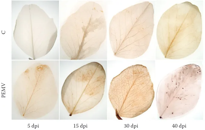
The symptoms of pea enation mosaic were not observed at 5 dpi, which indicates that the first studied phase of the disease progress preceded the systemic spreading of viral particles. Down-ward curling of young expanding leaves was first observed at 8 dpi. Later (at 15 dpi), a mosaic was formed, which corresponded to the peak of virus titre found by qRT-PCR (Figure 1). At 30 and 40 dpi, severe chlorotic spots became translucent and clearly delineated (Figure 2A) and enations developed above leaf veins (Figures 2B, C). At this disease stage we have also observed plant stunting.
Endogenous concentrations of abscisic, salicylic, and jasmonic acid
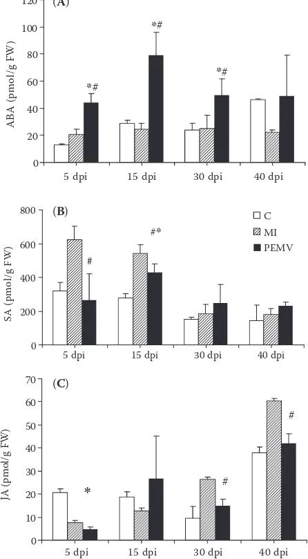
Changes in endogenous concentrations of ABA, SA, and JA are compared in Figure 3. In control plants, the ABA content found at 5 dpi (12.8 pmol/g FW) doubled at 15 and 30 dpi and was almost 4 times higher at 40 dpi (46.1 pmol/g FW). How-ever, in MI plants it remained almost the same during the whole experiment (20.8–25 pmol/g FW). PEMV infection caused an increase in ABA concentration, compared to MI and C plants, with significant differences observed at 5–30 dpi (Figure 3A). The maximal ABA content was found at 15 dpi (79 pmol/g FW), followed by a drop to 49 pmol/g FW at 30 and 40 dpi.
The highest endogenous concentration of SA, i.e.
622 pmol/g FW, was found in MI plants at 5 dpi, and this value was significantly different from both control
and PEMV plants (Figure 3B). There was a gradual decrease in the concentration of SA for C as well as for MI plants from 5 dpi, while in PEMV-infected plants the SA concentration peaked at 15 dpi, fol-lowed by a later decrease to the initial value.
The level of JA in control plants varied from ca 20 pmol/g FW at 5 and 15 dpi, to 9.4 pmol/g FW at 30 dpi and 37.8 pmol/g FW at 40 dpi. In PEMV-in-fected plants, the JA content at 5 dpi was significantly lower compared to the healthy plants. However, a similar decrease in JA appeared also in MI plants, which indicates that the initial decrease in JA was caused by the mechanical injury. From 15 dpi to 40 dpi the JA concentration in PEMV plants was higher and did not differ significantly from controls. While the JA content in MI plants was low at 5 dpi
0.0 0.2 0.4 0.6 0.8 1.0 1.2 1.4 1.6
5 dpi 15 dpi 30 dpi 40 dpi
Relative amount of viral RNA
[image:6.595.307.528.86.211.2]Inoculated Systemic
Figure 1. Relative content of viral RNA of PEMV-1 for inoculated and systemic leaves at 5–40 dpi, related to the RNA content in the inoculated leaves at 5 dpi. No viral RNA was detected in the systemic leaves at 5 dpi. The inoculated leaves dropped off after 15 dpi. Values represent means ± SD (n = 3–4)
[image:6.595.64.540.537.722.2](7.6 pmol/g FW), at 30 and 40 dpi it was significantly higher than in control and PEMV plants (Figure 3C).
Hsp70 content
Western-blotting followed by immunohisto-chemical staining visualised a single band with the molecular mass of 70 kDa (not shown) and enabled relative quantification of the Hsp70 content (Figure 4). In PEMV infected plants, the amount of Hsp70 was significantly higher than in
control plants at 5–30 dpi, with the maximum at 30 dpi. During the ontogeny, Hsp70 concentration gradually increased in all plants, thus there was no apparent difference between the inoculated and control samples at 40 dpi. Notably, the mock-inoculation induced an insignificant decrease in Hsp70 concentration at 5 dpi and a slight increase at 15–40 dpi compared to the healthy plants.
POX activity and localisation
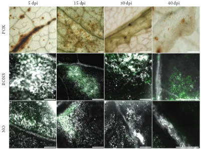
[image:7.595.311.532.85.226.2]The activity of soluble peroxidase (POX) was assayed spectrophotometrically, using guaiacol as a substrate. In all plants the maximal POX activity was found at 5 dpi, followed by a gradual decrease (Figure 5). In control plants the activity of POX ranged between 30–34 μkat/g FW up to 30 dpi and a moderate decrease was observed at 40 dpi (Figure 5). The highest activity of POX was detected in PEMV-infected plants; at 5 dpi it reached 49 μkat/g FW, which corresponds to 144% of the POX activity in control plants. At 15 dpi the POX activity in PEMV plants significantly differed not only from control, but also from MI plants. Later the POX activity in PEMV plants dropped to the values similar to those observed in MI and control plants. POX activity was also evaluated by histochemical staining with DAB and visualised by light microscopy. In situ localisation of POX activity correlated with the biochemical data in leaves of PEMV infected plants. The strongest signal was found at 5 dpi in leaf regions close to veins, Figure 3. Contents of abscisic acid (A), salicylic acid (B),
and jasmonic acid (C) in leaves of control, mock-inocu-lated, and PEMV-infected pea plants during 40 dpi. Data show means ± SD (n = 3). Values significantly different at
P = 0.05 from control (*) or mock-inoculated plants (MI) (#) are highlighted
0.0 1.0 2.0 3.0 4.0 5.0 6.0 7.0
5 dpi 15 dpi 30 dpi 40 dpi
H
sp
70
(r
el
. u
.)
C MI PEMV
*#
*#
*
Figure 4. Relative concentrations of Hsp70 in leaves of control (C), mock-inoculated (MI), and PEMV-infected (PEMV) pea plants. Data are normalised to the concentra-tions of the protein in control leaves at 5 dpi and expressed as means ± SD (n = 3–10), for details see Material and
Methods. Highlighted values significantly differ from C (*) or MI (#) at P = 0.05
0 20 40 60 80 100 120
5 dpi 15 dpi 30 dpi 40 dpi
ABA (pmol/g FW)
*
#*
#*
#0 200 400 600 800
5 dpi 15 dpi 30 dpi 40 dpi
SA (pmol/g FW)
C MI PEMV #*
#
#
0 10 20 30 40 50 60 70
JA
(p
m
ol
/g
F
W
)
∗
#5 dpi 15 dpi 30 dpi 40 dpi
# (A)
(B)
[image:7.595.67.290.90.494.2]where pathological changes might have been induced. During the systemic spread of PEMV at 15 dpi and later, the POX signal was weaker (Figure 5).
Localisation of hydrogen peroxide
[image:8.595.66.289.84.242.2]Hydrogen peroxide was localised in pea leaves as a red-brown precipitate after DAB staining (Figure 6). A weak signal was observed already
in control and mock-inoculated plants; however, in PEMV-infected plants the staining was much stronger and appeared mainly in mesophyll cells close to veins. At 5 dpi the signal for H2O2 was found mostly in the leaf tips, at 15 dpi it was local-iszed in the central part of the leaves and at 30 dpi it was spread all over the area of examined leaves. At 40 dpi, a strong signal for H2O2 corresponded with the area of the newly formed enations, but in the rest of the leaf the signal was weaker (Figure 6). No apparent difference was observed between the leaves of control and MI plants (data not shown).
Localisation of RONS by confocal microscopy
Cell-permeable fluorescence probes were used to track NO (DAF-FM DA) and RONS (H2DCF DA) in fresh leaves (green in Figure 7). Fluorescent signal for RONS was observed in tissue regions in the vi-cinity of veins and developing enations at 5–40 dpi. CLSM enabled also software 2.5D reconstructions from a series of optical sections in individual ob-jects, which revealed a presence of RONS within the volume of enations (Figure 8). The accumulation of tracked molecules was intensive from 15 dpi to 40 dpi, suggesting that in susceptible pea plants the local changes in RONS concentrations enable tissue reorganisation due to PEMV infection. The signal for NO was weak and was found namely in the initial phases of tissue reprogramming, i.e. at 5 and 15 dpi. Minor signal was detected also at 40 dpi.
15 20 25 30 35 40 45 50 55 60
5 dpi 15 dpi 30 dpi 40 dpi
POX (µkat/g FW)
C MI PEMV *
[image:8.595.121.457.509.723.2]*#
Figure 5. Variations in the activity of cytosolic peroxidase (POX) in leaves of control (C), mock-inoculated (MI), and infected (PEMV) pea plants during 40 dpi. Data are expressed as means ± SD (n = 3). Highlighted values sig-nificantly differ from C (*) or MI (#) at P = 0.05. The small graph highlights the effect of viral infection on changes in POX activity. The data were obtained as the difference between PEMV and MI plants
Figure 6. Accumulation of H2O2 within leaves of control (C) and PEMV-infected pea plants during 40 dpi was visu-alised by light microscopy as a deposition of a brown pigment after DAB staining
0 5 10 15
5 dpi 15 dpi 30 dpi 40 dpi
(PEMV-MI
)
5 dpi 15 dpi 30 dpi 40 dpi
PE
MV
DISCUSSION
Viruses, similarly to bacteria, cannot pass ac-tively through plant cell walls and require insect vectors (e.g. aphids for PEMV) or wounding to invade the cells of their hosts (Salgueiro & Hull
1999). There is growing evidence that even tiny wounding caused by aphids has a deleterious ef-fect on plant metabolism (Reymond et al. 2000). During artificial inoculation by leaf abrasion, the viral ingress is combined with injury (Smith et al. 2004), as evidenced by systemic increase in POX activity at 5 dpi (Figure 5). The comparison of the components of signalling pathways in in-tact, mock-inoculated, and PEMV-infected plants showed that the processes induced by infection differ from those caused by mechanical stress (best exhibited at 5 dpi) and from proceeding natural senescence. At 15 dpi, the maximal content of viral RNA corresponded with an increase in ABA and Hsp70 level, and in POX activity in systemic leaves.
Our previous study dealt with the alterations of photosynthetic parameters in Pisum sativum
[image:9.595.94.495.83.381.2]infected by PEMV. During 40 dpi, PEMV-accel-erated senescence took place in the symptomatic leaves, which was accompanied by a decrease in the efficiency of both CO2 assimilation and photosystem II photochemistry (ΦPSII), but was Figure 7. Changes in RONS localisation within leaf tissues of pea plants systemically infected by PEMV at 5, 15, 30, and 40 dpi. POX activity was localised by DAB staining protocol and light microscopy. Green signal for the RONS production was visualised by the reaction with H2DCF DA and with DAF-FM DA (especially nitric oxide). Both his-tochemical staining procedures were followed by confocal laser scanning microscopy. Note brighter parts in greyscale channel, corresponding to chlorotic cells within developing enations. Bar corresponds to 100 μm
Figure 8. Volume model of signal for RONS (green) was reconstructed from a series of eight optical sections through PEMV-infected pea leaf at 40 dpi. The highest intensity of signal corresponds to enations
5 dpi 15 dpi 30 dpi 40 dpi
N
O
RON
S
PO
[image:9.595.62.293.563.698.2]not linked to stomatal closure (Kyseláková et al. 2011). Additionally, the degradation of photo-synthetic pigments has been accelerated following the appearance of macroscopic disease symptoms (at 30 dpi). Local changes previously shown by chlorophyll fluorescence imaging (gradual de-crease of ΦPSII photochemical efficiency starting from the leaf tip) and detected oxidative damage of cell membranes (Kyseláková et al. 2011) are connected to oxidative processes and enation development presented here.
In pea-PEMV interactions, the virus starts to spread systemically at 5 dpi, but in the studied 3rd and 4th leaves the viral RNA was not detected
before 15 dpi (Figure 1). The activation of plant defence by viral attack was shown to be accom-panied by the changes in the content of molecu-lar chaperones, e.g. Hsp70 (Byth et al. 2001). However, in our study, Hsp70 expression was in-duced also by mechanical injury and senescence (Figure 4). Senescence caused a gradual increase in the amount of the protein in control plants. PEMV induced significant increase in the Hsp70 content from 5 dpi, with the maximum detected at 30 dpi. Wounding in MI plants led to the initial decrease in Hsp70 concentration at 5 dpi, while at 15–40 dpi it gradually increased and was not significantly different from control plants.
The metabolism of reactive oxygen and nitrogen species provides insight in disease dynamics and plant’s destiny. Prompt death due to unbalanced RONS accumulation is well described in several incompatible interactions, where the virus-attacked cells are sacrificed to limit the infection. Delayed cell death characterises susceptible plants, e.g. in potato-Potato virus Y pathosystem (Hinrichs-Berger 1999). Levels of ROS and the capacity of antioxidant systems have been investigated in several compatible plant-virus interactions, but contradictory results have been obtained (Rie-dle-Bauer 2000; Hernández et al. 2004). Both compatible (Solanum esculentum and Capsicum an-nuum) and incompatible (Nicotiana glutinosa and
N. tabacum) hosts during tobamovirus (TMV and
ToMV) infection were studied by Madhusudhan
et al. (2009). The increased SA, hydrogen peroxide,
lipid peroxidation, protein oxidation, POX activity, and decreased catalase activity were observed in the incompatible host-tobamovirus interaction. Minor changes in the activity of enzymes, lipid peroxidation, and hydrogen peroxide content were found in susceptible hosts (Madhusudhan et
al. 2009). Long-term infection by plum pox virus studied by Díaz-Vivancos et al. (2006) in peach and apricot showed that general increase in H2O2 concentration led to lipid hydroperoxidation of cell membranes and proteins in susceptible, but not in resistant hosts. In compatible interactions, ion leakage, a marker of damaged plasmalemma, was accompanied by a decrease in POX and superoxide dismutase activity, whereas in the incompatible interactions, the activities of these enzymes were increased. These results were opposite to data presented for other pathosystems. Milavec et al.
(2001) reported an inverse correlation between the chlorophyll content and the activity of soluble and ionic-bound peroxidases in 4-weeks old potatoes at 5–7 dpi with Potato virus Y. However, in our time-course study the decrease in the activity of soluble POX (Figure 5) was found to correspond with the previously reported decrease in the con-tent of chlorophylls and carotenoids (Kyseláková
et al. 2011).
Leaf enations, the specific PEMV symptom (Fig-ure 2), appear days to weeks following the infection. Enations are formed as a result of virus-altered regulation of cell cycle in leaf parenchyma, which then turns into meristem (Choi et al. 2011). This process seems to be related to the changes in hor-monal levels and affected by signalling molecules, e.g. RONS. For their cell wall reconstruction, the newly derived cells require H2O2, regulated by POX activity (Quiroga et al. 2000). During its systemic spreading, the PEMV induces oxidative stress in the infected plant, which is evidenced by the increase in lipid peroxidation and electrolyte leakage at 15 and 30 dpi (Kyseláková et al. 2011). In our experiments, strictly localised changes in H2O2 level were detected within PEMV-infected leaves (Figure 6). The H2O2 production was ob-served namely in mesophyll cells near the minor veins (Figure 7). Many exceptions in proposed patterns of oxidative processes can be found in the literature, e.g. in A. thaliana-CaMV pathosystem, H2O2 accumulated both locally and systemically in virus- but not in mock-inoculated plants (Love
et al. 2005). In several host-virus interactions, the
enhanced POX activity correlated with the sever-ity of symptoms, but this does not seem to be the case in pea-PEMV (Figure 5) or in lettuce-Lettuce
mosaic virus (LMV) pathosystems (El-Fahaam
et al. 1990). POX not only scavenges H2O2, but
oxidative stress in systemic plant-virus interac-tions (Riedle-Bauer 2000). Due to the PEMV inoculation, a remarkable enhancement of POX activity was recorded, which gradually decreased. The wounding alone increased POX activity at 5 dpi, later its values were comparable with those in control plants. The major influence of PEMV on POX activity is visible at 15 dpi (small graph in Figure 5), which is probably linked to systemic virus transmission. Induction of higher activity of antioxidant enzymes by pathogen may lead to the disruption of signals generated by ROS, which would have otherwise triggered defence reactions in the host (Arias et al. 2005).
There is limited information about the mecha-nisms that lead to the generation of ROS in plant cells following ABA signalling (Jiang & Zhang 2001). In guard cells of Arabidopsis, stomatal clo-sure is enabled by ABA-induced H2O2 production and the H2O2-activated Ca2+ channels (Pei et al.
2000). In such tissue, the availability of CO2 for photosynthesis is lower, which can lead to the formation of ROS due to the leakage of electrons from reduced photosystems to oxygen. However, we did not observe stomatal closure in our pathosys-tem (Kyseláková et al. 2011). In PEMV-infected plants, ABA levels peaked at 15 dpi (Figure 3A), which corresponds to the highest value of PEMV-1 RNA detected by q-RT-PCR in sampled leaves (Figure 1). It is well known that ABA up-regulates events that occur during senescence, such as in-creased lipid peroxidation, membrane permeabil-ity, and the induction of proteinase and RNAse activities (Panavas et al. 1998).
It has been suggested that SA induces resistance mechanisms against virus infection (Radwan et al. 2007). Increase in endogenous SA concentra-tion due to mechanical injury was observed at 5 and 15 dpi in both PEMV-infected and MI plants (Figure 3B). Interestingly, comparing these two, the SA level was lower in PEMV-infected plants, which suggests that the virus might be somehow involved in the down-regulation of SA signalling pathway. This would be in agreement with sug-gestions of other authors that SA could induce an inhibition of long-distance virus movement (reviewed by Jameson & Clarke 2002). Con-sidering temporal changes in SA levels in PEMV plants, an increase was recorded at 15 dpi, which coincided with virus multiplication and spread-ing. However, at that time PEMV is out of the control by the host plant. At later stages the SA
concentration decreased. Similarly, in compatible potato-Potato virus Y interaction, the dynamics of the responses was shown to be modulated by SA, which can delay viral multiplication and disease symptoms (Baebler et al. 2011). The enhanced SA levels were found to coincide with the rise in CaMV levels in A. thaliana (Love et al. 2005).
In general, we have observed antagonistic rela-tionship in temporal changes of SA and JA concen-trations, especially for MI plants (Figures 3B, C). Mechanical injury induced accumulation of SA at 5 dpi, which later gradually decreased. On the other hand, the concentration of JA increased from 5 dpi to 40 dpi, which suggests that there can be a crosstalk of both pathways during abiotic stress caused by inoculation. These results correspond with previous results of some other authors (e.g. Niki et al. 1998; Radwan et al. 2008). Experiments with exogenously applied SA and JA demonstrated this antagonism in various plant species, suggesting an evolutionary conserved process (Spoel & Dong 2008). Nevertheless, the outcome of interactions between SA and JA signalling were reported to be concentration-dependent, ranging from synergy to antagonism depending on particular concen-tration (Mur et al. 2006). It has been suggested that a positive regulator or transcription factor of one pathway also functions in the other hormone response pathway, thus facilitating the hormonal crosstalk (Robert-Seilaniantz et al. 2011). SA may also inactivate catalase, which results in higher ROS concentration and increased level of lipid peroxidation, which is the starting point of JA biosynthesis. Thus SA can participate in delayed JA accumulation (Preston et al. 1999). On the other hand, exogenous treatment with SA and JA enhanced activity of catalase and other enzymes which may hinder virus replication (Clarke et al.
Antagonistic interactions between multiple components of ABA and JA-ethylene signalling pathways modulate gene expression in response to biotic and abiotic stresses (Anderson et al.
2004). Increase in ABA is common in compatible interactions, suppressing SA signalling and HR (Cao et al. 2011). Smith et al. (2004) identified a specific group of genes that exhibited differential transcript abundance in response to a combination of wounding and systemic viral infection. However, genes implicated in early wound responses, such as JA-induced genes and transcription factors (Li et al. 2001), did not exhibit an increase in transcript abundance in response to wounding alone. JA in high concentration might be involved in the in-hibition of viral replication. It was shown that JA as well as SA do not prevent systemic spreading of the virus, but reduce its titre (Clarke et al.
1998). Clarke et al. (2000) reported two phases of increase in endogenous JA level, the first one occurring during several hours after inoculation and the second one taking place during the in-crease of virus titre in plant body and anthocyanin accumulation. In our study, the peak of JA was observed at 15 dpi and appears to be linked with host plasmalemma damage upon replication of virus (i.e. PEMV transmission) and with chlorophyll degradation (Kyseláková et al. 2011).
CONCLUSIONS
It has been suggested that the higher activity of antioxidant enzymes can interrupt defence signals generated by ROS in several pathosystems, and thus leads to compatible virus-plant interactions. In this type of interaction, also the hormone-triggered defence reactions have to be suppressed, which makes virus interaction even more complex. From our results we can conclude that oxidative processes linked to balancing of hormone levels affect the susceptibility of pea plants to PEMV. The interference of hormone levels suggests that the abiotic stress induced by inoculation might delay viral colonisation. The increased lipid peroxida-tion is not reflected in JA formaperoxida-tion. The systemic transmission of PEMV coincides with ABA and SA accumulation and POX activity. In PEMV-colonised pea plants, the decrease in the activity of POX and higher RONS levels enable the expression of symptoms. However, the heterogeneity of reactions within a leaf must be taken into account, e.g. the
accumulation of RONS was confirmed in altering tissue spots undergoing chlorosis. Localisation of oxidative processes to regions of developing ena-tions is quite unique and enables the development of distinctive symptoms; the significance of this process has not been answered yet. For the first time we have localised nitric oxide, an important signalling molecule, in plant tissues undergoing virus-triggered reorganisation.
Acknowledgements. PEMV isolate was provided by the Department of Cell Biology and Genetics, Faculty of Science, Palacky University in Olomouc, Czech Republic, where the inoculation and DASI-ELISA tests were performed. Authors highly appreciate the assistance of U. Sikorová (Western blotting), P. Ama- korová (extraction of hormones), and valuable com-ments on the manuscript by Dr. M. Špundová.
References
Adie B.A., Pérez-Pérez J., Pérez-Pérez M.M., Godoy M., Sánchez-Serrano J.J., Schmelz E.A., Solano R. (2007): ABA is an essential signal for plant resistance to pathogens affecting JA biosynthesis and the activation of defenses in Arabidopsis. The Plant Cell, 19: 1665–1681. Anderson J.P., Badruzsaufari E., Schenk P.M., Man-ners J.M., Desmond O.J., Ehlert Ch., Maclean D.J., Ebert P.R., Kazana K. (2004): Antagonistic interaction between abscisic acid and jasmonate-ethylene signaling pathways modulates defence gene expression and disease resistance in Arabidopsis. Plant Cell, 16: 3460–3479. Angeliny R., Manes F., Federico R. (1990): Spatial and
functional correlation between diamine-oxidase and peroxidase activities and their dependence upon de-etiolation and wounding in chick-pea stems. Planta, 182: 89–96.
Arias M. C., Luna C., Rodríguez M., Lenardon S., Taleisnik E. (2005): Sunflower chlorotic mottle virus in compatible interactions with sunflower: ROS generation and antioxidant response. European Journal of Plant Pathology, 113: 223–232.
Baebler Š., Stare K., Kovač M., Blejec A., Prezelj N., Stare T., Kogovšek P., Pompe-Novak M., Rosah S., Ravnikar M., Gruden K. (2011): Dynamics of re-sponses in compatible potato – Potato virus Y interaction are modulated by salicylic acid. PLoS ONE 6(12): e29009. doi:10.1371/journal.pone.0029009
Bell E., Creelman R.A., Mullet J.E. (1995): A chloroplast lipoxygenase is required for wound induced accumulation of jasmonic acid in Arabidopsis. Proceedings of National Academy of Science USA, 92: 8675–8679.
Bendahmane A., Kanyuka K., Baulcombe D.C. (1999): The Rx gene from potato controls separate virus resist-ance and cell death responses. Plant Cell, 11: 781–791. Bergougnoux V., Hlaváčková V., Plotzová R., Novák
O., Fellner M. (2009): The 7B-1 mutation in tomato (Solanum lycopersicum L.) confers a blue light-specific lower sensitivity to coronatine, a toxin produced by Pseu-domonas syringae pv. tomato. Journal of Experimental Botany, 60: 1219–1230.
Byth H.-A., Kuun K.G., Bornman L. (2001): Virulence-dependent induction of Hsp70/Hsc70 in tomato by Ral-stonia solanacearum. Plant Physiology and Biochemistry, 39: 697–705.
Cao F.Y., Yoshioka K., Desveaux D. (2011): The role of ABA in plant-pathogen interactions. Journal of Plant Research 124: 489–499.
Chen Z., Zhou T., Wu X., Hong Y., Fan Z., Li H. (2008): Influence of cytoplasmic heat shock protein 70 on viral infection of Nicotiana benthamiana. Molecular Plant Pathology, 9: 809–817.
Choi J., Choi D., Lee S., Ryu C.-M., Hwang I. (2011): Cytokinins and plant immunity: Old foes or new friends? Trends in Plant Science, 16: 388–394.
Clarke S.F., Burritt D.J., Jameson P.E., Guy P.L. (1998): Influence of plant hormones on virus replication and pathogenesis-related proteins in Phaseolus vulgaris L. infected with white clover mosaic potexvirus. Physiology and Molecular Plant Pathology, 53: 195–207.
Clarke S.F., Guy P.L, Jameson P.E., Schmierer D., Bur-ritt D.J. (2000): Influence of white clover mosaic potex-virus infection on the endogenous levels of jasmonic acid and related compounds in Phaseolus vulgaris L. seedlings. Journal of Plant Physiology, 156: 433–437.
Clarke S.F., Guy P.L, Burritt D.J., Jameson P.E. (2002): Changes in the activities of antioxidant enzymes in re-sponse to virus infection and hormone treatment. Physi-ologia Plantarum, 114: 157–164.
Cole A.B., Király L., Ross K., Schoelz J.E. (2001): Un-coupling resistance from cell death in the hypersensitive response of Nicotiana species to Cauliflower mosaic virus infection. Molecular Plant-Microbe Interactions, 14: 31–41.
Cooley M.B., Pathirana S., Wu H.-J., Kachroo P., Kles-sig D.F. (2000): Members of the Arabidopsis HRT/RPP8 family of resistance genes confer resistance to both viral and oomycete pathogens. Plant Cell, 12: 663–676. Demler S.A., de Zoeten G.A., Adam G., Harris K.F.
(1996): Polyhedral Virions and Bipartite RNA Genomes.
In: Harrison B.D., Murant A.F. (eds): The Plant Vi-ruses. Vol. 5. Plenum Press, New York: 303–304. Díaz-Vivancos P., Rubio M., Mesonero V., Periago
P.M., Ros Barceló A., Martínez-Gómez P., Hernán-dez J.A. (2006): The apoplastic antioxidant system in
Prunus: Response to plum pox virus. Journal of Experi-mental Botany, 57: 3813–3824.
El-Fahaam Y.M., Fegla G.I., Wagih E.E., El-Karyoni H.A. (1990): Biochemical changes in lettuce plants in-fected with lettuce mosaic virus. Journal of King Saud University, 2: 271–277.
El Rahman T.A., El Oirdi M., Gonzalez-Lamothe R., Bouarab K. (2012): Necrotrophic pathogens use the salicylic acid signaling pathway to promote disease development in tomato. Molecular Plant-Microbe Inter-actions, 25: 1584–1593.
Fujita M., Fujita Y., Noutoshi Y., Takahashi F., Narusa-ka Y., Yamaguchi-Shinozaki K., Shinozaki K.. (2006): Crosstalk between abiotic and biotic stress responses: a current view from the points of convergence in the stress signaling networks. Current Opinion in Plant Biology, 9: 436–442.
Glazebrook J. (2005): Contrasting mechanisms of defense against biotrophic and necrotrophic pathogens. Annual Review of Phytopathogy, 43: 205–227.
Grünwald N.J., Chen W., Larsen R.C. (2004): Pea dis-eases and their management. In: Naqvi S.A.M.H. (ed.): Diseases of Fruits and Vegetables. Volume II. Kluwer Academic Publishers, Dordrecht: 301–331.
Gundlach H., Muller M.J., Kutchan T.M., Zenk M.H. (1992): Jasmonic acid is a signal transducer in elicitor-induced plant cell cultures. Proceedings of the National Academy of Science USA, 89: 2389–2393.
Hernández J.A., Rubio M., Olmos E., Ros-Barceló A., Martínez-Gómez P. (2004): Oxidative stress induced by long-term plum pox virus infection in peach (Prunus persica). Physiologia Plantarum, 122: 486–495.
Hinrichs-Berger J., Harfold M., Berger S., Buchenauer H. (1999): Cytological responses of susceptible and ex-tremely resistant potato plants to inoculation with potato virus Y. Physiological and Molecular Plant Pathology, 55: 143–150.
Hlaváčková V., Krchňák P., Nauš J., Novák O., Špun- dová M., Strnad M. (2006): Electrical and chemical signals involved in short-term systemic photosynthetic responses of tobacco plants to local burning. Planta, 225: 235–244.
Jameson P.E., Clarke S.F. (2002): Hormone-virus inter-actions in plants. Critical Reviews in Plant Sciences, 21: 205–228.
oxida-tive damage in leaves of maize seedlings. Plant and Cell Physiology 42: 1265–1273.
Koornneef A., Pieterse C.M.J. (2008): Cross talk in de-fense signaling. Plant Physiology, 146: 839–844. Kyseláková H., Prokopová J., Nauš J., Novák O., Na-
vrátil M., Šafářová D., Špundová M., Ilík P. (2011): Photosynthetic alterations of pea leaves infected systemi-cally by pea enation mosaic virus: A coordinated decrease in efficiencies of CO2 assimilation and photosystem II photochemistry. Plant Physiology and Biochemistry, 49: 1279–1289.
Lamotte L., Courtois C., Barnavon L., Pugin A., Wen-dehenne D. (2005): Nitric oxide in plants: the biosynthe-sis and cell signalling properties of a fascinating molecule. Planta, 221: 1–4.
Li L., Li C., Howe G.A. (2001): Genetic analysis of wound signaling in tomato. Evidence for a dual role of jasmonic acid in defense and female fertility. Plant Physiology, 127: 1414–1417.
Love A.J., Yun B.W., Laval V., Loake G.J., Milner J.J. (2005):
Cauliflower mosaic virus, a compatible pathogen of
Arabidopsis, engages three distinct defense-signaling pathways and activates rapid systemic generation of re-active oxygen species. Plant Physiology, 139: 935–948. Madhusudhan K.N., Srikanta B.M., Shylaja M.D.,
Prakash H.S., Shetty H.S. (2009): Changes in antioxidant enzymes, hydrogen peroxide, salicylic acid and oxidative stress in compatible and incompatible host-tobamovirus interaction. Journal of Plant Interactions, 4: 157–166. Mauch-Mani B., Mauch F. (2005): The role of abscisic
acid in plant–pathogen interactions. Current Opinion in Plant Biology, 8: 409–414.
Maule A., Leh V., Lederer C. (2002): The dialogue be-tween viruses and hosts in compatible interactions. Cur-rent Opinion in Plant Biology, 5: 279–284.
Mayer M.P., Bakau B. (2005): Hsp70 chaperones: Cellular functions and molecular mechanism. Cell and Molecular Life Sciences, 62: 670–684.
Milavec M., Ravnikar M., Kovač M. (2001): Peroxi-dases and photosynthetic pigments in susceptible potato infected with potato virus YNTN. Plant Physiology and Biochemistry, 39: 891–898.
Mur L.A.J., Kenton P., Atzorn R., Miersch O., Waster-nack C. (2006): The outcomes of concentration-specific interactions between salicylate and jasmonate signaling include synergy, antagonism, and oxidative stress leading to cell death. Plant Physiology, 140: 249–262.
Niki T., Mitsuhara I., Seo S., Ohtsubo N., Ohashi Y. (1998): Antagonistic effect of salicylic acid and jasmonic acid on the expression of pathogenesis-related (PR) pro-tein genes in wounded mature tobacco leaves. Plant Cell Physiology 39: 500–507.
Panavas T., Walker E.L., Rubinstein B. (1998): Possible involvement of abscisic acid in senescence of daylily petals. Journal of Experimental Botany, 49: 1987–1997. Pei Z.-M., Murata Y., Benning G., Thomine S., Klusener
B., Allen G.J., Grill E., Schroeder J.I. (2000): Calcium channels activated by hydrogen peroxide mediate abscisic acid signalling in guard cells. Nature, 406: 731–734. Piterková J., Petřivalský M., Luhová L., Mieslerová
B., Sedlářová M., Lebeda A. (2009): Local and systemic production of nitric oxide in tomato responses to powdery mildew infection. Molecular Plant Pathology, 10: 501–513. Preston C.A., Lewandowski C., Enyedi A.J., Baldwin
I.T. (1999): Tobacco mosaic virus inoculation inhibits wound-induced jasmonic acid-mediated responses within but not between plants. Planta, 209: 87-95.
Prins M., Laimer M., Noris E., Schubert J., Wassenegger M., Tepfer M. (2008): Strategies for antiviral resistance in transgenic plants. Molecular Plant Pathology, 9: 73–83. Quiroga M., Guerrero C., Botella M.A., Ros Barceló
A., Amaya I., Medina M.I., Alonso F.J., de Forchetti S.M., Tigier H., Valpuesta V. (2000): A tomato peroxi-dase involved in the synthesis of lignin and suberin. Plant Physiology, 122: 1119–1127.
Radwan D.E.M., Fayez K.A., Mahmoud S.Y., Hamad A., Lu G. (2007): Physiological and metabolic changes of Cucurbita pepo leaves in response to zucchini yellow mosaic virus (ZYMV) infection and salicylic acid treat-ments. Plant Physiology and Biochemistry, 45: 480–489. Radwan D.E., Lu G., Fayez K.A., Mahmoud S.Y. (2008):
Protective action of salicylic acid against Bean yellow mosaic virus infection in Vicia faba leaves. Journal of Plant Physiology, 165: 845–857.
Riedle-Bauer M. (2000): Role of reactive oxygen species and antioxidant enzymes in systemic virus infections of plants. Journal of Phytopathology, 148: 297–302. Reymond P., Weber H., Damond M., Farmer E.E. (2000):
Differential gene expression in response to mechanical wounding and insect feeding in Arabidopsis. The Plant Cell, 12: 707–719.
Robert-Seilaniantz A., Grant M., Jones J.D.G. (2011): Hormone crosstalk in plant disease and defense: more than just jasmonate-salicylate antagonism. Annual Re-view of Phytopathology, 49: 317–343.
Salgueiro S., Hull R. (1999): Comparison of pea enation mosaic virus (PEMV) isolates: Sequence of coat protein and aphid transmission protein genes of a European iso-late. Journal of Phytopathology, 147: 391–396.
Smith C.M., Rodriquez-Buey M., Karlsson J., Camp-bell M.M. (2004): The response of the poplar transcrip-tome to wounding and subsequent infection by a viral pathogen. The New Physiologist, 164: 123–136. Smith J.L., De Morales C.M., Mescher M.C. (2009):
Jas-monate- and salicylate-mediated plant defense responses to insect herbivores, pathogens and parasitic plants. Pest Management Science, 65: 497–503.
Spoel S.H., Dong X. (2008): Making sense of hormone crosstalk during plant immune responses. Cell Host and Microbe, 3: 348–351.
Thordal-Christensen H., Zhang Z., Wei Y., Collinge D.B. (1997): Subcellular localization of H2O2 in plants in papillae and hypersensitive response during the
bar-ley-powdery mildew interaction. The Plant Journal, 11: 1187–1194.
von Bargen S., Salchert K., Paape M., Piechulla B., Kellmann J.W. (2001): Interactions between the tomato spotted wilt virus movement protein and plant proteins showing homologies to myosin, kinesin and DnaJ-like chaperones. Plant Physiology and Biochemistry, 39: 1083–1093.
Whitham S.A., Wang Y.Z. (2004): Roles for host factors in plant viral pathogenicity. Current Opinion in Plant Biology, 7: 365–371.
Received for publication July 16, 2012 Accepted after corrections January 12, 2013
Corresponding author: