Outdoor Environment as a Source
of Listeria monocytogenes in Food Chain
Tereza GelbíčoVá
1,3and Renáta KaRpíšKoVá
1,21National Institute of public Health prague, brno, Czech Republic;
2University of Veterinary and pharmaceutical Sciences brno, brno, Czech Republic;
3Faculty of Science, Masaryk University brno, brno, Czech Republic
Abstract
Gelbíčová T., Karpíšková R. (2012): Outdoor environment as a source of Listeria monocytogenes in
food chain. Czech J. Food Sci., 30: 83–88.
We monitored the presence of listeria monocytogenes in environmental sources and to evaluate the phenotypic and molecular characteristics of the isolates recovered. l. monocytogenes was isolated in 12 (11.2%) of the 107 samples from the wild, farm environment, and vegetation. Most isolates (83.3%) were of serotype 1/2a and the remainder (2) were of serotype 4b. All 12 isolates were susceptible to the whole range of antimicrobials tested. These12 strains were carriers of the virulence genes prfa, hlya, acta, plca, plcb, inla, inlb, inlC,and inlJ. The detection of the inla gene in 4 (33.3%) of 12 strains using the PCR-RFLP suggests the potential of some of these strains to penetrate into epithelial cells of the intestinal barrier. Macrorestriction analysis also confirmed clonal identity of some environmental isolates with food and human isolates. These results indicate that the external environment is a source of potentially pathogenic strains of l. monocytogenes.
Keywords: virulence; listeria; antimicrobial resistance; restriction fragment length polymorphism; pulsed-field gel
elec-trophoresis (PFGE)
listeria monocytogenes is a ubiquitous saprophytic bacterium, adapted to life in soil and decaying vegetation, but also in the cytosol of eukaryotic host cells (Freitag et al. 2009). l. monocytogenes
can also be isolated from the surface and under-ground waters, improperly fermented silage, sewage sludge, slaughter wastes, animal and human faeces, foodstuffs, and food industry plants (Farber & Peterkin 1991; Ivanek et al. 2006). In both ani-mals and susceptible humans, l. monocytogenes
causes serious invasive disease. The main source of listeria to animals is contaminated feed, and that to humans is food for direct consumption (Vázquez-Boland et al. 2001).
The presence of l. monocytogenes in farm animals is mainly associated with cattle and sheep farming, and less often with pig and poultry farming. In ruminants, the disease manifests itself primarily by neurological symptoms or abortion, and very uncommonly, by mastitis. However, as a rule,
l. monocytogenes passes asymptomatically through the gastrointestinal tract to be excreted in faeces (Ivanek et al. 2006; Esteban et al. 2009). Fecal contamination of soil, vegetation, and surface water is the major source of l. monocytogenes in primary food production (Ivanek et al. 2006).
Pathogenicity of l. monocytogenes is due to its capacity of penetrating into and proliferating in
various types of host cells, including non-phagocytic cells. The intracellular cycle of l. monocytogenes
starts with the invasion of host cells mediated by the surface proteins internalins A (encoded by inla
gene) and B (inlb) (Bierne et al. 2007). An essential prerequisite for l. monocytogenes proliferation and replication in the cytosol is the bacterial escape from phagolysosomes, mediated by listeriolysin O (hlya) in combination with phospholipases, phos-phatidylinositol (plca), and phosphatidylcholine (plcb) phospholipases C. The intracellular motility of l. monocytogenes and spread from cell to cell are due to an actin polymerisation protein (acta). The PrfA (positive regulatory factor A) protein is crucial to the transcriptional regulation of the above-men-tioned genes located in LIPI-1 (listeria pathogenicity island 1) and in part also of inla and inlb genes (Gray et al. 2006; Freitag et al. 2009).
Variation in the pathogenic potential of l. mono-cytogenes isolated from foodstuffs and the environ-ment has been reported. This may be due to the deletion of one or more genes encoding the key virulence factors (Doumith et al. 2004a). However, a number of studies have found such genes to be stable parts of the genome of l. monocytogenes
(Jaradat et al. 2002; Doumith et al. 2004a). The reduced virulence of l. monocytogenes may also be a result of point mutations of the above-mentioned genes. Sequencing analysis of the inla
gene has identified point mutations leading to the production of a truncated InlA. l. monocytogenes
strains producing a truncated form of InlA pro-tein have a reduced capacity of invading tissue culture epithelial cells. For a rapid screening of these potentially non-invasive l. monocytogenes
strains, PCR-RFLP (restriction fragment length polymorphism) can be used to identify the inla
gene polymorphism (Rousseaux et al. 2004).
MATERIAL AND METHODS
From February to July 2010, 107 environmental samples collected in the Czech Republic, particu-larly in Brno and its surroundings, were analysed (Table 1). The samples were obtained from the outdoor environment (soil, mud, surface water, silt, compost, sand), urban environment (soil, sandpits), animal farm environment (soil, bed-ding, feed, sand, faeces), vegetation (aquatic algae, leaves, grass, moss), and wild animals (wireworm, earthworms, leeches, grubs, dead fish).
Detection of L. monocytogenes. The samples were analysed in compliance with EN/ISO 11290-1:2004, using a modified protocol with the following cul-ture media: Buffered Peptone Water (BPW), Fraser broth, and ALOA (Bio-Rad, Paris, France). The modifications made to the protocol were as follows: (a) homogenisation of 5 g to 25 g of the test sample in BPW (at a ratio of 1 to 9), incubation at 37°C for 24 h, (b) subsequent inoculation of 1 ml of culture into 10 ml of Fraser broth, incubation at 37°C for 24 h, and (c) plating on ALOA (incubation at 37°C for 24 h). When water was analysed, 1 ml of sample was inoculated into 9 ml of BPW and cultured at 37°C for 24 hours. Other steps were identical to those specified in the standard protocol.
Serotyping. Serotyping was performed by the slide
agglutination method using commercially available antisera (Denka Seiken, Tokyo, Japan) and subse-quently confirmed by multiplex PCR (Doumith et al. 2004b) using PPP polymerase (Top-Bio, Prague, Czech Republic) and primers synthesised by Generi Biotech (Hradec Králové, Czech Republic).
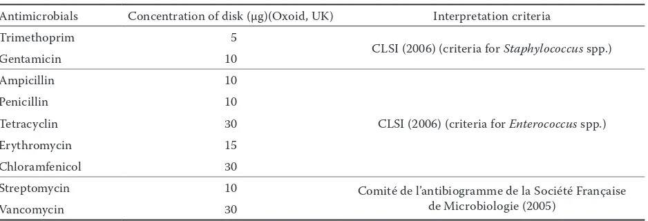
Resistance to antimicrobials. Phenotypic resist-ance testing was performed by the disk diffusion method on Mueller-Hinton agar (Bio-Rad, Paris, France). The antimicrobials tested and interpreta-tion criteria used are shown in Table 2.
Detection of virulence genes. Four PCR assays
were performed to detect the following virulence genes: (1) prfa (D’Agostino et al. 2004) and plca
(Jaradat et al. 2002), (2) hlya (Aurora et al.
2008) and acta (Jaradat et al. 2002), (3) plcb
(Jaradat et al. 2002), and (4) inla, inlC, inlJ
(Liu et al. 2007) and inlb (Jaradat et al. 2002). All assays used primers synthesised by Generi Biotech (Hradec Králové, Czech Republic), PPP polymerase (Top-Bio, Prague, Czech Republic) or a Qiagen Multiplex PCR Kit (Bio-Consult, Prague, Czech Republic).
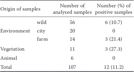
Table 1. Origin and number of analysed and positive
samples for l. monocytogenes
Origin of samples analysed samplesNumber of positive samplesNumber (%) of
Environment
wild 56 6 (10.7)
city 20 0
farm 14 3 (21.4)
Vegetation 11 3 (27.3)
Animal 6 0
[image:2.604.313.540.115.238.2]PCR-RFLP analysis of inlA polymorphism.
Potentially non-invasive l. monocytogenes strains were screened by PCR-RFLP based on inla poly-morphism (Rousseaux et al. 2004). The 733 bp
inla fragment was amplified using primers seq01 and seq02 (Rousseaux et al. 2004) (Generi Biotech, Hradec Králové, Czech Republic). The amplified product was cleaved with restriction endonuclease
aluI (BioLabs, Hitchin, UK) and PCR-RFLP frag-ments were separated by electrophoresis on 3.5% gel (SERVA Electrophoresis GmbH, Heidelberg, Germany).
Pulsed-field gel electrophoresis (PFGE).
Mac-rorestriction analysis using endonuclease ascI (BioLabs, Hitchin, UK) was performed according to the protocol PulseNet Europe (2002).
RESULTS AND DISCUSSION
l. monocytogenes was detected in 12 (11.2%) of 107 environmental samples analysed in this study. All positive samples were collected in areas characterised by high humidity or the presence of either farm or wild warm-blooded animals (Table 3), which is in line with the reports from other countries (Welshimer & Donket-Voet 1971; Weis & Seeliger 1975; Colburn et al.
1990; Lyautey et al. 2007). The highest proportion of l. monocytogenes isolates (66.7%) was found in the mud samples from the vicinity of water courses and in the soil samples from the areas where animals live.
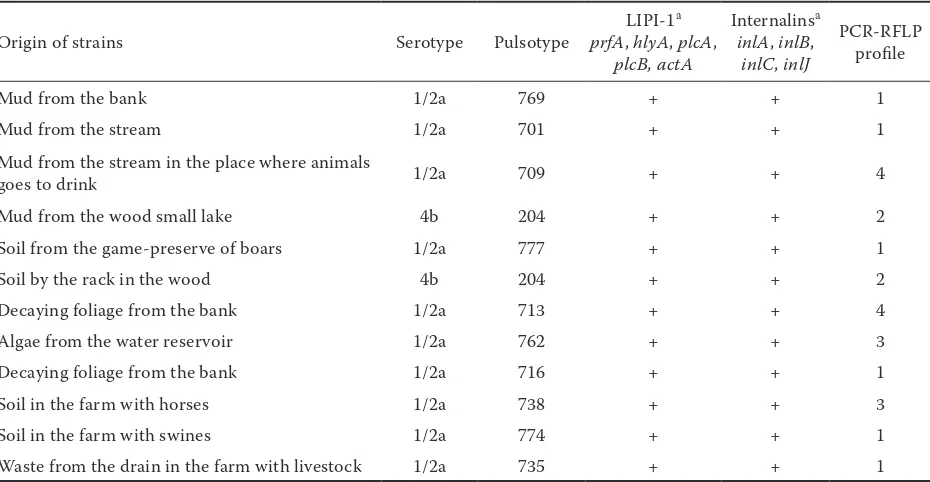
Most l. monocytogenes isolates (10/12) were of serotype 1/2a. Only two isolates from different sites of the same area were classified into serotype 4b. Both strains of this serotype were of the same
clonal type (pulsotype 204). Thus, it appears that 1/2a is the most widespread serotype in the Czech Republic, found not only in foodstuffs and humans (Karpíšková & Gelbíčová 2008) but also in the outdoor environment.
None of the 12 isolates recovered showed re-sistance to antimicrobials (ampicillin, penicillin, gentamicin, and trimetoprim) used in the treat-ment of human listeriosis. The environtreat-mental iso-lates were also susceptible to other antimicrobials tested. Although l. monocytogenes is commonly susceptible to a wide range of antimicrobials, some authors have reported tetracycline resist-ance in environmental strains of l. monocytogenes
(MacGowan et al. 1994) or multidrug resist-ance to ampicillin, erythromycin, tetracycline, dicloxacillin, and co-trimoxazole (Rodas-Suárez
et al. 2006). The monitoring of l. monocytogenes
resistance to antimicrobials is currently needed as well as the development of international criteria for the evaluation of resistant strains.
All genes involved in the pathogenicity of
l. monocytogenes (prfa, hlya, plca, plcb, acta,
inla, and inlb) were detected in the environmental isolates. However, some studies have also reported avirulent strains of l. monocytogenes recovered from foodstuffs (Chen et al. 2009a; Kaur et al.
2010) or from faeces of captive wild animals (Ka-lorey et al. 2006) that typically lack some of these virulence genes. Whether the strains lacking
prfa or hlya (Kalorey et al. 2006; Kaur et al.
[image:3.604.65.534.99.259.2]2010) that are commonly used for the detection of l. monocytogenes were correctly identified to the species level by phenotypic methods remains a question. Based on the results of this study and other studies (Chen et al. 2009a; Mammina et al. 2009), the inlC and inlJ genes are also stable
Table 2. Tested antimicrobials with their concentrations and interpretation criteria used
Antimicrobials Concentration of disk (µg)(Oxoid, UK) Interpretation criteria
Trimethoprim 5
CLSI (2006) (criteria for Staphylococcus spp.)
Gentamicin 10
Ampicillin 10
CLSI (2006) (criteria for enterococcus spp.)
Penicillin 10
Tetracyclin 30
Erythromycin 15
Chloramfenicol 30
Streptomycin 10 Comité de l’antibiogramme de la Société Française
de Microbiologie (2005)
parts of the genome of l. monocytogenes, but their roles in the virulence remain unclear (Bierne et al. 2007). Nevertheless, other authors (Chen et al.
2009a,b) have reported the absence of the inlC and
inlJ genes in l. monocytogenes strains of serotype 4a exhibiting a lower virulence. This is explained by a higher genetic relatedness between serotype 4a and the non-pathogenic species l. innocua that is not a producer of InlC and InlJ proteins either (Chen et al. 2009b).
An important role in the pathogenicity of l. mono-cytogenes is played by internalin A that mediates the interaction with E-cadherin of epithelial cells, thus enabling the passage through the intestinal barrier (Bierne et al. 2007). By the PCR-RFLP method for the detection of the inla gene polymorphism, the strains tested were classified into four of the five profiles described previously (Rousseaux
et al. 2004). Most strains tested were of profile 1 (6/12). The strains of profiles 1 and 4 have been found to produce the truncated form InlA and to have a lower capability of invading tissue culture epithelial cells (Caco-2) (Rousseaux et al. 2004; Lyautey et al. 2007). However, in our study, these profiles were also detected in the strains that are clonally identical to those recovered from human listeriosis cases, i.e. in a strain of pulsotype 713 and PCR-RFLP profile 4, and in strains of pulsotypes 716 and 735 and of PCR-RFLP profile 1. Therefore,
it can be stated that some strains of profiles 1 and 4 may also be producers of the functional form of InlA (Rousseaux et al. 2004). Nevertheless, from our results, it cannot be concluded unambiguously whether these strains actually produce an 80 kDa InlA or whether other factors also play a role in their invasiveness. On the other hand, 4 (33.3%) of the 12 strains classified into profiles 2 and 3 are potentially invasive strains in which functional internalin A was reported by others (Rousseaux
et al. 2004).
Macrorestriction analysis revealed 11 different pulsotypes among the isolates recovered. Some of them were detected for the first time (774, 777), while others have already been found not only in food chain isolates but also in human isolates (as documented by the database of the National Institute of Public Health, National Reference Laboratory for Listeria, Prague, Czech Republic).
[image:4.604.74.539.497.738.2]l. monocytogenes clones of serotype 1/2a and of pulsotypes 713, 716, and 738 as well as the clone of serotype 4b and pulsotype 204 were recovered from sporadic human listeriosis cases from 2007 through 2010. The l. monocytogenes strain of serotype 1/2a and pulsotype 735 detected in waste from a drain in the farm with livestock (sewer of livestock farm) belongs to the same clone as that implicated in the listeriosis outbreak related to vacuum packed ham in the South Bohemian Region at the turn of
Table 3. Characteristics of l. monocytogenes strains of environmental origin
Origin of strains Serotype Pulsotype LIPI-1
a
prfa, hlya, plca, plcb, acta
Internalinsa
inla, inlb, inlC, inlJ
PCR-RFLP profile
Mud from the bank 1/2a 769 + + 1
Mud from the stream 1/2a 701 + + 1
Mud from the stream in the place where animals
goes to drink 1/2a 709 + + 4
Mud from the wood small lake 4b 204 + + 2
Soil from the game-preserve of boars 1/2a 777 + + 1
Soil by the rack in the wood 4b 204 + + 2
Decaying foliage from the bank 1/2a 713 + + 4
Algae from the water reservoir 1/2a 762 + + 3
Decaying foliage from the bank 1/2a 716 + + 1
Soil in the farm with horses 1/2a 738 + + 3
Soil in the farm with swines 1/2a 774 + + 1
Waste from the drain in the farm with livestock 1/2a 735 + + 1
2008/2009 (Karpíšková & Gelbíčová 2009). In-terestingly, the strain of serotype 1/2a and pulsotype 713 is classified into the persistent clone which has long been detected in a Czech plant making soft-ripened cheeses (Gelbíčová et al. 2008). These results are in accordance with those reported in other countries (Fugett et al. 2007), showing that some l. monocytogenes clones are associated with specific sources, while others are widespread and can be found among the isolates from the farm environment, foodstuffs, environmental sources, or humans. Therefore, to facilitate epidemiological investigations and identification of l. monocytogenes
sources, it is necessary to maintain an extensive database with phenotypic and genotypic charac-teristics of strains of different origin.
CONCLUSION
The bacterium l. monocytogenes can be isolated from the external environment, in particular from wet areas or from those where animals are present. The predominance of serotype 1/2a and susceptibil-ity of the isolates to antimicrobials are in line with the available data on food and human isolates from all over the Czech Republic. The results obtained have confirmed that the environment is a source of potentially pathogenic l. monocytogenes strains that carry a basic set of virulence genes and have the potential for invading the host epithelial cells.
References
Aurora R., Prakash A., Prakash S., Rawool D.B., Bar-buddhe S.B. (2008): Comparison of PI-PLC based assays and PCR along with in vivo pathogenicity tests for rapid detection of pathogenic listeria monocytogenes. Food Control, 19: 641–647.
Bierne H., Sabet C., Personnic N., Cossart P. (2007): Internalins: a complex family of leucine-rich repeat-con-taining proteins in listeria monocytogenes. Microbes and Infection, 9: 1156–1166.
Chen J., Luo X., Jiang L., Jin P., Wei W., Liu D., Fang W. (2009a): Molecular characteristics and virulence potential of listeria monocytogenes isolates from Chinese food systems. Food Microbiology, 26: 103–111.
Chen J., Jiang L., Chen X., Lou X., Chen Y., Yu Y., Tian G., Liu D., Fang W. (2009b): listeria monocytogenes serovar 4a is a possible evolutionary intermediate be-tween l. monocytogenes serovars 1/2a and 4b and l.
in-nocua. Journal of Microbiology and Biotechnology, 19: 238–249.
Colburn K.G., Kaysner CH.A., Carlos Abeyta J.R., Wekell M.M. (1990): listeria species in a California coast estuarine environment. Applied and Environmental Microbiology, 56: 2007–2011.
CLSI (2006); Performance standards for antimicrobial sus-ceptibilty testing; sixteenth informational supplement. NCCLS document M100-S16. Clinical and Laboratory Standards Institute, Wayne: 44–55.
Comité de l’antibiogramme de la Société Française de Microbiologie (2005): Concentrations et diamètres cri-tiques pour les diverses classes d’antibiocri-tiques. Société Française de Microbiologie:11–12.
D’Agostino M., Wagner M., Vazquez-Boland J.A., Kuch- ta T., Karpiskova R., Hoorfar J., Novella S., Scortti M., Ellison J., Murray A., Fernandes I., Kuhn M., Pazla- rova J., Heuvelink A., Cook N.A. (2004): A validated PCR-based method to detect listeria monocytogenes us-ing raw milk as a food model-towards an international standard. Journal of Food Protection, 67: 1646–1655.
Doumith M., Cazalet Ch., Simoes N., Frangeul L., Jacquet Ch., Kunst F., Martin P., Cossart C., Glaser P., Buchrieser C. (2004a): New aspects regarding evolu-tion and virulence of listeria monocytogenes revealed by comparative genomics and DNA arrays. Infection and Immunity, 72: 1072–1083.
Doumith M., Buchrieser C., Glaser P., Jacquet C., Martin P. (2004b): Differentiation of the major listeria monocytogenes serovars by multiplex PCR. Journal of Clinical Microbiology, 42: 3819–3822.
Esteban J.I., Oporto B., Aduriz G., Juste R.A., Hurtado A. (2009): Faecal shedding and strain diversity of listeria monocytogenes in healthy ruminants and swine in North-ern Spain. BMC Veterinary Research, 5: 2.
EN ISO 11290-1:1996/Amd.1:2004: Microbiology of food and animal feeding stuffs-Horizonatl method for the detection and enumeration of listeria monocytogenes – Part 1: Detection Method.
Farber J.M., Peterkin P.I. (1991): listeria monocytogenes, a food-borne pathogen. Microbiological Reviews, 55: 476–511.
Freitag N.E., Port G.C., Miner M.D. (2009): listeria monocytogenes – from saprophyte to intracellular patho-gen. Nature Reviews Microbiology, 7: 623–628.
Gelbíčová T., Šťástková Z., Pospíšilová M., Kar- píšková R. (2008): Charakteristika izolátů listeria mono- cytogenes z mléčných výrobků. Veterinářství, 58: 324–326. Gray M.J., Freitag N.E., Boor K.J. (2006): How the bacte-rial pathogen listeria monocytogenes mediates the switch from environmental Dr. Jekyll to pathogenic Mr. Hyde. Infection and Immunity, 74: 2505–2512.
Ivanek R., Gröhn Y.T., Wiedmann M. (2006): listeria monocytogenes in multiple habitats and host populations: review of available data for mathematical modeling. Food-borne Pathogens and Disease, 3: 319–336.
Jaradat Z.W., Schutze G.E., Bhunia A.K. (2002): Genetic homogeneity among listeria monocytogenes strains from infected patients and meat products from two geographic locations determined by phenotyping, ribotyping and PCR analysis of virulence genes. International Journal of Food Microbiology, 76: 1–10.
Kalorey D.R., Kurkure N.V., Warke S.R., Rawool D.B., Malík S.V.S., Barbuddhe S.B. (2006): Isolation of patho-genic listeria monocytogenes in faeces of wild animals in captivity. Comparative Immunology, Microbiology and Infectious Diseases, 29: 295–300.
Karpíšková R., Gelbíčová T. (2008): Charakteristika a prevalence klonů listeria monocytogenes izolovaných od pacientů v letech 2001–2008 v České republice. Epidemio- logie, mikrobiologie, imunologie, 57: 137–140.
Karpíšková R., Gelbíčová T. (2009): The role of typing methods in epidemiological investigations of listeriosis. In: 3rd Congress of European Microbiologists FEMS.
Sweden: 232.
Kaur S., Malík S.V.S., Bhilegaonkar K.N., Vaidya V.M., Barbuddhe S.B. (2010): Use of phospholipase-C assay, in vivo pathogenicity assays and PCR in assessing the viru-lence of listeria spp. Veterinary Journal, 184: 366–370. Liu D., Lawrence M.L., Austin F.W., Ainsworth A.J.
(2007): A multiplex PCR for species – and virulence – spe-cific determination of listeria monocytogenes. Journal of Microbiological Methods, 71: 133–140.
Lyautey E., Lapen D.R., Wilkes G., McCleary K., Pagot-to F., Tyler K., Hartmann A., Piveteau P., Rieu A., Robertson W.J., Medeiros D.T., Edge T.A., Gannon V., Topp E. (2007): Distribution and characteristics of
listeria monocytogenes isolates from surface waters of the south nation river watershed, Ontario, Canada. Applied and Environmental Microbiology, 73: 5401–5410. MacGowan A.P., Bowker K., McLauchlin J., Bennett
P.M., Reeves D.S. (1994): The occurrence and seasonal changes in the isolation of listeria spp. in shop bought food stuffs, human faeces, sewage and soil from urban sources. International Journal of Food Microbiology, 21: 325–334.
Mammina C., Aleo A., Romani C., Pellissier N., Nicoletti P., Pecile P., Nastasi A., Pontello M.M. (2009): Characterization of listeria monocytogenes iso-lates from human listeriosis cases in Italy. Journal of Clinical Microbiology, 47: 2925–2930.
Pulse-net Europe (2002): Standardized protocol for molecu-lar subtyping of listeria monocytogenes by pulsed-field gel electrophoresis (PFGE): 10. Available at http://www. pulsenet-europe.org
Rodas-Suárez O.R., Flores-Pedroche J.F., Betan-court-Rule J.M., Quiñones-Ramírez E.I., Vázquez-Salinas C. (2006): Occurrence and antibiotic sensitivity of listeria monocytogenes strains isolated from oysters, fish, and estuarine water. Applied and Environmental Microbiology, 72: 7410–7412.
Rousseaux S., Olier M., Lemaître J.P., Piveteau P., Guzzo J. (2004): Use of PCR-restriction fragment length polymorphism of inla for rapid screening of listeria monocytogenes strains deficient in the ability to invade Caco-2 cells. Applied and Environmental Microbiology,
70: 2180–2185.
Vázquez-Boland J.A., Kuhn M., Berche P., Chakraborty T., Domínguez-Bernal G., Goebel W., González-Zorn B., Wehland J., Kreft J. (2001): listeria patho-genesis and molecular virulence determinants. Clinical Microbiological Reviews, 14: 584–640.
Weis J., Seeliger H.P.R. (1975): Incidence of listeria mono-cytogenes in nature. Applied Microbiology, 30: 29–32. Welshimer H.J., Donket-Voet J. (1971): listeria
monocy-togenes in nature. Applied Microbiology, 21: 516–519.
Received for publication January 7, 2011 Accepted after corrections March 7, 2011
Corresponding author:
Mgr. Tereza Gelbíčová, Ph.D., Masarykova univerzita, Přírodovědecká fakulta, Ústav experimentální biologie, Tvrdého 14, 602 00 Brno, Česká republika