N
-Methyl-2-(1-methyl-3-phenylprop-2-en-1-ylidene)hydrazinecarbothioamide
Fillipe Vieira Rocha,aAdelino Vieira de Godoy Netto,a Johannes Beck,bJo¨rg Danielsb and Adriano Bof de Oliveirac*
aInstituto de Quı´mica, Universidade Estadual Paulista, Rua Francisco Degni s/n, 14801-970 Araraquara-SP, Brazil,b
Institut fu¨r Anorganische Chemie, Universita¨t Bonn, Gerhard-Domagk-Strasse 1, D-53121 Bonn, Germany, andcDepartamento de Quı´mica, Universidade Federal de Sergipe, Av. Marechal Rondon s/n, Campus, 49100-000 Sa˜o Cristo´va˜o-SE, Brazil
Correspondence e-mail: adriano@daad-alumni.de
Received 30 May 2014; accepted 13 June 2014
Key indicators: single-crystal X-ray study;T= 123 K; mean(C–C) = 0.002 A˚; Rfactor = 0.031;wRfactor = 0.080; data-to-parameter ratio = 13.6.
In the title compound, C12H15N3S, the molecule deviates
slightly from planarity, with a maximum deviation from the mean plane of the non-H atoms of 0.2756 (6) A˚ for the S atom and a torsion angle for the N—N—C—N fragment of 7.04 (16). In the crystal, molecules are linked by N— H S hydrogen-bond interactions, forming centrosymmetric dimers. Additionally, one weak intramolecular N—H N hydrogen-bond interaction is observed. The crystal packing shows a herringbone arrangement viewed along thecaxis.
Related literature
For one of the first reports of the synthesis of thio-semicarbazone derivatives, see: Freund & Schander (1902). For a report of the antifungal activity of the title compound, see: Nishimuraet al.(1979).
Experimental
Crystal data
C12H15N3S Mr= 233.33
a= 10.5832 (2) A˚
b= 7.9509 (2) A˚
c= 28.9259 (5) A˚
V= 2434.00 (9) A˚3
MoKradiation
= 0.24 mm1
T= 123 K
0.440.310.27 mm
Data collection
Nonius KappaCCD diffractometer Absorption correction: multi-scan
(Blessing, 1995)
Tmin= 0.904,Tmax= 0.955
26770 measured reflections 2783 independent reflections 2414 reflections withI> 2(I)
Rint= 0.046
Refinement
R[F2> 2(F2)] = 0.031
wR(F2) = 0.080
S= 1.05 2783 reflections
205 parameters
All H-atom parameters refined
max= 0.27 e A˚
3
min=0.20 e A˚
[image:1.610.313.564.282.322.2]3
Table 1
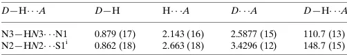
Hydrogen-bond geometry (A˚ ,).
D—H A D—H H A D A D—H A
N3—HN3 N1 0.879 (17) 2.143 (16) 2.5877 (15) 110.7 (13) N2—HN2 S1i
0.862 (18) 2.663 (18) 3.4296 (12) 148.7 (15)
Symmetry code: (i)xþ1;y;z.
Data collection: COLLECT (Nonius, 1998); cell refinement: SCALEPACK (Otwinowski & Minor, 1997); data reduction:
DENZO (Otwinowski & Minor, 1997) and SCALEPACK;
program(s) used to solve structure: SHELXS97 (Sheldrick, 2008); program(s) used to refine structure:SHELXL97(Sheldrick, 2008); molecular graphics:DIAMOND(Brandenburg, 2006); software used to prepare material for publication:publCIF(Westrip, 2010).
We gratefully acknowledge financial support by the German Research Foundation (DFG) through the Collaborative Research Center SFB 813, Chemistry at Spin Centers and by FAPITEC/SE/FUNTEC/CNPq through the PPP Program 04/ 2011. FVR acknowledges FAPESP for the Post-Doctoral scholarship, Proc. No. 2013/20156–5.
Supporting information for this paper is available from the IUCr electronic archives (Reference: BX2460).
References
Blessing, R. H. (1995).Acta Cryst.A51, 33–38.
Brandenburg, K. (2006).DIAMOND. Crystal Impact GbR, Bonn, Germany. Freund, M. & Schander, A. (1902).Chem. Ber.35, 2602–2606.
Nishimura, T., Toku, H., Matsumoto, K., Iwata, M. & Watanabe, T. (1979). Jpn Patent No. 54119029 A.
Nonius (1998).COLLECT. Nonius BV, Delft, The Netherlands.
Otwinowski, Z. & Minor, W. (1997). Methods in Enzymology, Vol. 276, Macromolecular Crystallography, Part A, edited by C. W. Carter Jr & R. M. Sweet, pp. 307–326. New York: Academic Press, United States.
Sheldrick, G. M. (2008).Acta Cryst.A64, 112–122. Westrip, S. P. (2010).J. Appl. Cryst.43, 920–925.
Structure Reports Online
supporting information
Acta Cryst. (2014). E70, o800 [https://doi.org/10.1107/S1600536814013889]
N-Methyl-2-(1-methyl-3-phenylprop-2-en-1-ylidene)hydrazinecarbothioamide
Fillipe Vieira Rocha, Adelino Vieira de Godoy Netto, Johannes Beck, J
ö
rg Daniels and Adriano
Bof de Oliveira
S1. Comment
Thiosemicarbazone derivatives have a wide range of biological properties. For example, some thiosemicarbazones similar
to the title compound show antifungal activity (Nishimura et al., 1979). As part of our study on synthesis and structural
chemistry of thiosemicarbazone derivatives from natural products, we report herein the crystal structure of a derivative of
the essential oil of cinnamon bark (benzylideneacetone, a methyl derivative of the cinnamaldehyde).
In the crystal structure of the title compound the central N–N–C–N unit is not planar with an torsion angle along N1–
N2–C10–N3 of -7.04 (16)° and the maximum deviation from the mean plane of the non-H atoms amounting to 0.2756 (6)
Å for S1. The molecule, shows a trans conformation at the C7—C8 and N1—N2 bonds (Fig. 1).
In the crystal the molecules are linked by N—H···S hydrogen bonds interactions forming centrosymmetric dimers.
Additionally, one weak N—H···N intramolecular H-interaction is observed.The crystal packing shows a herringbone
arrangement viewed along the c-axis.(Fig. 3).
S2. Experimental
Starting materials were commercially available and were used without further purification. The title compound synthesis
was adapted to a procedure reported previously (Freund & Schander, 1902). The hydrochloric acid catalyzed reaction, a
mixture of benzylideneacetone (10 mmol) and 4-methyl-3-thiosemicarbazide (10 mmol) in ethanol (80 ml) was refluxed
for 5 h. After cooling and filtering, the title compound was obtained. Crystals suitable for X-ray diffraction were obtained
in ethanol by the slow evaporation of solvent.
S3. Refinement
All hydrogen atoms were localized in a difference density Fourier map. Their positions and isotropic displacement
Figure 1
The molecular structure of the title compound with labeling and displacement ellipsoids drawn at the 50% probability
level.
Figure 2
Part of the crystal structure of the title compound showing the inter- and intramolecular hydrogen bonding as dashed
[image:3.610.129.475.312.500.2]Figure 3
Crystal structure of the title compound viewed along the c-axis. The herringbone pattern of the crystal packing along the
a-axis is observed.
N-Methyl-2-(1-methyl-3-phenylprop-2-en-1-ylidene)hydrazinecarbothioamide
Crystal data
C12H15N3S Mr = 233.33
Orthorhombic, Pbca
Hall symbol: -P 2ac 2ab
a = 10.5832 (2) Å
b = 7.9509 (2) Å
c = 28.9259 (5) Å
V = 2434.00 (9) Å3 Z = 8
F(000) = 992
Dx = 1.273 Mg m−3
Mo Kα radiation, λ = 0.71073 Å Cell parameters from 31577 reflections
θ = 2.9–27.5°
µ = 0.24 mm−1 T = 123 K Fragment, yellow 0.44 × 0.31 × 0.27 mm
Data collection
Nonius KappaCCD diffractometer
Radiation source: fine-focus sealed tube, Nonius KappaCCD
Graphite monochromator
Detector resolution: 9 pixels mm-1
CCD rotation images, thick slices scans Absorption correction: multi-scan
(Blessing, 1995)
Tmin = 0.904, Tmax = 0.955
26770 measured reflections 2783 independent reflections 2414 reflections with I > 2σ(I)
Rint = 0.046
θmax = 27.5°, θmin = 3.3° h = −13→13
k = −10→10
l = −37→37
Refinement
Refinement on F2
Least-squares matrix: full
R[F2 > 2σ(F2)] = 0.031 wR(F2) = 0.080 S = 1.05 2783 reflections 205 parameters
Primary atom site location: structure-invariant direct methods
Secondary atom site location: difference Fourier map
Hydrogen site location: inferred from neighbouring sites
where P = (Fo2 + 2Fc2)/3
(Δ/σ)max = 0.001
Δρmin = −0.20 e Å−3
Special details
Geometry. All e.s.d.'s (except the e.s.d. in the dihedral angle between two l.s. planes) are estimated using the full covariance matrix. The cell e.s.d.'s are taken into account individually in the estimation of e.s.d.'s in distances, angles and torsion angles; correlations between e.s.d.'s in cell parameters are only used when they are defined by crystal symmetry. An approximate (isotropic) treatment of cell e.s.d.'s is used for estimating e.s.d.'s involving l.s. planes.
Refinement. Refinement of F2 against ALL reflections. The weighted R-factor wR and goodness of fit S are based on F2,
conventional R-factors R are based on F, with F set to zero for negative F2. The threshold expression of F2 > σ(F2) is used
only for calculating R-factors(gt) etc. and is not relevant to the choice of reflections for refinement. R-factors based on F2
are statistically about twice as large as those based on F, and R- factors based on ALL data will be even larger.
Fractional atomic coordinates and isotropic or equivalent isotropic displacement parameters (Å2)
x y z Uiso*/Ueq
Atomic displacement parameters (Å2)
U11 U22 U33 U12 U13 U23
S1 0.02483 (17) 0.02489 (17) 0.01390 (16) −0.00102 (12) 0.00080 (11) 0.00161 (11) N1 0.0220 (5) 0.0225 (5) 0.0138 (5) −0.0007 (4) −0.0006 (4) 0.0000 (4) N2 0.0226 (5) 0.0224 (5) 0.0140 (5) −0.0047 (4) −0.0002 (4) 0.0002 (4) N3 0.0236 (5) 0.0208 (5) 0.0183 (5) −0.0042 (4) 0.0058 (4) −0.0026 (4) C1 0.0228 (6) 0.0204 (6) 0.0163 (6) 0.0009 (5) 0.0004 (5) 0.0005 (5) C2 0.0241 (6) 0.0282 (7) 0.0191 (6) −0.0035 (5) 0.0001 (5) 0.0012 (5) C3 0.0304 (7) 0.0275 (7) 0.0217 (7) −0.0049 (6) −0.0065 (5) 0.0002 (5) C4 0.0407 (8) 0.0288 (7) 0.0154 (6) −0.0017 (6) −0.0031 (5) 0.0007 (5) C5 0.0336 (8) 0.0356 (8) 0.0185 (6) −0.0045 (6) 0.0046 (5) 0.0030 (6) C6 0.0253 (6) 0.0292 (7) 0.0201 (6) −0.0052 (5) 0.0005 (5) 0.0011 (5) C7 0.0214 (6) 0.0216 (6) 0.0179 (6) −0.0013 (5) −0.0012 (5) −0.0014 (5) C8 0.0217 (6) 0.0212 (6) 0.0180 (6) −0.0009 (5) −0.0009 (5) −0.0013 (5) C9 0.0197 (6) 0.0183 (6) 0.0170 (6) 0.0019 (5) 0.0002 (4) 0.0010 (5) C10 0.0168 (5) 0.0176 (5) 0.0177 (6) 0.0035 (4) 0.0001 (4) 0.0019 (4) C11 0.0245 (6) 0.0258 (7) 0.0252 (7) −0.0062 (5) 0.0086 (5) −0.0030 (6) C12 0.0212 (6) 0.0267 (7) 0.0161 (6) −0.0029 (5) 0.0004 (5) 0.0000 (5)
Geometric parameters (Å, º)
S1—C10 1.6875 (12) C4—H4 0.964 (17)
N1—C9 1.2994 (16) C5—C6 1.3896 (19)
N1—N2 1.3769 (14) C5—H5 0.950 (18)
N2—C10 1.3708 (15) C6—H6 0.967 (17)
N2—HN2 0.862 (18) C7—C8 1.3402 (18)
N3—C10 1.3261 (16) C7—H7 0.950 (16)
N3—C11 1.4518 (16) C8—C9 1.4616 (16)
N3—HN3 0.879 (17) C8—H8 0.948 (16)
C1—C6 1.3980 (18) C9—C12 1.4981 (17)
C1—C2 1.4029 (18) C11—H11A 0.96 (2)
C1—C7 1.4716 (17) C11—H11B 0.92 (2)
C2—C3 1.3917 (18) C11—H11C 0.94 (2)
C2—H2 0.949 (18) C12—H12A 0.965 (18)
C3—C4 1.388 (2) C12—H12B 0.964 (18)
C3—H3 0.965 (17) C12—H12C 0.977 (17)
C4—C5 1.387 (2)
C9—N1—N2 117.87 (10) C8—C7—C1 125.58 (12)
C10—N2—N1 117.43 (10) C8—C7—H7 120.2 (9)
C10—N2—HN2 117.2 (11) C1—C7—H7 114.3 (9)
N1—N2—HN2 121.0 (11) C7—C8—C9 126.19 (12)
C10—N3—C11 123.89 (11) C7—C8—H8 121.4 (10)
C10—N3—HN3 115.2 (11) C9—C8—H8 112.4 (10)
C11—N3—HN3 120.8 (11) N1—C9—C8 113.27 (11)
C3—C2—C1 120.46 (12) N3—C10—S1 124.41 (9)
C3—C2—H2 119.3 (10) N2—C10—S1 119.73 (9)
C1—C2—H2 120.3 (10) N3—C11—H11A 110.6 (12)
C4—C3—C2 120.56 (13) N3—C11—H11B 111.8 (13)
C4—C3—H3 120.8 (10) H11A—C11—H11B 108.3 (17)
C2—C3—H3 118.7 (10) N3—C11—H11C 109.8 (13)
C5—C4—C3 119.49 (12) H11A—C11—H11C 105.7 (18)
C5—C4—H4 121.0 (10) H11B—C11—H11C 110.4 (18)
C3—C4—H4 119.5 (10) C9—C12—H12A 111.0 (10)
C4—C5—C6 120.18 (13) C9—C12—H12B 112.4 (10)
C4—C5—H5 119.6 (11) H12A—C12—H12B 105.3 (14)
C6—C5—H5 120.2 (11) C9—C12—H12C 111.2 (10)
C5—C6—C1 121.07 (13) H12A—C12—H12C 110.1 (14)
C5—C6—H6 119.2 (10) H12B—C12—H12C 106.6 (14)
C1—C6—H6 119.7 (10)
N1—N1—N2—C10 0.00 (9) N2—N1—C9—N1 0 (100)
C9—N1—N2—C10 178.27 (11) N1—N1—C9—C8 0.00 (7)
C9—N1—N2—N1 0 (100) N2—N1—C9—C8 178.31 (10)
C6—C1—C2—C3 −0.9 (2) N1—N1—C9—C12 0.000 (19)
C7—C1—C2—C3 178.99 (13) N2—N1—C9—C12 −2.44 (18)
C1—C2—C3—C4 −0.8 (2) C7—C8—C9—N1 −176.06 (12)
C2—C3—C4—C5 1.4 (2) C7—C8—C9—N1 −176.06 (12)
C3—C4—C5—C6 −0.3 (2) C7—C8—C9—C12 4.7 (2)
C4—C5—C6—C1 −1.4 (2) C11—N3—C10—N2 −178.42 (12)
C2—C1—C6—C5 2.0 (2) C11—N3—C10—S1 0.53 (18)
C7—C1—C6—C5 −177.89 (13) N1—N2—C10—N3 −7.04 (16) C6—C1—C7—C8 168.26 (13) N1—N2—C10—N3 −7.04 (16)
C2—C1—C7—C8 −11.7 (2) N1—N2—C10—S1 173.96 (9)
C1—C7—C8—C9 177.50 (12) N1—N2—C10—S1 173.96 (9)
Hydrogen-bond geometry (Å, º)
D—H···A D—H H···A D···A D—H···A
N3—HN3···N1 0.879 (17) 2.143 (16) 2.5877 (15) 110.7 (13) N2—HN2···S1i 0.862 (18) 2.663 (18) 3.4296 (12) 148.7 (15)