4-(2
H
-1,3-Benzodioxol-5-yl)-1-(4-methyl-phenyl)-1
H
-pyrazol-5-amine
Nilesh N. Gajera,aMukesh C. Patel,aMukesh M. Jotanib‡ and Edward R. T. Tiekinkc*
aP.S. Science and H.D. Patel Arts College, S.V. Campus, Kadi, Gujarat 382 715, India,bDepartment of Physics, Bhavan’s Sheth R.A. College of Science, Ahmedabad, Gujarat 380 001, India, andcDepartment of Chemistry, University of Malaya, 50603 Kuala Lumpur, Malaysia
Correspondence e-mail: edward.tiekink@gmail.com
Received 10 April 2013; accepted 10 April 2013
Key indicators: single-crystal X-ray study;T= 293 K; mean(C–C) = 0.002 A˚; Rfactor = 0.045;wRfactor = 0.136; data-to-parameter ratio = 16.1.
In the title compound, C17H15N3O2, two independent
mol-ecules (A andB) comprise the asymmetric unit. The major conformational difference arises in the relative orientation of the pyrazole ring amine and dioxole substituents which are antiinAandsyninB. The five-membered dioxole ring in each molecule has an envelope conformation with the methylene C atom as the flap. The mean plane through the benzodioxole and benzene groups make dihedral angles of 31.67 (8) and 68.22 (9), respectively, with the pyrazole ring in A; the equivalent values for B are 47.18 (7) and 49.08 (9). In the crystal, supramolecular zigzag chains along the b-axis direc-tion arise as a result of N—H N hydrogen bonding. These are consolidated into supramolecular double chains via C— H O and C—H interactions.
Related literature
For background to the biological activity of amino substituted pyrazole derivatives, see: Tanitameet al.(2004); Chimentiet al. (2006); Dinget al.(2009); Shenet al.(2011); Denget al.(2012). For a related structure, see: Murugananthamet al.(2007).
Experimental
Crystal data
C17H15N3O2 Mr= 293.32 Triclinic,P1
a= 9.7690 (7) A˚
b= 10.4250 (7) A˚
c= 14.283 (1) A˚
= 96.626 (2) = 91.903 (2)
= 91.164 (2)
V= 1443.67 (17) A˚3 Z= 4
MoKradiation
= 0.09 mm1 T= 293 K
0.400.250.20 mm
Data collection
Bruker APEXII CCD diffractometer
Absorption correction: multi-scan (SADABS; Bruker, 2004)
Tmin= 0.965,Tmax= 0.982
29089 measured reflections 6620 independent reflections 4674 reflections withI> 2(I)
Rint= 0.031
Refinement
R[F2> 2(F2)] = 0.045 wR(F2) = 0.136 S= 1.02 6620 reflections 412 parameters 4 restraints
H atoms treated by a mixture of independent and constrained refinement
max= 0.19 e A˚
3
min=0.17 e A˚
[image:1.610.312.567.392.485.2]3
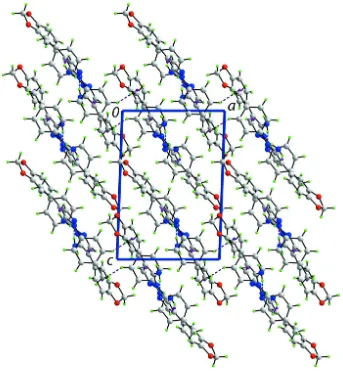
Table 1
Hydrogen-bond geometry (A˚ ,).
Cg1–Cg3 are the centroids of the C28–C33, C19–C24 and C2–C7 rings, respectviely.
D—H A D—H H A D A D—H A
N3—H1N N5i 0.89 (2) 2.20 (2) 3.059 (2) 161 (2) N6—H3N N2ii
0.89 (1) 2.11 (1) 2.9914 (19) 170 (2) C1—H1C O3ii
0.96 2.54 3.479 (2) 164
C3—H3 Cg1iii 0.93 2.83 3.5365 (19) 133 C10—H10 Cg2ii
0.93 2.88 3.6055 (17) 135 C27—H27 Cg3i
0.93 2.94 3.5903 (18) 128
Symmetry codes: (i) xþ1;y;zþ1; (ii) xþ1;yþ1;zþ1; (iii)
xþ1;y;zþ1.
Data collection:APEX2(Bruker, 2004); cell refinement:APEX2
andSAINT(Bruker, 2004); data reduction:SAINT; program(s) used to solve structure:SHELXS97(Sheldrick, 2008); program(s) used to refine structure:SHELXL97(Sheldrick, 2008); molecular graphics:
ORTEP-3 for Windows(Farrugia, 2012),QMol(Gans & Shalloway, 2001) and DIAMOND (Brandenburg, 2006); software used to prepare material for publication:publCIF(Westrip, 2010).
The authors are grateful to the Department of Science and Technology (DST), and SAIF, IIT Madras, Chennai, India, for the X-ray data collection. MCP is thankful to the University Grant Commission, New Delhi, India, for research funding under research project No. 39–715/2010(SR). We also thank the Ministry of Higher Education, Malaysia, for funding structural studies through the High-Impact Research scheme (UM.C/HIR-MOHE/SC/03).
Supplementary data and figures for this paper are available from the IUCr electronic archives (Reference: SU2585).
Acta Crystallographica Section E Structure Reports
Online
References
Brandenburg, K. (2006).DIAMOND. Crystal Impact GbR, Bonn, Germany. Bruker (2004).APEX2,SADABSandSAINT, Bruker AXS Inc., Madison,
Wisconsin, USA.
Chimenti, F., Bolasco, A., Manna, F., Secci, D., Chimenti, P., Granese, A., Befani, O., Turini, P., Cirilli, R., La Torre, F., Alcaro, S., Ortuso, F. & Langer, T. (2006).Curr. Med. Chem.13, 1411–1428.
Deng, H., Yu, Z., Shi, G., Chen, M., Tao, K. & Hou, T. (2012).Chem. Biol. Drug Des.79, 279–289.
Ding, X.-L., Zhang, H.-Y., Qi, L., Zhao, B.-X., Lian, S., Lv, H.-S. & Miao, J.-Y. (2009).Bioorg. Med. Chem. Lett.19, 5325–5328.
Farrugia, L. J. (2012).J. Appl. Cryst.45, 849–854.
Gans, J. & Shalloway, D. (2001).J. Mol. Graph. Model.19, 557–559. Muruganantham, R., Mobin, S. M. & Namboothiri, I. N. N. (2007).Org. Lett.9,
1125–1128.
Sheldrick, G. M. (2008).Acta Cryst.A64, 112–122.
Shen, D.-M., Brady, E. J., Candelore, M. R., Dallas-Yang, Q., Ding, V. D.-H., Feeney, W. P., Jiang, G., McCann, M. E., Mock, S., Qureshi, S. A., Saperstein, R., Shen, X., Tong, X., Tota, L. M., Wright, M. J., Yang, X., Zheng, S., Chapman, K. T., Zhang, B. B., Tata, J. R. & Parmee, E. R. (2011).Bioorg. Med. Chem. Lett.21, 76–81.
Tanitame, A., Oyamada, Y., Ofuji, K., Fujimoto, M., Iwai, N., Hiyama, Y., Suzuki, K., Ito, H., Terauchi, H., Kawasaki, M., Nagai, K., Wachi, M. & Yamagishi, J. (2004).J. Med. Chem.47, 3693–3696.
supporting information
Acta Cryst. (2013). E69, o736–o737 [https://doi.org/10.1107/S1600536813009914]
4-(2
H
-1,3-Benzodioxol-5-yl)-1-(4-methylphenyl)-1
H
-pyrazol-5-amine
Nilesh N. Gajera, Mukesh C. Patel, Mukesh M. Jotani and Edward R. T. Tiekink
S1. Comment
The amino substituted pyrazole unit is an important backbone in the area of synthetic as well as medicinal chemistry due to the broad range of biological activities of such compounds, such as depressant, anxiety, fungal, anti-bacterial, anti-diabetic and anti-cancer (Tanitame et al., 2004; Chimenti et al., 2006; Ding et al.,2009; Shen et al., 2011; Deng et al., 2012). In this connection, the title compound, (I), was synthesized and its crystal structure is reported on herein.
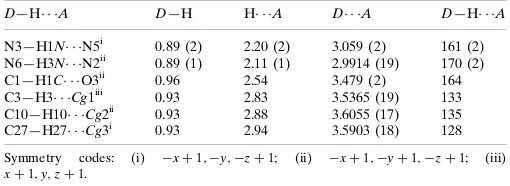
Two independent molecules (A and B), comprise the asymmetric unit of (I), see Fig. 1. As seen from the overlay diagram, Fig. 2, different conformations are observed for both the benzodioxole and benzene substituents. The five-membered dioxole rings in each molecule has an envelope conformation with the methylene-C17 or C34 atoms being the flap atoms. The r.m.s. deviation of the five non-hydrogen atoms = 0.049 Å for the N1-containing molecule which is considerably less than 0.129 Å for the second independent molecule, where the C34 atom lies 0.115 (2) Å out of the mean plane. For the N1-containing molecule, with respect to the pyrazole ring (r.m.s. deviation = Å), the benzodioxole and benzene groups make dihedral angles of 31.67 (8) and 68.22 (9)°, respectively. The equivalent values for the N2-containing molecule are 47.18 (7)° and 49.08 (9)°, respectively. From a conformational point of view, the dioxole ring in the N1-containing molecule is anti to the amine substituent whereas it is syn for the second molecule. The observed conformations in (I) are similar to those in a closely related structure, i.e. diethyl 4-(benzo[d][1,3]dioxol-5-yl)-1H -pyrazol-3-yl-3-phosphonate (Muruganantham et al., 2007).
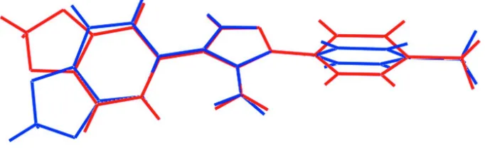
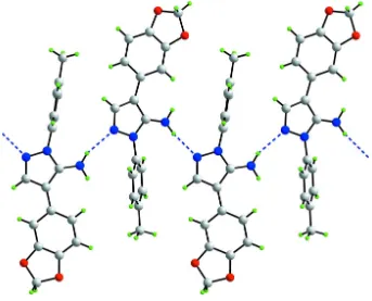
The presence of N—H···N hydrogen bonding leads to supramolecular zigzag chains along the b axis in the crystal structure of (I), see Fig. 3 and Table 1. These are consolidated into supramolecular double chains via C—H···O and C— H···π interactions (Table 1). These stack with no specific intermolecular interactions between them (Fig. 4).
S2. Experimental
A mixture of 3,4-methyleneoxyphenyl acetonitrile (2 g, 0.012 mol) and N,N-dimethylformamide dimethylacetal (4.89 ml, 0.037 mol) was stirred at 355 K; progress of the reaction was monitored by TLC. At the end of the reaction, the solvent was removed under vacuum. The residual crude mass was mixed with 4-methyl phenyl hydrazine hydrochloride (1.96 g, 0.012 mol) in methanol (20 ml) at room temperature. The mixture was refluxed and the reaction progress was monitored by TLC. At the end of the reaction, the solvent was removed under reduced pressure. The residue was dissolved in water and NaHCO3 solution was added until basic pH was obtained. The product was extracted in ethyl acetate (200 ml × 2),
and this ethyl acetate layer passed through Na2SO4 and concentrated to dryness. The crude mass was purified by silica gel
S3. Refinement
The C-bound H atoms were geometrically placed (C—H = 0.93–0.97 Å) and refined as riding with Uiso(H) = 1.2–
1.5Ueq(C). The N-bound H-atom was refined with the distance restraint N—H = 0.89 (1) Å, and with Uiso(H) = 1.2Ueq(N).
[image:4.610.131.475.137.282.2]Being affected by the beam-stop, the (0 0 1) reflection was removed from the final cycles of refinement.
Figure 1
The molecular structure of the two independent molecules comprising the asymmetric unit of the title compound (I), showing the atom labelling. Displacement ellipsoids are drawn at the 35% probability level.
Figure 2
[image:4.610.136.477.341.445.2]Figure 3
Figure 4
A view in projection along the b axis of the crystal packing of compound (I). The N—H···N, C—H···O (obscured) and C —H···π interactions are shown as dashed lines (see Table 1 for details).
4-(2H-1,3-Benzodioxol-5-yl)-1-(4-methylphenyl)-1H-pyrazol-5-amine
Crystal data
C17H15N3O2 Mr = 293.32
Triclinic, P1 Hall symbol: -P 1
a = 9.7690 (7) Å
b = 10.4250 (7) Å
c = 14.283 (1) Å
α = 96.626 (2)°
β = 91.903 (2)°
γ = 91.164 (2)°
V = 1443.67 (17) Å3
Z = 4
F(000) = 616
Dx = 1.350 Mg m−3
Mo Kα radiation, λ = 0.71069 Å Cell parameters from 5105 reflections
θ = 2.5–28.1°
µ = 0.09 mm−1 T = 293 K
Data collection
Bruker APEXII CCD diffractometer
Radiation source: fine-focus sealed tube Graphite monochromator
Detector resolution: 10.0 pixels mm-1 ω and φ scan
Absorption correction: multi-scan (SADABS; Bruker, 2004)
Tmin = 0.965, Tmax = 0.982
29089 measured reflections 6620 independent reflections 4674 reflections with I > 2σ(I)
Rint = 0.031
θmax = 27.5°, θmin = 2.0° h = −12→12
k = −13→13
l = −17→18
Refinement
Refinement on F2
Least-squares matrix: full
R[F2 > 2σ(F2)] = 0.045 wR(F2) = 0.136 S = 1.02 6620 reflections 412 parameters 4 restraints
Primary atom site location: structure-invariant direct methods
Secondary atom site location: difference Fourier map
Hydrogen site location: inferred from neighbouring sites
H atoms treated by a mixture of independent and constrained refinement
w = 1/[σ2(F
o2) + (0.0657P)2 + 0.2775P]
where P = (Fo2 + 2Fc2)/3
(Δ/σ)max = 0.001
Δρmax = 0.19 e Å−3
Δρmin = −0.17 e Å−3
Extinction correction: SHELXL97 (Sheldrick, 2008), Fc*=kFc[1+0.001xFc2λ3/sin(2θ)]-1/4
Extinction coefficient: 0.0100 (15)
Special details
Geometry. All s.u.'s (except the s.u. in the dihedral angle between two l.s. planes) are estimated using the full covariance matrix. The cell s.u.'s are taken into account individually in the estimation of s.u.'s in distances, angles and torsion angles; correlations between s.u.'s in cell parameters are only used when they are defined by crystal symmetry. An approximate (isotropic) treatment of cell s.u.'s is used for estimating s.u.'s involving l.s. planes.
Refinement. Refinement of F2 against ALL reflections. The weighted R-factor wR and goodness of fit S are based on F2,
conventional R-factors R are based on F, with F set to zero for negative F2. The threshold expression of F2 > 2σ(F2) is
used only for calculating R-factors(gt) etc. and is not relevant to the choice of reflections for refinement. R-factors based on F2 are statistically about twice as large as those based on F, and R- factors based on ALL data will be even larger.
Fractional atomic coordinates and isotropic or equivalent isotropic displacement parameters (Å2)
x y z Uiso*/Ueq
C28 0.21039 (14) 0.32219 (14) −0.05457 (10) 0.0424 (3) C29 0.14039 (16) 0.43657 (15) −0.06373 (11) 0.0480 (4) H29 0.1429 0.5044 −0.0153 0.058* C30 0.06824 (16) 0.44450 (15) −0.14665 (12) 0.0500 (4) C31 0.06488 (15) 0.34652 (17) −0.21981 (11) 0.0487 (4) C32 0.13435 (16) 0.23582 (17) −0.21447 (11) 0.0524 (4) H32 0.1332 0.1702 −0.2645 0.063* C33 0.20731 (15) 0.22566 (15) −0.13039 (11) 0.0484 (4) H33 0.2561 0.1511 −0.1247 0.058* C34 −0.0848 (2) 0.4919 (2) −0.25516 (15) 0.0751 (6) H34A −0.1776 0.4686 −0.2404 0.090* H34B −0.0891 0.5551 −0.2999 0.090*
Atomic displacement parameters (Å2)
U11 U22 U33 U12 U13 U23
C23 0.0564 (10) 0.0600 (10) 0.0450 (9) −0.0057 (8) 0.0047 (7) 0.0055 (7) C24 0.0777 (12) 0.0623 (10) 0.0360 (8) 0.0017 (9) 0.0020 (8) 0.0058 (7) C25 0.0510 (8) 0.0396 (7) 0.0340 (7) 0.0004 (6) 0.0014 (6) 0.0005 (6) C26 0.0402 (8) 0.0420 (8) 0.0440 (8) 0.0027 (6) −0.0014 (6) −0.0018 (6) C27 0.0508 (9) 0.0422 (8) 0.0648 (11) −0.0065 (7) −0.0122 (8) 0.0041 (7) C28 0.0353 (7) 0.0480 (8) 0.0425 (8) −0.0029 (6) 0.0001 (6) 0.0000 (6) C29 0.0497 (9) 0.0464 (8) 0.0459 (8) 0.0007 (7) −0.0007 (7) −0.0025 (6) C30 0.0478 (9) 0.0492 (9) 0.0535 (9) 0.0018 (7) −0.0012 (7) 0.0088 (7) C31 0.0394 (8) 0.0666 (10) 0.0399 (8) −0.0067 (7) −0.0023 (6) 0.0070 (7) C32 0.0455 (9) 0.0631 (10) 0.0444 (9) −0.0042 (7) 0.0002 (7) −0.0098 (7) C33 0.0403 (8) 0.0503 (9) 0.0517 (9) 0.0030 (6) −0.0020 (7) −0.0061 (7) C34 0.0674 (12) 0.0865 (14) 0.0729 (13) 0.0056 (11) −0.0168 (10) 0.0199 (11)
Geometric parameters (Å, º)
C13—C14 1.361 (3) C30—C31 1.374 (2) C13—H13 0.9300 C31—C32 1.359 (2) C14—C15 1.369 (3) C32—C33 1.392 (2) C15—C16 1.361 (2) C32—H32 0.9300 C16—H16 0.9300 C33—H33 0.9300 C17—H17A 0.9700 C34—H34A 0.9700 C17—H17B 0.9700 C34—H34B 0.9700
N2—C10—H10 123.1 N5—C27—H27 123.0 C9—C10—H10 123.1 C26—C27—H27 123.0 C12—C11—C16 118.51 (14) C33—C28—C29 118.87 (14) C12—C11—C9 122.89 (13) C33—C28—C26 119.78 (13) C16—C11—C9 118.54 (13) C29—C28—C26 121.19 (13) C11—C12—C13 122.64 (15) C30—C29—C28 117.52 (14) C11—C12—H12 118.7 C30—C29—H29 121.2 C13—C12—H12 118.7 C28—C29—H29 121.2 C14—C13—C12 116.94 (16) C29—C30—C31 122.34 (15) C14—C13—H13 121.5 C29—C30—O3 128.16 (15) C12—C13—H13 121.5 C31—C30—O3 109.50 (14) C13—C14—C15 121.39 (15) C32—C31—C30 121.80 (15) C13—C14—O1 128.86 (17) C32—C31—O4 128.42 (15) C15—C14—O1 109.75 (16) C30—C31—O4 109.78 (15) C16—C15—C14 122.54 (15) C31—C32—C33 116.63 (14) C16—C15—O2 127.73 (17) C31—C32—H32 121.7 C14—C15—O2 109.74 (15) C33—C32—H32 121.7 C15—C16—C11 117.97 (15) C28—C33—C32 122.80 (15) C15—C16—H16 121.0 C28—C33—H33 118.6 C11—C16—H16 121.0 C32—C33—H33 118.6 O1—C17—O2 109.53 (17) O4—C34—O3 107.51 (14) O1—C17—H17A 109.8 O4—C34—H34A 110.2 O2—C17—H17A 109.8 O3—C34—H34A 110.2 O1—C17—H17B 109.8 O4—C34—H34B 110.2 O2—C17—H17B 109.8 O3—C34—H34B 110.2 H17A—C17—H17B 108.2 H34A—C34—H34B 108.5
N3—C8—C9—C10 174.19 (18) N6—C25—C26—C27 177.51 (18) N1—C8—C9—C11 179.89 (14) N4—C25—C26—C28 177.61 (14) N3—C8—C9—C11 −5.4 (3) N6—C25—C26—C28 −5.2 (3) N1—N2—C10—C9 −0.85 (19) N4—N5—C27—C26 −0.59 (19) C8—C9—C10—N2 0.89 (19) C25—C26—C27—N5 0.20 (19) C11—C9—C10—N2 −179.51 (14) C28—C26—C27—N5 −177.22 (14) C8—C9—C11—C12 −34.1 (2) C25—C26—C28—C33 138.92 (16) C10—C9—C11—C12 146.40 (18) C27—C26—C28—C33 −44.3 (2) C8—C9—C11—C16 148.63 (16) C25—C26—C28—C29 −45.8 (2) C10—C9—C11—C16 −30.9 (2) C27—C26—C28—C29 130.89 (17) C16—C11—C12—C13 0.2 (3) C33—C28—C29—C30 2.3 (2) C9—C11—C12—C13 −177.07 (17) C26—C28—C29—C30 −172.96 (14) C11—C12—C13—C14 0.1 (3) C28—C29—C30—C31 −1.2 (2) C12—C13—C14—C15 0.4 (3) C28—C29—C30—O3 178.57 (16) C12—C13—C14—O1 −179.75 (19) C34—O3—C30—C29 −167.77 (18) C17—O1—C14—C13 −174.4 (2) C34—O3—C30—C31 11.99 (19) C17—O1—C14—C15 5.4 (2) C29—C30—C31—C32 −0.6 (3) C13—C14—C15—C16 −1.3 (3) O3—C30—C31—C32 179.63 (15) O1—C14—C15—C16 178.84 (17) C29—C30—C31—O4 179.92 (15) C13—C14—C15—O2 178.86 (19) O3—C30—C31—O4 0.15 (19) O1—C14—C15—O2 −1.0 (2) C34—O4—C31—C32 168.27 (18) C17—O2—C15—C16 176.4 (2) C34—O4—C31—C30 −12.29 (19) C17—O2—C15—C14 −3.8 (3) C30—C31—C32—C33 1.1 (2) C14—C15—C16—C11 1.6 (3) O4—C31—C32—C33 −179.54 (15) O2—C15—C16—C11 −178.62 (18) C29—C28—C33—C32 −1.9 (2) C12—C11—C16—C15 −1.0 (2) C26—C28—C33—C32 173.45 (14) C9—C11—C16—C15 176.39 (15) C31—C32—C33—C28 0.2 (2) C14—O1—C17—O2 −7.8 (3) C31—O4—C34—O3 19.6 (2) C15—O2—C17—O1 7.2 (3) C30—O3—C34—O4 −19.6 (2)
Hydrogen-bond geometry (Å, º)
Cg1–Cg3 are the centroids of the C28–C33, C19–C24 and C2–C7 rings, respectviely.
D—H···A D—H H···A D···A D—H···A
N3—H1N···N5i 0.89 (2) 2.20 (2) 3.059 (2) 161 (2)
N6—H3N···N2ii 0.89 (1) 2.11 (1) 2.9914 (19) 170 (2)
C1—H1C···O3ii 0.96 2.54 3.479 (2) 164
C3—H3···Cg1iii 0.93 2.83 3.5365 (19) 133
C10—H10···Cg2ii 0.93 2.88 3.6055 (17) 135
C27—H27···Cg3i 0.93 2.94 3.5903 (18) 128