6-Phenyloxane-2,4-dione
Kara A. Slater,a,bBrad Andersh,aEdward B. Flinta* and Gregory M. Ferrencec
a
Mund-Lagowski Department of Chemistry & Biochemistry, Bradley University, Peoria, IL 61625, USA,bDepartment of Chemistry, University of Iowa, Iowa City, IA 52242, USA, andcCB 4160, Department of Chemistry, Illinois State University,
Normal, IL 61790, USA
Correspondence e-mail: [email protected]
Received 30 November 2012; accepted 4 December 2012
Key indicators: single-crystal X-ray study;T= 100 K; mean(C–C) = 0.003 A˚; Rfactor = 0.047;wRfactor = 0.114; data-to-parameter ratio = 14.2.
The title compound, C11H10O3, is a phenyl-subsituted dihydro-pyrandione in which the heterocycle adopts a boat conforma-tion with the phenyl substituent canted 72.14 (5)relative to
the mean plane of the heterocycle.
Related literature
For the crystal structure of methyl 4-methyl-3,5-dioxo-1-phenyl-2-oxaspiro[5.5]-4-carboxylate, see: Kirillovet al.(2010) and of trans-5,6-diphenylperhydropyran-2,4-dione, see: de Souza et al. (2009). For the synthesis, see: Andersh et al. (2008). For the biological activity of the title compound and its derivatives, see: Aguiar Amaral et al. (2005); Souza et al. (2004); Taitet al.(1997); Wanget al.(1999). For a description of the Cambridge Structural Database, see: Allen (2002). A geometry check was performed usingMogul, see: Brunoet al. (2004). For puckering parameters, see: Cremer & Pople (1975).
Experimental
Crystal data
C11H10O3
Mr= 190.19 Orthorhombic,Pbca a= 16.9888 (6) A˚
b= 5.4501 (2) A˚
c= 19.7350 (8) A˚
V= 1827.28 (12) A˚3
Z= 8
MoKradiation = 0.10 mm 1
T= 100 K
0.170.140.03 mm
Data collection
Bruker APEXII CCD diffractometer
Absorption correction: multi-scan (SADABS; Bruker, 2008)
Tmin= 0.662,Tmax= 0.746
17960 measured reflections 1804 independent reflections 1322 reflections withI> 2(I)
Rint= 0.071
Refinement
R[F2> 2(F2)] = 0.047
wR(F2) = 0.114
S= 1.06 1804 reflections
127 parameters
H-atom parameters constrained max= 0.29 e A˚ 3
min= 0.25 e A˚ 3
Data collection:APEX2(Bruker, 2008); cell refinement:APEX2
andSAINT(Bruker, 2008); data reduction:SAINT; program(s) used to solve structure: SUPERFLIP (Palatinus & Chapuis, 2007); program(s) used to refine structure:SHELXL97(Sheldrick, 2008); molecular graphics: ORTEP-3 for Windows (Farrugia, 2012) and
Mercury(Macraeet al., 2008); software used to prepare material for publication:WinGX (Farrugia, 2012),PLATON (Spek, 2009) and
publCIF(Westrip, 2010).
The authors thank the NSF–CHE (grant No. 1039689) for funding the X-ray diffractometer.
Supplementary data and figures for this paper are available from the IUCr electronic archives (Reference: BX2433).
References
Aguiar Amaral, P., Bergold, A. M. & Eifler-Lima, V. L. (2005).J. Pharm. Pharm. Sci.8, 69–75.
Allen, F. H. (2002).Acta Cryst.B58, 380–388.
Andersh, B., Gereg, J., Amanuel, M. & Stanley, C. (2008).Synth. Commun.38, 482–488.
Bruker (2008).APEX2,SAINTandSADABS. Bruker AXS Inc., Madison, Wisconsin, USA.
Bruno, I. J., Cole, J. C., Kessler, M., Luo, J., Motherwell, W. D. S., Purkis, L. H., Smith, B. R., Taylor, R., Cooper, R. I., Harris, S. E. & Orpen, A. G. (2004).J. Chem. Inf. Comput. Sci.44, 2133–2144.
Cremer, D. & Pople, J. A. (1975).J. Am. Chem. Soc.97, 1354–1358. Farrugia, L. J. (2012).J. Appl. Cryst.45, 849–854.
Kirillov, N. F., Melekhin, V. S. & Aliev, Z. G. (2010).J. Struct. Chem,51, 996– 997.
Macrae, C. F., Bruno, I. J., Chisholm, J. A., Edgington, P. R., McCabe, P., Pidcock, E., Rodriguez-Monge, L., Taylor, R., van de Streek, J. & Wood, P. A. (2008).J. Appl. Cryst.41, 466–470.
Palatinus, L. & Chapuis, G. (2007).J. Appl. Cryst.40, 786–790. Sheldrick, G. M. (2008).Acta Cryst.A64, 112–122.
Souza, L. C. de, Imbroisi, D. de O., De Simone, C. A., Pereira, M. A. & Malta, V. R. S. (2009).Acta Cryst.E65, o250.
Souza, L. C., Soares de Araujo, A., Sant’Ana, A. E. G. & Oliveira Imbroisi, D. (2004).Bioorg. Med. Chem.12, 865–869.
Spek, A. L. (2009).Acta Cryst.D65, 148–155.
Tait, B. D., Hagen, S., Domagala, J., Ellsworth, E. L., Gajda, C., Hamilton, H. W., Vara Prasad, J. V. N., Ferguson, D., Graham, N., Hupe, D., Nouhan, C., Tummino, P. J., Humblet, C., Lunney, E. A., Pavlovsky, A., Rubin, J., Gracheck, S. J., Baldwin, E. T., Bhat, T. N., Erickson, J. W., Gulnik, S. V. & Liu, B. (1997).J. Med. Chem.40, 3782–3791.
Wang, Y., Li, Z., Li, J., Li, S. & Zhang, S. (1999).Gaodeng Xuexiao Huaxue Xuebao,20, 1559–1563.
Westrip, S. P. (2010).J. Appl. Cryst.43, 920–925.
Acta Crystallographica Section E
Structure Reports
Online
supporting information
Acta Cryst. (2013). E69, o69 [https://doi.org/10.1107/S1600536812049781]
6-Phenyloxane-2,4-dione
Kara A. Slater, Brad Andersh, Edward B. Flint and Gregory M. Ferrence
S1. Comment
The title compound has a diverse array of biological effects, including reducing sensitivity to pain (Aguiar Amaral et al.,
2005) and killing mollusks (Souza et al., 2004). Derivatives of this compound have anti-fungal properties (Wang et al.,
1999) and are effective HIV protease inhibitors (Tait et al., 1997).
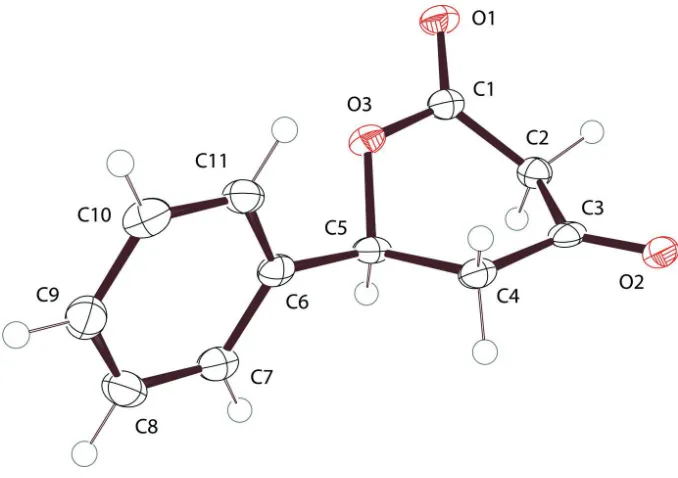
The molecular structure (Fig. 1.) is the singular moiety in the asymmetric unit. A Mogul (Bruno et al., 2004) geometry
check showed all non-H bond angles and distances to be normal. Ring puckering analysis of the dihydropyrandione ring
using PLATON (Spek, 2009; Cremer & Pople, 1975) indicates Φ = 297.5 (2)° and θ = 84.76 (18)° for the O3—C1—C2—
C3—C4—C5 ring. These parameters are consistent with a formal conformational assignment close to an idealized BC2,C5
boat with C2 at the bow and C5 at the stern. The plane of the phenyl ring attached to C5 may be described as a rudder
canted 72.14 (5)° relative to the mean plane of the six core atoms of the heterocycle. The 106.6 (2)° C6—C5—O3 bond
angle compared to the 112.8 (2)° C6—C5—C4 bond angle indicates a small steer to said rudder; however, whether it is to
port or starboard depends upon which enantiomer is considered.
Based upon a CSD search (Allen, 2002), two structures containing similar lactone ring motifs have been reported in the
crystallographic literature. These include the spiro compound methyl
4-methyl-3,5-dioxo-1-phenyl-2-oxaspiro[5.5]-4-carboxylate with CSD refcode IRITIN (Kirillov et al., 2010) and trans-5,6-diphenylperhydropyran-2,4-dione with CSD
refcode PONVAQ (de Souza et al., 2009). In all three cases the pyran rings adopt the boat conformation.
S2. Experimental
The title compound 6-(phenyl)-dihydro-2H-pyran-2,4-(3H)-dione, (also named 5-phenyl-3-oxo-delta-lactone), was
prepared by the literature method (Andersh et al., 2008). Benzaldehyde (2 mmol), ethanol (2 ml), ethylacetoacetate (2
mmol), and potassium carbonate (4 mmol) were heated overnight under nitrogen at 318 K. The solution was diluted with
ethylacetate, treated with 1 M HCl, and the combined organic layer extracts were dried, filtered, concentrated, and
purified by flash chromatography.
Crystals suitable for X-Ray analysis were grown by vapor diffusion of pentane into a concentrated solution of the
lactone in dichloromethane.
S3. Refinement
All non-H atoms were refined anisotropically. All H atoms were included in the refinement in the riding-model
Figure 1
The molecular structure of the compound with the atomic numbering scheme. Displacement ellipsoids are drawn at the
50% probability level.
6-Phenyloxane-2,4-dione
Crystal data
C11H10O3
Mr = 190.19
Orthorhombic, Pbca
Hall symbol: -P 2ac 2ab
a = 16.9888 (6) Å
b = 5.4501 (2) Å
c = 19.7350 (8) Å
V = 1827.28 (12) Å3
Z = 8
F(000) = 800
Dx = 1.383 Mg m−3
Mo Kα radiation, λ = 0.71073 Å Cell parameters from 2535 reflections
θ = 2.4–23.5°
µ = 0.10 mm−1
T = 100 K Prism, colourless 0.17 × 0.14 × 0.03 mm
Data collection
Bruker APEXII CCD diffractometer
Graphite monochromator
φ and ω scans
Absorption correction: multi-scan
(SADABS; Bruker, 2008)
Tmin = 0.662, Tmax = 0.746 17960 measured reflections
1804 independent reflections 1322 reflections with I > 2σ(I)
Rint = 0.071
θmax = 26.0°, θmin = 2.1°
h = −20→20
k = −6→6
l = −24→24
Refinement
Refinement on F2 Least-squares matrix: full
R[F2 > 2σ(F2)] = 0.047
wR(F2) = 0.114
S = 1.06 1804 reflections
127 parameters 0 restraints
Primary atom site location: structure-invariant direct methods
Hydrogen site location: inferred from neighbouring sites
H-atom parameters constrained
w = 1/[σ2(F
o2) + (0.039P)2 + 1.8272P] where P = (Fo2 + 2Fc2)/3
(Δ/σ)max < 0.001 Δρmax = 0.29 e Å−3 Δρmin = −0.25 e Å−3
Special details
Geometry. All s.u.'s (except the s.u. in the dihedral angle between two l.s. planes) are estimated using the full covariance matrix. The cell s.u.'s are taken into account individually in the estimation of s.u.'s in distances, angles and torsion angles; correlations between s.u.'s in cell parameters are only used when they are defined by crystal symmetry. An approximate (isotropic) treatment of cell s.u.'s is used for estimating s.u.'s involving l.s. planes.
Refinement. Refinement of F2 against ALL reflections. The weighted R-factor wR and goodness of fit S are based on F2, conventional R-factors R are based on F, with F set to zero for negative F2. The threshold expression of F2 > 2σ(F2) is used only for calculating R-factors(gt) etc. and is not relevant to the choice of reflections for refinement. R-factors based on F2 are statistically about twice as large as those based on F, and R- factors based on ALL data will be even larger.
Fractional atomic coordinates and isotropic or equivalent isotropic displacement parameters (Å2)
x y z Uiso*/Ueq
C1 0.15992 (11) 0.2655 (4) 0.22819 (11) 0.0198 (5)
C2 0.13487 (12) 0.0646 (4) 0.27556 (11) 0.0208 (5)
H2A 0.1203 0.1376 0.3198 0.025*
H2B 0.1799 −0.0474 0.2833 0.025*
C3 0.06630 (12) −0.0808 (4) 0.24883 (12) 0.0200 (5)
C4 0.06699 (12) −0.1241 (4) 0.17377 (11) 0.0203 (5)
H4A 0.0222 −0.0357 0.1529 0.024*
H4B 0.0598 −0.3015 0.1649 0.024*
C5 0.14301 (12) −0.0391 (4) 0.14093 (11) 0.0197 (5)
H5 0.1869 −0.1489 0.1557 0.024*
C6 0.13875 (12) −0.0384 (4) 0.06490 (11) 0.0196 (5)
C7 0.17318 (12) −0.2284 (4) 0.02863 (12) 0.0240 (5)
H7 0.2002 −0.3554 0.052 0.029*
C8 0.16830 (13) −0.2336 (4) −0.04141 (12) 0.0289 (6)
H8 0.1921 −0.364 −0.0659 0.035*
C9 0.12897 (13) −0.0501 (4) −0.07580 (12) 0.0283 (5)
H9 0.1257 −0.0542 −0.1238 0.034*
C10 0.09416 (13) 0.1406 (4) −0.03986 (12) 0.0284 (6)
H10 0.0672 0.2673 −0.0634 0.034*
C11 0.09869 (12) 0.1464 (4) 0.03021 (11) 0.0242 (5)
H11 0.0745 0.2763 0.0546 0.029*
O1 0.17979 (9) 0.4702 (3) 0.24675 (8) 0.0240 (4)
O2 0.01417 (8) −0.1540 (3) 0.28609 (8) 0.0237 (4)
O3 0.16073 (8) 0.2144 (3) 0.16170 (7) 0.0211 (4)
Atomic displacement parameters (Å2)
U11 U22 U33 U12 U13 U23
C1 0.0139 (10) 0.0164 (11) 0.0290 (13) 0.0004 (8) −0.0010 (9) −0.0005 (10)
C2 0.0213 (10) 0.0181 (11) 0.0230 (11) 0.0013 (8) 0.0009 (9) −0.0004 (9)
C4 0.0183 (10) 0.0147 (10) 0.0279 (12) −0.0013 (9) 0.0001 (9) −0.0002 (9)
C5 0.0193 (10) 0.0125 (10) 0.0274 (12) 0.0011 (8) −0.0004 (9) −0.0019 (9)
C6 0.0151 (10) 0.0168 (10) 0.0269 (12) −0.0038 (8) 0.0001 (9) −0.0007 (10)
C7 0.0221 (11) 0.0184 (11) 0.0314 (13) −0.0003 (9) 0.0002 (9) −0.0002 (10)
C8 0.0305 (12) 0.0234 (12) 0.0327 (14) −0.0010 (10) 0.0030 (10) −0.0079 (11)
C9 0.0332 (13) 0.0285 (13) 0.0232 (12) −0.0066 (10) −0.0002 (10) −0.0006 (11)
C10 0.0315 (13) 0.0210 (12) 0.0326 (14) −0.0021 (10) −0.0039 (10) 0.0030 (11)
C11 0.0258 (11) 0.0179 (11) 0.0289 (13) −0.0008 (9) 0.0017 (10) −0.0029 (10)
O1 0.0257 (8) 0.0159 (7) 0.0303 (9) −0.0028 (6) −0.0007 (7) −0.0015 (7)
O2 0.0230 (8) 0.0158 (7) 0.0324 (9) −0.0014 (6) 0.0062 (7) 0.0014 (7)
O3 0.0229 (7) 0.0152 (7) 0.0253 (9) −0.0048 (6) −0.0002 (6) 0.0004 (7)
Geometric parameters (Å, º)
C1—O1 1.222 (3) C5—H5 1
C1—O3 1.342 (2) C6—C7 1.388 (3)
C1—C2 1.501 (3) C6—C11 1.395 (3)
C2—C3 1.505 (3) C7—C8 1.385 (3)
C2—H2A 0.99 C7—H7 0.95
C2—H2B 0.99 C8—C9 1.381 (3)
C3—O2 1.218 (2) C8—H8 0.95
C3—C4 1.500 (3) C9—C10 1.391 (3)
C4—C5 1.517 (3) C9—H9 0.95
C4—H4A 0.99 C10—C11 1.385 (3)
C4—H4B 0.99 C10—H10 0.95
C5—O3 1.472 (2) C11—H11 0.95
C5—C6 1.502 (3)
O1—C1—O3 118.69 (19) C6—C5—H5 109.2
O1—C1—C2 123.9 (2) C4—C5—H5 109.2
O3—C1—C2 117.43 (18) C7—C6—C11 119.4 (2)
C1—C2—C3 112.66 (18) C7—C6—C5 119.53 (19)
C1—C2—H2A 109.1 C11—C6—C5 121.03 (19)
C3—C2—H2A 109.1 C8—C7—C6 120.3 (2)
C1—C2—H2B 109.1 C8—C7—H7 119.8
C3—C2—H2B 109.1 C6—C7—H7 119.8
H2A—C2—H2B 107.8 C9—C8—C7 120.3 (2)
O2—C3—C4 123.38 (19) C9—C8—H8 119.9
O2—C3—C2 121.6 (2) C7—C8—H8 119.9
C4—C3—C2 115.03 (18) C8—C9—C10 119.8 (2)
C3—C4—C5 112.35 (17) C8—C9—H9 120.1
C3—C4—H4A 109.1 C10—C9—H9 120.1
C5—C4—H4A 109.1 C11—C10—C9 120.2 (2)
C3—C4—H4B 109.1 C11—C10—H10 119.9
C5—C4—H4B 109.1 C9—C10—H10 119.9
H4A—C4—H4B 107.9 C10—C11—C6 120.0 (2)
O3—C5—C6 106.59 (17) C10—C11—H11 120
C6—C5—C4 112.73 (17) C1—O3—C5 117.72 (16)
O3—C5—H5 109.2
O1—C1—C2—C3 139.8 (2) C11—C6—C7—C8 0.4 (3)
O3—C1—C2—C3 −40.6 (3) C5—C6—C7—C8 178.5 (2)
C1—C2—C3—O2 −141.94 (19) C6—C7—C8—C9 −0.1 (3)
C1—C2—C3—C4 37.3 (2) C7—C8—C9—C10 0.0 (3)
O2—C3—C4—C5 −173.43 (19) C8—C9—C10—C11 −0.2 (3)
C2—C3—C4—C5 7.4 (2) C9—C10—C11—C6 0.5 (3)
C3—C4—C5—O3 −50.7 (2) C7—C6—C11—C10 −0.6 (3)
C3—C4—C5—C6 −169.46 (17) C5—C6—C11—C10 −178.68 (19)
O3—C5—C6—C7 137.70 (18) O1—C1—O3—C5 174.97 (17)
C4—C5—C6—C7 −101.5 (2) C2—C1—O3—C5 −4.6 (3)
O3—C5—C6—C11 −44.2 (2) C6—C5—O3—C1 173.64 (17)