400
doi:10.1107/S1600536814023265 Acta Cryst.(2014).E70, 400–402research communications
Received 4 October 2014 Accepted 22 October 2014
Edited by P. C. Healy, Griffith University, Australia
†
Keywords:crystal structure; phenyl benzoates; hydrogen bonding
CCDC reference:1030398
Supporting information:this article has supporting information at journals.iucr.org/e
Crystal structure of
3-({[(morpholin-4-yl)carbono-thioyl]sulfanyl}acetyl)phenyl benzoate
Sachin P. Ambekar,aK. Mahesh Kumar,aArun Kumar M. Shirahatti,aO. Kotresha and G. N. Anil Kumarb*
aDepartment of Chemistry, Karnatak University’s Karnatak Science College, Dharwad 580 001, Karanataka, India, and bDepartment of Physics, M S Ramaiah Institute of Technology, Bangalore 560 054, Karnataka, India. *Correspondence
e-mail: [email protected]
In the title compound, C20H19NO4S2, the morpholine ring adopts the expected
chair conformation. The central phenyl ring makes dihedral angles of 67.97 (4) and 7.74 (3), respectively, with the benzoate phenyl ring and the morpholine
mean plane. In the crystal, molecules are linked by C—H O hydrogen bonds, forming zigzag chains along the b-axis direction. C—H interactions link centrosymmetrically related molecules, reinforcing the three-dimensional structure.
1. Chemical context
The title compound is a dithiocarbamate ester derivative of 3-(2-bromacetyl) phenyl benzoate, a key starting material used in the synthesis of phenylephrine, (R)-3-[ 1-hydroxy-2-(methylamino)ethyl] phenol, which is a selective 1-adre-nergic receptor agonist used primarily as a decongestant and as an agent to dilate the pupil and to increase blood pressure. Our current research work is aimed at the synthesis of a series of 3-(2-bromacetyl) phenyl benzoate dithiocarbamate ester derivatives. Dithiocarbamate acid esters exhibit a range of biological effects, including anti-bacterial, anti-fungal and anti-oxidant activity (Hirschelmanet al., 2002) and inhibition of cardiac hypertrophy (Naoto et al. 2008). Recently, it was found that 5-oxohexyl dithiocarbamic acid methyl esters are potent phase II enzyme inducers, which could be used as cancer chemo-preventive agents (Scozzafavaet al., 2000).
2. Structural commentary
In the molecular structure of the title compound, the morpholine ring adopts the expected chair conformation. The phenyl ring makes dihedral angles of 67.97 (4) and 7.74 (3)
respectively with phenyl ring and the morpholine mean plane. This is also reflected in the deviation of the torsion angles C5—S1—C6—C7 = 175.32 (2) and C12—O3—C14—C15 = 178.91 (3). Weak intramolecular C—H S hydrogen bonds
exist within the morpholyl dithiocarbamate moiety (Table 1).
3. Supramolecular features
In the crystal, molecules are linked by weak C—H O hydrogen bonds, forming zigzag chains along thebaxis. C— H interactions link centrosymmetrically related mol-ecules, reinforcing the three-dimensional structure (Fig. 2)
4. Database survey
A search of the Cambridge Structural Database (Version 5.35, updates February 2014; Groom & Allen, 2014) for 2-(4-methoxyphenyl)-2-oxoethyl dithiocarbamate gave one hit, namely GEGGUV01 (Jianet al., 2006). A search for 2-oxoe-thyl dithioformate gave two related structures, viz. 2-oxo-2-(2-oxo-2H-chromen-3-yl)ethyl pyrrolidine-1-carbodithioate (Kumar et al., 2013) and (6-methoxy-2-oxo-2H -chromen-4-yl)methyl morpholine-4-carbodithioate (Devarajegowdaet al., 2013). Interestingly, dimer formationviaC—H O hydrogen bonds is a feature of the packing in these structures.
5. Synthesis and Crystallization
To a solution of NaOH (1 mmol) in 3 ml water was added to a mixture of morpholine (1 mmol) in ethanol (25 ml). After stirring at room temperature for about 20 min, carbon di-sulfide (1.2 mmol) was added dropwise and the resulting mixture was further stirred at room temperature for 90 min. Then 3-(2-bromacetyl) phenyl benzoate (1 mmol) was added and stirring was continued. After completion of the reaction (monitored by TLC), the solvent was removed under vacuum and the residue was extracted with dichloromethane (2 25 ml) and dried over anhydrous MgSO4. The solvent was
evaporated and the compound recrystallized from an ethanol– chloroform mixture (3:1) to give the title compound as colourless crystals in 81% yield.
Off-white solid, IR (KBr) max/cm
1
: 2857, 3073 (C—H aliphatic and aromatic), 1732 (C O), 1421, 1680 (C C),
research communications
Acta Cryst.(2014).E70, 400–402 Ambekaret al. C
20H19NO4S2
401
Figure 1 [image:2.610.46.298.69.247.2] [image:2.610.313.565.107.167.2]The molecular structure of the title compound, showing 50% displace-ment ellipsoids.
Figure 2
Packing diagram of the title compound, with C—H O and C—H
interactions indicated by dashed lines.
Table 1
Hydrogen-bond geometry (A˚ ,).
Cgis the centroid of the C15–C20 ring.
D—H A D—H H A D A D—H A
C2—H2B S1 0.97 2.41 2.938 (2) 114
C3—H3A S2 0.97 2.56 3.052 (5) 111
C13—H13 O4i 0.93 2.43 3.224 (3) 143
C11—H11 Cgii 0.93 2.88 3.629 (2) 138
[image:2.610.314.562.441.717.2]Symmetry codes: (i)x;yþ1;z; (ii)xþ1;y;zþ1.
Table 2
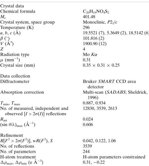
Experimental details.
Crystal data
Chemical formula C20H19NO4S2
Mr 401.48
Crystal system, space group Monoclinic,P21/c
Temperature (K) 296
a,b,c(A˚ ) 19.5521 (7), 5.3649 (2), 18.5142 (6) (
) 101.816 (2)
V(A˚3) 1900.90 (12)
Z 4
Radiation type MoK
(mm1) 0.31
Crystal size (mm) 0.350.310.25
Data collection
Diffractometer BrukerSMARTCCD area
detector
Absorption correction Multi-scan (SADABS; Sheldrick, 1996)
Tmin,Tmax 0.887, 0.934
No. of measured, independent and observed [I> 2(I)] reflections
12830, 3539, 2613
Rint 0.024
(sin/ )max(A˚
1) 0.606
Refinement
R[F2> 2(F2)],wR(F2),S 0.042, 0.122, 1.06
No. of reflections 3539
No. of parameters 244
H-atom treatment H-atom parameters constrained
max,min(e A˚
3) 0.31,0.22
Computer programs:SMARTandSAINT(Bruker, 1998),SIR92(Altomareet al., 1993),
SHELXL97(Sheldrick, 2008),ORTEP-3 for Windows(Farrugia, 2012),CAMERON
[image:2.610.44.299.532.713.2]1264 (C—O), 1228 (C S), 1061 (C—N).1H NMR (300 MHz, CDCl3):3.77–3.80 (t, 4H), 4.23–4.43 (t, 4H), 4.91(s, 2H), 7.26–
7.47 (m, 1H), 7.48–7.51 (m, 2H), 7.53–7.60 (m, 1H), 7.63–768 (m, 1H), 7.90–7.91 (t, 1H), 7.99–8.02 (d, 1H), 8.20–8.22 (d, 2H);
13
C NMR (75 MHz, CDCl3):44.6 (C6), 49.5 (C2, C3), 65.6
(C1, C4), 121.83 (C13), 126.0 (C9), 127.1 (C11), 128.6 (C10), 129.8 (C17, C19), 130.2 (C15, C16, C20), 137.6 (C18), 151.2 (C8), 154.93 (C12), 182.82 (C14), 192.15 (C7), 195.75 (C5); MS m/z: 402.10 [M + H]+. Analysis calculated (%) for C20H19NO4S2: C 59.83, H 4.77, N 3.49, S 15.97%; found: C
59.72, H 4.85, N 3.61, S 15.94.
6. Refinement
Crystal data, data collection and structure refinement details are summarized in Table 2. The C-bound H atoms were positioned geometrically and allowed to ride on their parent atoms, with C—H = 0.93–0.97A˚ andUiso(H) = 1.2Ueq(C).
Acknowledgements
The authors thank the University’s Sophisticated Instru-mentation Centre (USIC), Karnatak University, Dharwad, for the CCD X-ray facilities, X-ray data collection, GCMS, IR, CHNS and NMR data. SPA is grateful to Karnatak Science College, Dharwad, for providing laboratory facilities.
References
Altomare, A., Cascarano, G., Giacovazzo, C. & Guagliardi, A. (1993).
J. Appl. Cryst.26, 343–350.
Bruker (1998). SMART and SAINT. Bruker AXS Inc., Madison, Wisconsin, USA.
Devarajegowda, H. C., Kumar, K. M., Seenivasa, S., Arunkashi, H. K. & Kotresh, O. (2013).Acta Cryst.E69, o192.
Farrugia, L. J. (2012).J. Appl. Cryst.45, 849–854.
Groom, C. R. & Allen, F. H. (2014).Angew. Chem. Int. Ed.53, 662– 671.
Hirschelman, W. H., Kosmeder, J. W. II, Song, L. S., Park, E. J., Moriarty, R. M. & Pezzuto, J. M. (2002). 224th ACS National Meeting: Division ofMedicinal Chemistry,178.
Jian, F., Xiao, H., Zhu, C. & Xu, L. (2006).J. Heterocycl. Chem.43, 925–929.
Kumar, K. M., Mahabhaleshwaraiah, N. M., Kotresh, O., Roopashree, K. R. & Devarajegowda, H. C. (2013).Acta Cryst.E69, o1382. Naoto, O., Mariko, O., Takashi, S., Satoshi, K., Atsuko, M., Noriaki,
U., Yoshisuke, N., Keishi, K., Masamori, S. & Yushi, K. (2008). PCT Int. Appl. WO 2008029825/A1 20080313.
Nardelli, M. (1995).J. Appl. Cryst.28, 659.
Scozzafava, A., Mastrolorenzo, A. & Supuran, C. T. (2000).Bioorg. Med. Chem. Lett.10, 1887–1891.
Sheldrick, G. M. (1996). SADABS. University of Go¨ttingen, Germany.
Sheldrick, G. M. (2008).Acta Cryst.A64, 112–122. Spek, A. L. (2009).Acta Cryst.D65, 148–155.
Watkin, D. M., Pearce, L. & Prout, C. K. (1993). CAMERON. Chemical Crystallography Laboratory, University of Oxford, England.
402
Ambekaret al. C20H19NO4S2 Acta Cryst.(2014).E70, 400–402
supporting information
sup-1
Acta Cryst. (2014). E70, 400-402supporting information
Acta Cryst. (2014). E70, 400-402 [doi:10.1107/S1600536814023265]
Crystal structure of 3-({[(morpholin-4-yl)carbonothioyl]sulfanyl}acetyl)phenyl
benzoate
Sachin P. Ambekar, K. Mahesh Kumar, Arun Kumar M. Shirahatti, O. Kotresh and G. N. Anil
Kumar
Computing details
Data collection: SMART (Bruker, 1998); cell refinement: SMART (Bruker, 1998); data reduction: SAINT (Bruker, 1998);
program(s) used to solve structure: SIR92 (Altomare et al., 1993); program(s) used to refine structure: SHELXL97
(Sheldrick, 2008); molecular graphics: ORTEP-3 for Windows (Farrugia, 2012) and CAMERON (Watkin et al., 1993);
software used to prepare material for publication: PARST (Nardelli, 1995) and PLATON (Spek, 2009).
3-({[(Morpholin-4-yl)carbonothioyl]sulfanyl}acetyl)phenyl benzoate
Crystal data
C20H19NO4S2 Mr = 401.48 Monoclinic, P21/c
Hall symbol: -P 2ybc
a = 19.5521 (7) Å
b = 5.3649 (2) Å
c = 18.5142 (6) Å
β = 101.816 (2)°
V = 1900.90 (12) Å3
Z = 4
F(000) = 840
Dx = 1.403 Mg m−3
Mo Kα radiation, λ = 0.71073 Å
θ = 1.5°
µ = 0.31 mm−1 T = 296 K Block, colourless 0.35 × 0.31 × 0.25 mm
Data collection
Bruker SMART CCD area-detector diffractometer
Radiation source: fine-focus sealed tube Graphite monochromator
φ and ω scans
Absorption correction: multi-scan (SADABS; Sheldrick, 1996)
Tmin = 0.887, Tmax = 0.934
12830 measured reflections 3539 independent reflections 2613 reflections with I > 2σ(I)
Rint = 0.024
θmax = 25.5°, θmin = 1.1° h = −23→21
k = −6→6
l = −22→22
Refinement
Refinement on F2
Least-squares matrix: full
R[F2 > 2σ(F2)] = 0.042 wR(F2) = 0.122 S = 1.06 3539 reflections 244 parameters 0 restraints
Primary atom site location: structure-invariant direct methods
Secondary atom site location: difference Fourier map
Hydrogen site location: inferred from neighbouring sites
supporting information
sup-2
Acta Cryst. (2014). E70, 400-402w = 1/[σ2(F
o2) + (0.0581P)2 + 0.4768P]
where P = (Fo2 + 2Fc2)/3
(Δ/σ)max = 0.001
Δρmax = 0.31 e Å−3
Δρmin = −0.22 e Å−3
Special details
Geometry. All e.s.d.'s (except the e.s.d. in the dihedral angle between two l.s. planes) are estimated using the full covariance matrix. The cell e.s.d.'s are taken into account individually in the estimation of e.s.d.'s in distances, angles and torsion angles; correlations between e.s.d.'s in cell parameters are only used when they are defined by crystal symmetry. An approximate (isotropic) treatment of cell e.s.d.'s is used for estimating e.s.d.'s involving l.s. planes.
Refinement. Refinement of F2 against ALL reflections. The weighted R-factor wR and goodness of fit S are based on F2,
conventional R-factors R are based on F, with F set to zero for negative F2. The threshold expression of F2 > σ(F2) is used
only for calculating R-factors(gt) etc. and is not relevant to the choice of reflections for refinement. R-factors based on F2
are statistically about twice as large as those based on F, and R-factors based on ALL data will be even larger.
Fractional atomic coordinates and isotropic or equivalent isotropic displacement parameters (Å2)
x y z Uiso*/Ueq
supporting information
sup-3
Acta Cryst. (2014). E70, 400-402C13 0.30942 (10) 0.5091 (4) 0.50962 (11) 0.0414 (5) H13 0.3214 0.616 0.5498 0.05* C14 0.43558 (11) 0.1310 (5) 0.59189 (11) 0.0420 (5) C15 0.50633 (10) 0.1476 (4) 0.63896 (11) 0.0372 (5) C16 0.52631 (11) −0.0360 (5) 0.69145 (12) 0.0453 (6) H16 0.4962 −0.1674 0.695 0.054* C17 0.59067 (12) −0.0250 (5) 0.73859 (12) 0.0504 (6) H17 0.604 −0.149 0.7738 0.061* C18 0.63503 (12) 0.1690 (5) 0.73352 (13) 0.0513 (6) H18 0.6783 0.1772 0.7656 0.062* C19 0.61594 (12) 0.3512 (5) 0.68132 (14) 0.0541 (7) H19 0.6464 0.4817 0.678 0.065* C20 0.55158 (11) 0.3418 (5) 0.63371 (12) 0.0469 (6) H20 0.5388 0.4654 0.5983 0.056*
Atomic displacement parameters (Å2)
U11 U22 U33 U12 U13 U23
supporting information
sup-4
Acta Cryst. (2014). E70, 400-402Geometric parameters (Å, º)
S1—C5 1.774 (2) C6—H6B 0.97 S1—C6 1.800 (2) C7—C8 1.487 (3) S2—C5 1.653 (2) C8—C9 1.387 (3) O1—C1 1.401 (3) C8—C13 1.394 (3) O1—C4 1.407 (3) C9—C10 1.374 (3) O2—C7 1.214 (2) C9—H9 0.93 O3—C14 1.354 (3) C10—C11 1.381 (3) O3—C12 1.410 (2) C10—H10 0.93 O4—C14 1.191 (3) C11—C12 1.367 (3) N1—C5 1.338 (3) C11—H11 0.93 N1—C3 1.472 (3) C12—C13 1.374 (3) N1—C2 1.480 (3) C13—H13 0.93 C1—C2 1.493 (4) C14—C15 1.479 (3) C1—H1A 0.97 C15—C16 1.382 (3) C1—H1B 0.97 C15—C20 1.383 (3) C2—H2A 0.97 C16—C17 1.378 (3) C2—H2B 0.97 C16—H16 0.93 C3—C4 1.474 (4) C17—C18 1.370 (3) C3—H3A 0.97 C17—H17 0.93 C3—H3B 0.97 C18—C19 1.371 (3) C4—H4A 0.97 C18—H18 0.93 C4—H4B 0.97 C19—C20 1.382 (3) C6—C7 1.507 (3) C19—H19 0.93 C6—H6A 0.97 C20—H20 0.93
supporting information
sup-5
Acta Cryst. (2014). E70, 400-402N1—C3—H3B 109.5 O3—C14—C15 112.34 (19) C4—C3—H3B 109.5 C16—C15—C20 119.5 (2) H3A—C3—H3B 108.1 C16—C15—C14 117.8 (2) O1—C4—C3 113.0 (2) C20—C15—C14 122.6 (2) O1—C4—H4A 109 C17—C16—C15 120.3 (2) C3—C4—H4A 109 C17—C16—H16 119.8 O1—C4—H4B 109 C15—C16—H16 119.8 C3—C4—H4B 109 C18—C17—C16 119.8 (2) H4A—C4—H4B 107.8 C18—C17—H17 120.1 N1—C5—S2 124.08 (17) C16—C17—H17 120.1 N1—C5—S1 113.60 (15) C17—C18—C19 120.3 (2) S2—C5—S1 122.32 (13) C17—C18—H18 119.8 C7—C6—S1 108.88 (15) C19—C18—H18 119.8 C7—C6—H6A 109.9 C18—C19—C20 120.3 (2) S1—C6—H6A 109.9 C18—C19—H19 119.9 C7—C6—H6B 109.9 C20—C19—H19 119.9 S1—C6—H6B 109.9 C19—C20—C15 119.7 (2) H6A—C6—H6B 108.3 C19—C20—H20 120.2 O2—C7—C8 121.2 (2) C15—C20—H20 120.2 O2—C7—C6 120.8 (2)
Hydrogen-bond geometry (Å, º)
Cg is the centroid of the C15–C20 ring.
D—H···A D—H H···A D···A D—H···A
C2—H2B···S1 0.97 2.41 2.938 (2) 114 C3—H3A···S2 0.97 2.56 3.052 (5) 111 C13—H13···O4i 0.93 2.43 3.224 (3) 143
C11—H11···Cgii 0.93 2.88 3.629 (2) 138