Crystal structure of (
E
)-2-(4-methoxy-styryl)-2,3-dihydro-1
H
-perimidine
acetonitrile monosolvate
A. Manimekalai,a* N. Vijayalakshmiaand S. Selvanayagamb
aDepartment of Chemistry, Faculty of Science, Annamalai University,
Annamalainagar 608 002, India, andbDepartment of Physics, Kalasalingam University, Krishnankoil 626 126, India. *Correspondence e-mail: profmeka1@gmail.com
Received 22 July 2014; accepted 23 July 2014
Edited by H. Stoeckli-Evans, University of Neuchaˆtel, Switzerland
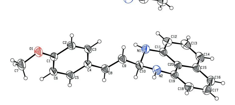
The title compound, C20H18N2OCH3CN, a perimidine
deriv-ative, crystallized as an acetonitrile monosolvate. The planes of the naphthalene ring system and the methoxyphenyl ring are oriented almost perpendicular to one another, with a dihedral angle of 87.61 (6). The conformation about the
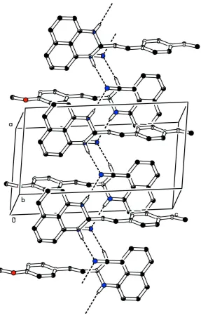
C C bond is E. The hexahydropyrimidine ring has an envelope conformation, with the methine C atom as the flap. In the crystal, the molecules are linked by N—H N hydrogen bonds involving the acetonitrile solvent molecule as acceptor, forming zigzag chains propagating along [100].
Keywords:crystal structure; perimidine derivative; bifurcated hydrogen bonding.
CCDC reference:1015577
1. Related literature
For the diverse range of biological activities of perimidines, see: Buet al.(2001); Ivicaet al.(2008); Azeez & Salih (2014). For a related structure, see: Maloneyet al.(2013).
2. Experimental
2.1. Crystal data
Triclinic,P1 a= 7.8128 (7) A˚ b= 8.4641 (7) A˚ c= 14.7427 (14) A˚
= 79.513 (7) = 83.861 (7) = 77.076 (7)
V= 932.11 (15) A˚3 Z= 2
CuKradiation
= 0.60 mm1 T= 292 K
0.300.300.19 mm
2.2. Data collection
Oxford Diffraction Gemini/EOS CCD diffractometer
Absorption correction: multi-scan (CrysAlis PRO; Oxford Diffrac-tion, 2008)
Tmin= 0.840,Tmax= 0.890
5843 measured reflections 3569 independent reflections 2940 reflections withI> 2(I) Rint= 0.023
2.3. Refinement
R[F2> 2(F2)] = 0.048 wR(F2) = 0.142 S= 1.03 3569 reflections 246 parameters
H atoms treated by a mixture of independent and constrained refinement
max= 0.19 e A˚
3
min=0.20 e A˚
3
Table 1
Hydrogen-bond geometry (A˚ ,).
D—H A D—H H A D A D—H A
N1—H1 N3i
0.88 (2) 2.49 (2) 3.305 (3) 154.1 (15) N2—H2 N3ii
0.84 (2) 2.44 (2) 3.234 (2) 156.8 (16)
Symmetry codes: (i)xþ1;yþ1;zþ1; (ii)x;yþ1;zþ1.
Data collection: CrysAlis PRO (Oxford Diffraction, 2008); cell refinement: CrysAlis PRO; data reduction: CrysAlis PRO; program(s) used to solve structure: SHELXS97 (Sheldrick, 2008); program(s) used to refine structure:SHELXL2013(Sheldrick, 2008); molecular graphics: ORTEP-3 for Windows (Farrugia, 2012) and PLATON (Spek, 2009); software used to prepare material for publication:PLATON.
Acknowledgements
We are grateful to IICT, Hyderabad, for support of the instrumentation facility.
Supporting information for this paper is available from the IUCr electronic archives (Reference: SU2762).
References
Azeez, H. J. & Salih, K. M. (2014).Res. Pharm. Biotechnol.3, 1–6. Bu, X., Deady, W. L., Finlay, J. G., Bagwey, C. B. & Denny, A. W. (2001).J.
Med. Chem.44, 2004–2014.
Farrugia, L. J. (2012).J. Appl. Cryst.45, 849–854.
Ivica, D., Mirta, R., Visnja, V., Sandra, P. K., Marijeta, K., Ivvo, D. & Marina, C. (2008). Bioorg. Med. Chem. 16, 5189–5198.
Maloney, S., Slawin, A. M. Z. & Woollins, J. D. (2013).Acta Cryst.E69, o246. Oxford Diffraction (2008).CrysAlis PRO. Oxford Diffraction Ltd, Yarnton,
England.
supporting information
Acta Cryst. (2014). E70, o959 [doi:10.1107/S1600536814017000]
Crystal structure of (
E
)-2-(4-methoxystyryl)-2,3-dihydro-1
H
-perimidine
aceto-nitrile monosolvate
A. Manimekalai, N. Vijayalakshmi and S. Selvanayagam
S1. Synthesis and crystallization
A mixture of 4-methoxy-trans-cinnamaldehyde (0.01 mol), 1,8-diaminonaphthalene (0.01 mol) and a pinch of MgSO4.7H2O was finely grinded in a mortor. The mixture was then kept under microwave irradiation (LG Grill,
Intellowave 160-800 W, consumption 800 W, output power 320 W and frequency 2450 MHz) operating in a cyclic mode to prevent intense boiling of the sample as well as aggregation of 320 W at 12 min. A brown colour solid separated out which was repeatedly recrystallized using CH2Cl2:CH3CN (1:5) to obtain brown crystals.
S2. Refinement
[image:2.610.112.498.422.595.2]Crystal data, data collection and structure refinement details are summarized in Table 2. Atoms H1 and H2 were located from a difference Fourier map and freely refined. The C-bound H atoms were positioned geometrically and allowed to ride on their parent atoms: C—H = 0.93-0.96 Å with Uiso(H) = 1.5Ueq(C-methyl) and Uiso(H) = 1.2Ueq for other C atoms.
Figure 1
Figure 2
Crystal packing of the title compound, viewed along the b axis, showing the hydrogen bonds as dashed lines (see Table 1 for details; H atoms not involved in hydrogen bonding have been omitted for clarity).
(E)-2-(4-Methoxystyryl)-2,3-dihydro-1H-perimidine acetonitrile monosolvate
Crystal data
C20H18N2O·C2H3N
Mr = 343.42
Triclinic, P1
a = 7.8128 (7) Å
b = 8.4641 (7) Å
c = 14.7427 (14) Å
α = 79.513 (7)°
β = 83.861 (7)°
γ = 77.076 (7)°
V = 932.11 (15) Å3
Z = 2
F(000) = 364
Dx = 1.224 Mg m−3
Cu Kα radiation, λ = 1.54184 Å Cell parameters from 2420 reflections
µ = 0.60 mm−1
T = 292 K
Block, colourless 0.30 × 0.30 × 0.19 mm
Data collection
Oxford Diffraction Gemini/EOS CCD diffractometer
Radiation source: fine-focus sealed tube
φ and ω scans
Absorption correction: multi-scan
(CrysAlis PRO; Oxford Diffraction, 2008)
Tmin = 0.840, Tmax = 0.890
5843 measured reflections
3569 independent reflections 2940 reflections with I > 2σ(I)
Rint = 0.023
θmax = 72.0°, θmin = 3.1°
h = −8→9
k = −9→10
l = −18→18
Refinement
Refinement on F2
Least-squares matrix: full
R[F2 > 2σ(F2)] = 0.048
wR(F2) = 0.142
S = 1.03 3569 reflections 246 parameters 0 restraints
Primary atom site location: structure-invariant direct methods
Secondary atom site location: difference Fourier map
Hydrogen site location: mixed
H atoms treated by a mixture of independent and constrained refinement
w = 1/[σ2(F
o2) + (0.0768P)2 + 0.0999P]
where P = (Fo2 + 2Fc2)/3
(Δ/σ)max < 0.001
Δρmax = 0.19 e Å−3
Δρmin = −0.20 e Å−3
Extinction correction: SHELXL, Fc*=kFc[1+0.001xFc2λ3/sin(2θ)]-1/4
Extinction coefficient: 0.038 (2)
Special details
Geometry. All esds (except the esd in the dihedral angle between two l.s. planes) are estimated using the full covariance matrix. The cell esds are taken into account individually in the estimation of esds in distances, angles and torsion angles; correlations between esds in cell parameters are only used when they are defined by crystal symmetry. An approximate (isotropic) treatment of cell esds is used for estimating esds involving l.s. planes.
Fractional atomic coordinates and isotropic or equivalent isotropic displacement parameters (Å2)
x y z Uiso*/Ueq
C7 0.2448 (3) 0.5081 (3) −0.05893 (11) 0.0848 (6) H7A 0.3710 0.4773 −0.0645 0.127* H7B 0.2041 0.5813 −0.1137 0.127* H7C 0.1961 0.4116 −0.0514 0.127* C8 0.29403 (18) 0.25000 (17) 0.38074 (9) 0.0518 (3) H8 0.3531 0.1408 0.3858 0.062* C9 0.2431 (2) 0.30587 (18) 0.45874 (10) 0.0599 (4) H9 0.1876 0.4158 0.4560 0.072* C10 0.26927 (19) 0.20271 (18) 0.55165 (9) 0.0550 (4) H10 0.3362 0.0925 0.5446 0.066* C11 0.37511 (18) 0.20595 (16) 0.70001 (9) 0.0495 (3) C12 0.5038 (2) 0.22395 (19) 0.75213 (11) 0.0616 (4) H12 0.5903 0.2804 0.7247 0.074* C13 0.5045 (2) 0.1575 (2) 0.84619 (11) 0.0668 (4) H13 0.5919 0.1710 0.8805 0.080* C14 0.3811 (2) 0.0739 (2) 0.88851 (10) 0.0625 (4) H14 0.3848 0.0307 0.9511 0.075* C15 0.24639 (18) 0.05219 (16) 0.83788 (9) 0.0526 (3) C16 0.1127 (2) −0.03118 (19) 0.87873 (11) 0.0644 (4) H16 0.1144 −0.0795 0.9407 0.077* C17 −0.0188 (2) −0.0412 (2) 0.82776 (12) 0.0684 (4) H17 −0.1062 −0.0964 0.8555 0.082* C18 −0.02515 (19) 0.03028 (19) 0.73418 (11) 0.0608 (4) H18 −0.1172 0.0235 0.7009 0.073* C19 0.10345 (17) 0.10989 (16) 0.69160 (9) 0.0489 (3) C20 0.24349 (16) 0.12050 (15) 0.74293 (9) 0.0460 (3) C21 0.2512 (3) 0.6594 (2) 0.67886 (14) 0.0835 (5) H21A 0.3480 0.5668 0.6877 0.125* H21B 0.1430 0.6246 0.7000 0.125* H21C 0.2655 0.7410 0.7134 0.125* C22 0.2473 (2) 0.7278 (2) 0.58287 (14) 0.0749 (5)
Atomic displacement parameters (Å2)
U11 U22 U33 U12 U13 U23
C10 0.0624 (8) 0.0534 (8) 0.0449 (7) −0.0015 (6) −0.0043 (6) −0.0096 (6) C11 0.0507 (7) 0.0481 (7) 0.0464 (7) −0.0012 (5) −0.0034 (5) −0.0108 (5) C12 0.0558 (8) 0.0629 (9) 0.0677 (9) −0.0096 (7) −0.0083 (7) −0.0157 (7) C13 0.0647 (9) 0.0715 (10) 0.0651 (9) 0.0021 (8) −0.0226 (7) −0.0230 (8) C14 0.0692 (9) 0.0649 (9) 0.0459 (7) 0.0092 (7) −0.0135 (7) −0.0127 (6) C15 0.0569 (8) 0.0471 (7) 0.0455 (7) 0.0080 (6) −0.0027 (6) −0.0097 (5) C16 0.0738 (10) 0.0573 (8) 0.0506 (8) −0.0005 (7) 0.0061 (7) −0.0015 (6) C17 0.0660 (9) 0.0624 (9) 0.0722 (10) −0.0144 (7) 0.0123 (8) −0.0081 (8) C18 0.0531 (8) 0.0610 (9) 0.0688 (9) −0.0100 (6) −0.0021 (7) −0.0154 (7) C19 0.0499 (7) 0.0450 (7) 0.0481 (7) 0.0011 (5) −0.0038 (5) −0.0119 (5) C20 0.0474 (7) 0.0419 (6) 0.0443 (7) 0.0033 (5) −0.0029 (5) −0.0111 (5) C21 0.0940 (13) 0.0738 (11) 0.0794 (12) −0.0137 (10) 0.0011 (10) −0.0126 (9) C22 0.0647 (10) 0.0877 (12) 0.0764 (12) −0.0204 (9) −0.0083 (8) −0.0161 (10)
Geometric parameters (Å, º)
O1—C1 1.3581 (16) C9—C10 1.4917 (19)
O1—C7 1.422 (2) C9—H9 0.9300
N1—C11 1.3902 (17) C10—H10 0.9800 N1—C10 1.4630 (19) C11—C12 1.379 (2) N1—H1 0.882 (18) C11—C20 1.4170 (19) N2—C19 1.3869 (17) C12—C13 1.398 (2) N2—C10 1.4489 (19) C12—H12 0.9300 N2—H2 0.842 (18) C13—C14 1.355 (2) N3—C22 1.130 (2) C13—H13 0.9300 C1—C6 1.381 (2) C14—C15 1.415 (2) C1—C2 1.3913 (19) C14—H14 0.9300 C2—C3 1.3685 (19) C15—C16 1.412 (2) C2—H2A 0.9300 C15—C20 1.4144 (18) C3—C4 1.3983 (19) C16—C17 1.362 (2)
C3—H3 0.9300 C16—H16 0.9300
C4—C5 1.3830 (18) C17—C18 1.403 (2) C4—C8 1.4666 (18) C17—H17 0.9300 C5—C6 1.3852 (19) C18—C19 1.368 (2)
C5—H5 0.9300 C18—H18 0.9300
C6—H6 0.9300 C19—C20 1.4226 (19) C7—H7A 0.9600 C21—C22 1.429 (3)
C7—H7B 0.9600 C21—H21A 0.9600
C7—H7C 0.9600 C21—H21B 0.9600
C8—C9 1.312 (2) C21—H21C 0.9600
C8—H8 0.9300
C10—N2—H2 114.7 (12) C11—C12—C13 120.18 (15) O1—C1—C6 125.11 (13) C11—C12—H12 119.9 O1—C1—C2 115.59 (13) C13—C12—H12 119.9 C6—C1—C2 119.30 (13) C14—C13—C12 121.58 (14) C3—C2—C1 120.63 (13) C14—C13—H13 119.2 C3—C2—H2A 119.7 C12—C13—H13 119.2 C1—C2—H2A 119.7 C13—C14—C15 120.31 (14) C2—C3—C4 121.10 (12) C13—C14—H14 119.8 C2—C3—H3 119.5 C15—C14—H14 119.8 C4—C3—H3 119.5 C16—C15—C20 118.76 (13) C5—C4—C3 117.41 (12) C16—C15—C14 122.73 (14) C5—C4—C8 119.73 (12) C20—C15—C14 118.49 (14) C3—C4—C8 122.85 (12) C17—C16—C15 120.21 (14) C4—C5—C6 122.15 (12) C17—C16—H16 119.9 C4—C5—H5 118.9 C15—C16—H16 119.9 C6—C5—H5 118.9 C16—C17—C18 121.29 (15) C1—C6—C5 119.41 (13) C16—C17—H17 119.4 C1—C6—H6 120.3 C18—C17—H17 119.4 C5—C6—H6 120.3 C19—C18—C17 120.33 (14) O1—C7—H7A 109.5 C19—C18—H18 119.8 O1—C7—H7B 109.5 C17—C18—H18 119.8 H7A—C7—H7B 109.5 C18—C19—N2 123.57 (13) O1—C7—H7C 109.5 C18—C19—C20 119.56 (13) H7A—C7—H7C 109.5 N2—C19—C20 116.74 (12) H7B—C7—H7C 109.5 C15—C20—C11 120.15 (12) C9—C8—C4 127.69 (13) C15—C20—C19 119.81 (13) C9—C8—H8 116.2 C11—C20—C19 119.98 (12) C4—C8—H8 116.2 C22—C21—H21A 109.5 C8—C9—C10 123.58 (14) C22—C21—H21B 109.5 C8—C9—H9 118.2 H21A—C21—H21B 109.5 C10—C9—H9 118.2 C22—C21—H21C 109.5 N2—C10—N1 106.95 (11) H21A—C21—H21C 109.5 N2—C10—C9 110.23 (12) H21B—C21—H21C 109.5 N1—C10—C9 110.56 (12) N3—C22—C21 179.1 (2)
C3—C4—C8—C9 −7.4 (2) C14—C15—C20—C11 1.10 (19) C4—C8—C9—C10 177.61 (13) C16—C15—C20—C19 2.48 (18) C19—N2—C10—N1 −54.88 (16) C14—C15—C20—C19 −175.97 (12) C19—N2—C10—C9 −175.12 (11) C12—C11—C20—C15 −1.08 (19) C11—N1—C10—N2 51.33 (16) N1—C11—C20—C15 −177.36 (11) C11—N1—C10—C9 171.35 (12) C12—C11—C20—C19 175.98 (12) C8—C9—C10—N2 −116.90 (17) N1—C11—C20—C19 −0.30 (19) C8—C9—C10—N1 125.05 (16) C18—C19—C20—C15 −1.48 (19) C10—N1—C11—C12 157.87 (14) N2—C19—C20—C15 174.65 (11) C10—N1—C11—C20 −25.97 (18) C18—C19—C20—C11 −178.55 (12) N1—C11—C12—C13 176.53 (13) N2—C19—C20—C11 −2.42 (18) C20—C11—C12—C13 0.4 (2)
Hydrogen-bond geometry (Å, º)
D—H···A D—H H···A D···A D—H···A
N1—H1···N3i 0.88 (2) 2.49 (2) 3.305 (3) 154.1 (15)
N2—H2···N3ii 0.84 (2) 2.44 (2) 3.234 (2) 156.8 (16)