Received 3 October 2019 Accepted 16 October 2019
Edited by M. Zeller, Purdue University, USA
Keywords:crystal structure; metal–organic framework; naphthalenedicarboxylic acid;N,N -diethylformamide; zinc carboxylates.
CCDC reference:1959604
Supporting information:this article has supporting information at journals.iucr.org/e
Crystal structure of a two-dimensional coordination
polymer of formula [Zn(NDC)(DEF)] (H
2NDC
is naphthalene-2,6-dicarboxylic acid and DEF is
N
,
N
-diethylformamide)
Nathalie Saffon-Merceron, Alain Vigroux and Pascal Hoffmann*
Universite´ de Toulouse, UPS, Institut de Chimie de Toulouse, ICT FR 2599, 118 route de Narbonne, F-31062 Toulouse, France. *Correspondence e-mail: pascal.hoffmann@univ-tlse3.fr
A zinc metal–organic framework, namely poly[bis(N,N-diethylformamide)(
4-naphthalene-2,6-dicarboxylato)(2-naphthalene-2,6-dicarboxylato)dizinc(II)],
[Zn(C12H6O4)(C15H11NO)]n, built from windmill-type secondary building
units and forming zigzag shaped two-dimensional stacked layers, has been solvothermally synthesized from naphthalene-2,6-dicarboxylic acid and
zinc(II) acetate as the metal source in N,N-diethylformamide containing
small amounts of formic acid.
1. Chemical context
In a preceding study, we showed how the presence of a small
amount of added organic acids in the solvent N,N
-diethyl-formamide (DEF), under solvothermal conditions, can be crucial in steering the production of new MOF (metal–organic framework) structures, as exemplified by the formation of two new zinc–terephthalate MOFs based on the trinuclear
Zn3(O2CR)6 secondary building unit (SBU) and containing
the formate anion, solvothermally obtained from the well-studied MOF-5 system Zn/H2BDC/DEF (H2BDC = benzene-1,4-dicarboxylic acid) in the presence of small amounts of
added formic acid (Saffon-Merceron et al., 2015). Here,
another ligand, NDC2 (H2NDC =
naphthalene-2,6-dicarb-oxylic acid) is considered to further study the influence of
added formic acid in DEF in MOF synthesis. The NDC2
ligand has been widely used previously to prepare a number of
MOFs (Ganguet al., 2017), including IRMOF-8 belonging to
the isoreticular MOF series IRMOF-1-16, which have the
[image:1.610.98.455.576.725.2]ISSN 2056-9890
Figure 1
same underlying topology as MOF-5 with oxygen-centred Zn4O tetrahedra as nodes but linked by different organic molecules (Rosiet al., 2003). As a control, we first successfully synthesized IRMOF-8, as already described, from H2NDC and
Zn(NO3)26H2O in DEF using a common solvothermal route
(Rowsell et al., 2004). Under the same experimental
condi-tions but in DEF containing ca 1.8% added formic acid, an
unidentified crystalline powder was obtained, seemingly in a pure phase, that did not correspond to any known NDC-based MOF. However, in the presence of zinc(II) acetate as the metal source instead of zinc(II) nitrate, we successfully
isolated a new 2D coordination network, [Zn(NDC)(DEF)]n
(1), identified by satisfactory elemental analysis and single-crystal X-ray diffraction.
2. Structural commentary
Complex1crystallizes in the triclinicP1 space group, with an
asymmetric unit containing one Zn2+ cation, one fully
deprotonated NDC2 ligand and a Zn-coordinated DEF
molecule. Each ZnIIion is pentacoordinated by five O atoms
[Zn1—O1 = 2.543 (5) A˚ , Zn1—O2 = 1.949 (2) A˚, Zn1—O3 =
2.026 (2) A˚ , Zn1—O4(DEF) = 1.979 (2) A˚ and Zn1—O5 =
1.980 (2) A˚ ] from three individual NDC2 anions and one
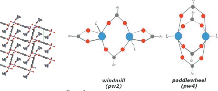
DEF molecule in a tetragonal pyramidal configuration. The
SBU consists of doubly-bridged dinuclear units of ZnIIatoms
in a ‘windmill’ fashion (Fig. 1), with a Zn Zn distance of
3.652 (1) A˚ , where each pair of Zn atoms is linked by two
NDC2anions and each Zn atom is linked by a further NDC2
anion and a DEF molecule (Fig. 2). The two carboxylate
groups of the same NDC2anion adopt either a1-1:1(O1
and O2) or a2-1:1(O3 and O5) coordination mode.
3. Supramolecular features
The structure of1shows a three-dimensional (3D)
supramo-lecular framework built of zigzag-shaped two-dimensional (2D) stacked layers. Neighbouring 2D layers are connected through nonclassical hydrogen-bonding
inter-actions between carboxylate O atoms (O1 and O3) and-H
atoms of NDC2ligands with COO H—C—NDC distances
1760
Saffon-Merceronet al. [Zn(C12H6O4)(C15H11NO)] Acta Cryst.(2019). E75, 1759–1762 [image:2.610.47.297.119.176.2] [image:2.610.315.565.220.368.2]research communications
Table 1
Hydrogen-bond geometry (A˚ ,).
Cg1 andCg2 are the centroids of the C2–C5/C5vii/C6viiand C5/C6/C2vii–C5vii
rings, respectively.
D—H A D—H H A D A D—H A
C4—H4 O1iv 0.95 2.39 3.307 (4) 161
C12—H12 O3v 0.95 2.63 3.548 (4) 156
C16—H16 Cg1vi 0.95 2.99 3.520 (17) 114
C16—H16 Cg2vii 0.95 2.99 3.520 (17) 114
Symmetry codes: (iv) x1;y;z; (v) xþ1;y;z; (vi) xþ1;yþ1;z; (vii)
x;yþ1;zþ2.
Figure 2
[image:2.610.89.516.495.726.2]of 3.307 (4) (O1—C4) and 3.548 (4) A˚ (O3—C12). Other interactions contributing to the stability of the framework
involve Hcentroid– interactions of H16—C16 (DEF
hydro-gens) and the centroids [Cg1iiiis the centroid of the C2–C5/ C5v/C6vring andCg2ivis the centroid of the C5/C6/C2v–C5v ring; symmetry codes: (iii)x+ 1,y+ 1,z; (iv)x,y+ 1,z+ 2;
(v)x1,y,z+ 2] of the aromatic rings of the NDC2
ligands, withCg H distances of 2.99 A˚ (Fig. 3 and Table 1). The layers are stacked in a self-locking fashion in a 3D supramolecular framework (Fig. 4), which has open channels
with dimensions of approximately 7.85 12.55 A˚2 largely
occupied by the Zn-coordinated DEF molecules (Fig. 5). It is
noteworthy that since1has been obtained in a DEF solution
containing small amounts of formic acid, formate ligands are not present in the framework.
4. Database survey
Naphthalene dicarboxylic acid derivatives (H2NDCs),
including 1,4-, 1,8- and 2,6-NDC, have been, due to their stability, richness in coordination modes and structural rigidity, widely used as organic molecules in the synthesis of novel MOF structures with a variety of metal ions, such as ZnII, CdII, CoII, NiII, MnIIor AgI. Among all the
2,6-NDC/Zn-based MOFs, two are closely related to MOF1,i.e.a MOF of
formula [Zn2(2,6-NDC)2(DMF)2]n(Yanget al., 2013), in which
the two carboxylate groups of all the NDC ligands have two
different coordination modes (1-1
:1
and 2-1
:1 ), and MOF-105 and its derivatives of generic formula
[image:3.610.46.297.71.278.2][Zn2(2,6-NDC)2(DMF)2] (Eddaoudi et al., 2002; Devi et al., 2004;
Figure 3
Hcentroid–interaction found in MOF1with DEF H atoms (H16) located
[image:3.610.308.562.74.244.2]near the centroid of the NDC2aromatic ring (all H atoms have been omitted for clarity, except for the DEF-H16 H atoms involved in the interactions).
Figure 4
View of the two-dimensional layers in MOF1stacked in a self-locking fashion yielding the three-dimensional supramolecular framework.
Figure 5
[image:3.610.44.563.534.731.2]Shahangi Shirazi et al., 2015; Yue et al., 2015), in which all
NDC-carboxylates have a2-1
:1
coordination mode, with a
typical pw4 paddle-wheel structure motif, [M2(CO2)4]. For
MOF1, the two carboxylate groups of the same NDC2ligand
adopt either a1-1 :1
(O1 and O2) or a2-1
:1
(O3 and O5) coordination mode, giving an uncommon pw2 paddle-wheel (‘windmill’) structural feature, [M2(CO2)2].
5. Synthesis and crystallization
MOF 1 was synthesized from naphthalene-2,6-dicarboxylic
acid and zinc(II) acetate. 2,6-H2NDC (87.3 mg, 0.4 mmol,
1.0 equiv.) and Zn(OAc)22H2O (224 mg, mol, 2.5 equiv.) were
dissolved in DEF (10 ml) containing formic acid (185ml,
12 equiv.) and sealed in a glass vial. The vial was heated in an
oven to 110C for 17 h. After cooling to room temperature,
the reaction was allowed to stand until colorless crystals suitable for X-ray diffraction formed. For further character-izations, the crystals were collected by filtration, washed with DEF several times, and dried at 373 K under vacuum. Ele-mental analysis (%) for C17H17NO5Zn based on the formula [Zn(NDC)(DEF)] found (calculated): C 53.00 (53.63), H 4.47
(4.50), N 3.39 (3.68), Zn 17.51 (17.17). FT–IR (cm1): 2979,
2938, 1647, 1602, 1586, 1557, 1494, 1460, 1406, 1385, 1361, 1348. The identity of the as-synthesized bulk material was confirmed by comparing the powder X-ray diffraction (PXRD) pattern with that simulated from the crystal structure (Fig. 6). After
heating a sample of1at 463 K under vaccum for 8 h,
coordi-nated DEF molecules were elimicoordi-nated, as evidenced by FT–
IR (loss of bands at 2979, 2938 and 1647 cm1). Elemental
analysis (%) for C12H6O4Zn based on the formula [Zn(NDC)] found (calculated): C 48.85 (51.56), H 2.75 (2.16), N 0.22 (0.00), Zn 21.47 (23.39). It should be noted that after removal
of DEF, MOF 1 lost its crystallinity, as evidenced by the
PXRD pattern.
6. Refinement
The ethyl groups of DEF were disordered over two positions, for which the occupancies were refined, converging to 0.51 and 0.49. The SAME, DELU and SIMU restraints were applied to model the disorder (Sheldrick, 2008). All H atoms were fixed geometrically and treated as riding, with C—H = 0.95
(aromatic), 0.98 (CH3), 0.99 (CH2) or 1.0 A˚ (CH), with
Uiso(H) = 1.5Ueq(C) for methyl H atoms or 1.2Ueq(C) other-wise. Crystal data, data collection and structure refinement details are summarized in Table 2.
References
Bruker (2008).APEX2,SAINT,SADABSandSHELXTL. Bruker AXS Inc., Madison, Wisconsin, USA.
Devi, R. N., Edgar, M., Gonzalez, J., Slawin, A. M. Z., Tunstall, D. P., Grewal, P., Cox, P. A. & Wright, P. A. (2004).J. Phys. Chem. B,108, 535–543.
Eddaoudi, M., Kim, J., Vodak, D., Sudik, A., Wachter, J., O’Keeffe, M. & Yaghi, O. M. (2002).Proc. Natl Acad. Sci. USA,99, 4900–4904. Gangu, K. K., Maddila, S. & Jonnalagadda, S. B. (2017).Inorg. Chim.
Acta,466, 308–323.
Rosi, N. L., Eckert, J., Eddaoudi, M., Vodak, D. T., Kim, J., O’Keeffe, M. & Yaghi, O. M. (2003).Science,300, 1127–1129.
Rowsell, J. L. C., Millward, A. R., Park, K. S. & Yaghi, O. M. (2004).J.
Am. Chem. Soc.126, 5666–5667.
Saffon-Merceron, N., Barthe´le´my, M.-C., Laurent, C., Fabing, I., Hoffmann, P. & Vigroux, A. (2015).Inorg. Chim. Acta,426, 15–19. Shahangi Shirazi, F. & Akhbari, K. (2015).Inorg. Chim. Acta,436, 1–
6.
Sheldrick, G. M. (2008).Acta Cryst.A64, 112–122. Sheldrick, G. M. (2015).Acta Cryst.C71, 3–8. Westrip, S. P. (2010).J. Appl. Cryst.43, 920–925.
Yang, S. Y., Yuan, H. B., Xu, X. B. & Huang, R. B. (2013).Inorg.
Chim. Acta,403, 53–62.
Yue, H., Shi, Z., Wang, Q., Du, T., Ding, Y., Zhang, J., Huo, N. & Yang, S. (2015).RSC Adv.5, 75653–75658.
1762
Saffon-Merceronet al. [Zn(C12H6O4)(C15H11NO)] Acta Cryst.(2019). E75, 1759–1762research communications
Figure 6
PXRD patterns (a) simulated from the single-crystal data of1and (b) measured from a sample of1prepared from 2,6-H2NDC and Zn(OAc)2in
[image:4.610.45.295.70.183.2] [image:4.610.313.562.88.374.2]DEF containing formic acid.
Table 2
Experimental details.
Crystal data
Chemical formula [Zn(C12H6O4)(C15H11NO)]
Mr 380.68
Crystal system, space group Triclinic,P1
Temperature (K) 193
a,b,c(A˚ ) 7.9134 (5), 8.3006 (5), 12.6413 (8)
,,() 97.873 (4), 91.620 (4), 91.991 (5)
V(A˚3) 821.57 (9)
Z 2
Radiation type MoK
(mm1) 1.52
Crystal size (mm) 0.100.040.04
Data collection
Diffractometer Bruker SMART APEXII CCD
area detector
Absorption correction Multi-scan (SADABS; Bruker, 2008)
Tmin,Tmax 0.863, 0.942
No. of measured, independent and observed [I> 2(I)] reflections
13141, 3336, 2436
Rint 0.075
(sin /)max(A˚1) 0.625
Refinement
R[F2> 2(F2)],wR(F2),S 0.042, 0.081, 1.00
No. of reflections 3336
No. of parameters 237
No. of restraints 41
H-atom treatment H-atom parameters constrained max,min(e A˚3) 0.33,0.37
Computer programs: APEX2 (Bruker, 2008), SAINT (Bruker, 2008), SHELXS97
(Sheldrick, 2008), SHELXL2017(Sheldrick, 2015), SHELXTL(Bruker, 2008) and
sup-1 Acta Cryst. (2019). E75, 1759-1762
supporting information
Acta Cryst. (2019). E75, 1759-1762 [https://doi.org/10.1107/S2056989019014142]
Crystal structure of a two-dimensional coordination polymer of formula
[Zn(NDC)(DEF)] (H
2NDC is naphthalene-2,6-dicarboxylic acid and DEF is
N
,
N
-diethylformamide)
Nathalie Saffon-Merceron, Alain Vigroux and Pascal Hoffmann
Computing details
Data collection: APEX2 (Bruker, 2008); cell refinement: APEX2 (Bruker, 2008); data reduction: SAINT (Bruker, 2008); program(s) used to solve structure: SHELXS97 (Sheldrick, 2008); program(s) used to refine structure: SHELXL2017
(Sheldrick, 2015); molecular graphics: SHELXTL (Bruker, 2008); software used to prepare material for publication:
publCIF (Westrip, 2010).
Poly[bis(N,N-diethylformamide)(µ4-naphthalene-2,6-dicarboxylato)(µ2-naphthalene-2,6-dicarboxylato)dizinc(II)]
Crystal data
[Zn(C12H6O4)(C15H11NO)]
Mr = 380.68
Triclinic, P1 Hall symbol: -P 1
a = 7.9134 (5) Å
b = 8.3006 (5) Å
c = 12.6413 (8) Å
α = 97.873 (4)°
β = 91.620 (4)°
γ = 91.991 (5)°
V = 821.57 (9) Å3
Z = 2
F(000) = 392
Dx = 1.539 Mg m−3
Mo Kα radiation, λ = 0.71073 Å Cell parameters from 1701 reflections
θ = 2.5–21.5°
µ = 1.52 mm−1
T = 193 K Block, colourless 0.10 × 0.04 × 0.04 mm
Data collection
Bruker SMART APEXII CCD area detector diffractometer
Radiation source: fine-focus selaed tube Detector resolution: 8.333 pixels mm-1 phi and ω scans
Absorption correction: multi-scan (SADABS; Bruker, 2008)
Tmin = 0.863, Tmax = 0.942
13141 measured reflections 3336 independent reflections 2436 reflections with I > 2σ(I)
Rint = 0.075
θmax = 26.4°, θmin = 2.8°
h = −9→9
k = −10→10
l = −15→15
Refinement
Refinement on F2 Least-squares matrix: full
R[F2 > 2σ(F2)] = 0.042
wR(F2) = 0.081
S = 1.00 3336 reflections
237 parameters 41 restraints 0 constraints
supporting information
sup-2 Acta Cryst. (2019). E75, 1759-1762
Secondary atom site location: difference Fourier map
Hydrogen site location: inferred from neighbouring sites
H-atom parameters constrained
w = 1/[σ2(F
o2) + (0.0318P)2] where P = (Fo2 + 2Fc2)/3 (Δ/σ)max = 0.001
Δρmax = 0.33 e Å−3 Δρmin = −0.37 e Å−3
Special details
Geometry. All esds (except the esd in the dihedral angle between two l.s. planes) are estimated using the full covariance matrix. The cell esds are taken into account individually in the estimation of esds in distances, angles and torsion angles; correlations between esds in cell parameters are only used when they are defined by crystal symmetry. An approximate (isotropic) treatment of cell esds is used for estimating esds involving l.s. planes.
Refinement. Refinement of F2 against ALL reflections. The weighted R-factor wR and goodness of fit S are based on F2, conventional R-factors R are based on F, with F set to zero for negative F2. The threshold expression of F2 > 2sigma(F2) is used only for calculating R-factors(gt) etc. and is not relevant to the choice of reflections for refinement. R-factors based on F2 are statistically about twice as large as those based on F, and R- factors based on ALL data will be even larger.
Fractional atomic coordinates and isotropic or equivalent isotropic displacement parameters (Å2)
x y z Uiso*/Ueq Occ. (<1)
Zn1 0.02528 (5) 0.13460 (5) 0.62683 (3) 0.02329 (12)
O1 −0.0138 (3) 0.0539 (3) 0.8118 (2) 0.0495 (7)
O2 −0.1959 (3) 0.1010 (3) 0.68613 (17) 0.0311 (5)
O3 0.0472 (2) −0.1661 (2) 0.52376 (16) 0.0249 (5)
O4 0.1080 (3) 0.3581 (3) 0.68413 (19) 0.0376 (6)
O5 0.2353 (2) 0.0121 (2) 0.60995 (16) 0.0267 (5)
C1 −0.1604 (4) 0.0679 (4) 0.7800 (3) 0.0292 (8)
C2 −0.3060 (4) 0.0478 (4) 0.8506 (2) 0.0229 (7)
C3 −0.4729 (4) 0.0766 (4) 0.8157 (3) 0.0259 (7)
H3 −0.491459 0.106685 0.746526 0.031*
C4 −0.6065 (4) 0.0621 (4) 0.8792 (2) 0.0253 (7)
H4 −0.716770 0.083839 0.854527 0.030*
C5 −0.5832 (3) 0.0151 (3) 0.9818 (2) 0.0204 (7)
C6 −0.7197 (4) −0.0032 (4) 1.0501 (2) 0.0247 (7)
H6 −0.831308 0.016042 1.026474 0.030*
C7 0.1971 (4) −0.1257 (4) 0.5574 (2) 0.0232 (7)
C8 0.3342 (4) −0.2464 (4) 0.5386 (2) 0.0231 (7)
C9 0.2888 (4) −0.4062 (4) 0.5012 (2) 0.0266 (7)
H9 0.173329 −0.437095 0.484564 0.032*
C10 0.4136 (4) −0.5243 (4) 0.4875 (2) 0.0241 (7)
C11 0.5046 (4) −0.1977 (4) 0.5621 (3) 0.0281 (8)
H11 0.534020 −0.086805 0.587033 0.034*
C12 0.6283 (4) −0.3091 (4) 0.5492 (3) 0.0285 (8)
H12 0.743193 −0.274861 0.564835 0.034*
C13 0.2535 (5) 0.4026 (4) 0.7188 (3) 0.0369 (9)
H13 0.341309 0.327989 0.705702 0.044*
C14 0.1574 (6) 0.6627 (5) 0.7929 (4) 0.0628 (13)
H14A 0.078313 0.652571 0.729974 0.075*
H14B 0.208053 0.774681 0.803717 0.075*
C15 0.0611 (7) 0.6341 (6) 0.8898 (4) 0.0970 (18)
sup-3 Acta Cryst. (2019). E75, 1759-1762
H15B −0.030364 0.710761 0.899858 0.145*
H15C 0.137829 0.650364 0.952935 0.145*
N1 0.2922 (4) 0.5454 (4) 0.7719 (2) 0.0448 (8)
C16 0.4576 (17) 0.6215 (19) 0.815 (2) 0.061 (4) 0.516 (8)
H16A 0.487379 0.713730 0.775639 0.074* 0.516 (8)
H16B 0.447852 0.665014 0.890959 0.074* 0.516 (8)
C17 0.5911 (12) 0.5053 (11) 0.8035 (8) 0.065 (3) 0.516 (8)
H17A 0.563272 0.415235 0.843378 0.098* 0.516 (8)
H17B 0.698301 0.559271 0.831750 0.098* 0.516 (8)
H17C 0.601884 0.462995 0.727791 0.098* 0.516 (8)
C16′ 0.4774 (18) 0.575 (2) 0.8054 (19) 0.062 (4) 0.484 (8)
H16C 0.544874 0.496333 0.760003 0.074* 0.484 (8)
H16D 0.513676 0.685674 0.792220 0.074* 0.484 (8)
C17′ 0.5149 (13) 0.5600 (11) 0.9178 (7) 0.074 (3) 0.484 (8)
H17D 0.426986 0.611745 0.962205 0.110* 0.484 (8)
H17E 0.625043 0.613689 0.939854 0.110* 0.484 (8)
H17F 0.517878 0.444637 0.926528 0.110* 0.484 (8)
Atomic displacement parameters (Å2)
U11 U22 U33 U12 U13 U23
Zn1 0.0191 (2) 0.0234 (2) 0.0272 (2) 0.00401 (14) 0.00315 (15) 0.00116 (15)
O1 0.0203 (14) 0.086 (2) 0.0477 (17) 0.0087 (13) 0.0085 (12) 0.0236 (15)
O2 0.0277 (13) 0.0404 (14) 0.0263 (13) 0.0025 (10) 0.0067 (11) 0.0068 (11)
O3 0.0184 (12) 0.0277 (12) 0.0302 (13) 0.0040 (9) 0.0037 (10) 0.0087 (10)
O4 0.0358 (15) 0.0268 (13) 0.0474 (16) 0.0019 (10) 0.0016 (12) −0.0052 (11)
O5 0.0258 (12) 0.0249 (13) 0.0286 (13) 0.0078 (9) 0.0039 (10) −0.0010 (10)
C1 0.0237 (19) 0.0315 (19) 0.033 (2) 0.0043 (14) 0.0064 (16) 0.0037 (16)
C2 0.0208 (17) 0.0233 (17) 0.0232 (18) 0.0008 (13) 0.0028 (14) −0.0022 (14)
C3 0.0250 (18) 0.0267 (18) 0.0270 (19) 0.0046 (14) 0.0008 (15) 0.0064 (15)
C4 0.0172 (17) 0.0308 (19) 0.0277 (19) 0.0021 (13) −0.0044 (14) 0.0042 (15)
C5 0.0170 (16) 0.0211 (16) 0.0222 (17) 0.0014 (12) −0.0003 (13) 0.0003 (13)
C6 0.0143 (16) 0.0288 (18) 0.0308 (19) 0.0031 (13) −0.0004 (14) 0.0027 (15)
C7 0.0287 (19) 0.0256 (18) 0.0176 (17) 0.0068 (14) 0.0075 (14) 0.0079 (14)
C8 0.0220 (17) 0.0257 (18) 0.0224 (18) 0.0067 (13) 0.0039 (14) 0.0046 (14)
C9 0.0188 (17) 0.0334 (19) 0.0284 (19) 0.0058 (14) 0.0032 (14) 0.0050 (15)
C10 0.0215 (17) 0.0276 (18) 0.0241 (17) 0.0035 (13) 0.0036 (14) 0.0049 (14)
C11 0.0274 (19) 0.0260 (19) 0.031 (2) 0.0038 (14) 0.0026 (15) 0.0018 (15)
C12 0.0233 (18) 0.0271 (18) 0.034 (2) −0.0003 (14) 0.0039 (15) 0.0011 (15)
C13 0.042 (2) 0.032 (2) 0.037 (2) 0.0006 (16) −0.0029 (18) 0.0063 (17)
C14 0.089 (4) 0.027 (2) 0.067 (3) −0.006 (2) 0.010 (3) −0.012 (2)
C15 0.111 (5) 0.084 (4) 0.087 (4) −0.011 (3) 0.037 (4) −0.023 (3)
N1 0.057 (2) 0.0383 (19) 0.0367 (19) −0.0130 (16) −0.0097 (16) 0.0030 (15)
C16 0.077 (6) 0.051 (8) 0.053 (6) −0.018 (5) −0.018 (5) 0.006 (6)
C17 0.063 (6) 0.069 (6) 0.066 (6) −0.014 (4) −0.011 (5) 0.022 (5)
C16′ 0.076 (6) 0.052 (9) 0.055 (6) −0.028 (6) −0.023 (6) 0.013 (7)
supporting information
sup-4 Acta Cryst. (2019). E75, 1759-1762
Geometric parameters (Å, º)
Zn1—O2 1.949 (2) C11—C12 1.368 (4)
Zn1—O4 1.979 (2) C11—H11 0.9500
Zn1—O5 1.980 (2) C12—H12 0.9500
Zn1—O3i 2.026 (2) C13—N1 1.302 (4)
Zn1—C1 2.571 (3) C13—H13 0.9500
O1—C1 1.231 (4) C14—N1 1.474 (5)
O2—C1 1.280 (4) C14—C15 1.503 (6)
O3—C7 1.267 (3) C14—H14A 0.9900
O4—C13 1.246 (4) C14—H14B 0.9900
O5—C7 1.264 (4) C15—H15A 0.9800
C1—C2 1.496 (4) C15—H15B 0.9800
C2—C6ii 1.368 (4) C15—H15C 0.9800
C2—C3 1.419 (4) N1—C16 1.488 (11)
C3—C4 1.358 (4) N1—C16′ 1.517 (11)
C3—H3 0.9500 C16—C17 1.452 (17)
C4—C5 1.413 (4) C16—H16A 0.9900
C4—H4 0.9500 C16—H16B 0.9900
C5—C6 1.420 (4) C17—H17A 0.9800
C5—C5ii 1.424 (5) C17—H17B 0.9800
C6—H6 0.9500 C17—H17C 0.9800
C7—C8 1.504 (4) C16′—C17′ 1.47 (2)
C8—C9 1.378 (4) C16′—H16C 0.9900
C8—C11 1.406 (4) C16′—H16D 0.9900
C9—C10 1.413 (4) C17′—H17D 0.9800
C9—H9 0.9500 C17′—H17E 0.9800
C10—C12iii 1.422 (4) C17′—H17F 0.9800
C10—C10iii 1.426 (6)
O2—Zn1—O4 107.22 (9) C8—C11—H11 119.8
O2—Zn1—O5 136.08 (9) C11—C12—C10iii 120.5 (3)
O4—Zn1—O5 103.46 (9) C11—C12—H12 119.7
O2—Zn1—O3i 99.73 (8) C10iii—C12—H12 119.7
O4—Zn1—O3i 100.55 (9) O4—C13—N1 123.8 (3)
O5—Zn1—O3i 104.71 (8) O4—C13—H13 118.1
O2—Zn1—C1 28.93 (9) N1—C13—H13 118.1
O4—Zn1—C1 100.44 (10) N1—C14—C15 111.5 (4)
O5—Zn1—C1 115.09 (9) N1—C14—H14A 109.3
O3i—Zn1—C1 128.55 (9) C15—C14—H14A 109.3
C1—O2—Zn1 103.60 (19) N1—C14—H14B 109.3
C7—O3—Zn1i 119.47 (19) C15—C14—H14B 109.3
C13—O4—Zn1 127.3 (2) H14A—C14—H14B 108.0
C7—O5—Zn1 107.70 (19) C14—C15—H15A 109.5
O1—C1—O2 122.1 (3) C14—C15—H15B 109.5
O1—C1—C2 121.0 (3) H15A—C15—H15B 109.5
O2—C1—C2 116.8 (3) C14—C15—H15C 109.5
sup-5 Acta Cryst. (2019). E75, 1759-1762
O2—C1—Zn1 47.47 (15) H15B—C15—H15C 109.5
C2—C1—Zn1 163.8 (2) C13—N1—C14 118.8 (3)
C6ii—C2—C3 119.1 (3) C13—N1—C16 131.2 (8)
C6ii—C2—C1 120.6 (3) C14—N1—C16 110.0 (8)
C3—C2—C1 120.3 (3) C13—N1—C16′ 115.1 (9)
C4—C3—C2 121.2 (3) C14—N1—C16′ 126.1 (9)
C4—C3—H3 119.4 C17—C16—N1 111.5 (12)
C2—C3—H3 119.4 C17—C16—H16A 109.3
C3—C4—C5 120.7 (3) N1—C16—H16A 109.3
C3—C4—H4 119.7 C17—C16—H16B 109.3
C5—C4—H4 119.7 N1—C16—H16B 109.3
C4—C5—C6 122.4 (3) H16A—C16—H16B 108.0
C4—C5—C5ii 119.0 (3) C16—C17—H17A 109.5
C6—C5—C5ii 118.6 (3) C16—C17—H17B 109.5
C2ii—C6—C5 121.4 (3) H17A—C17—H17B 109.5
C2ii—C6—H6 119.3 C16—C17—H17C 109.5
C5—C6—H6 119.3 H17A—C17—H17C 109.5
O5—C7—O3 122.1 (3) H17B—C17—H17C 109.5
O5—C7—C8 118.2 (3) C17′—C16′—N1 114.1 (15)
O3—C7—C8 119.6 (3) C17′—C16′—H16C 108.7
C9—C8—C11 120.8 (3) N1—C16′—H16C 108.7
C9—C8—C7 118.7 (3) C17′—C16′—H16D 108.7
C11—C8—C7 120.5 (3) N1—C16′—H16D 108.7
C8—C9—C10 120.1 (3) H16C—C16′—H16D 107.6
C8—C9—H9 119.9 C16′—C17′—H17D 109.5
C10—C9—H9 119.9 C16′—C17′—H17E 109.5
C9—C10—C12iii 121.8 (3) H17D—C17′—H17E 109.5
C9—C10—C10iii 119.2 (4) C16′—C17′—H17F 109.5
C12iii—C10—C10iii 118.9 (3) H17D—C17′—H17F 109.5
C12—C11—C8 120.4 (3) H17E—C17′—H17F 109.5
C12—C11—H11 119.8
Symmetry codes: (i) −x, −y, −z+1; (ii) −x−1, −y, −z+2; (iii) −x+1, −y−1, −z+1.
Hydrogen-bond geometry (Å, º)
Cg1 and Cg2 are the centroids of the C2–C5/C5ii/C6ii and C5/C6/C2ii–C5ii rings, respectively. [Symmetry code: (ii) -x-1, -y, -z+2.]
D—H···A D—H H···A D···A D—H···A
C4—H4···O1iv 0.95 2.39 3.307 (4) 161
C12—H12···O3v 0.95 2.63 3.548 (4) 156
C16—H16···Cg1vi 0.95 2.99 3.520 (17) 114
C16—H16···Cg2vii 0.95 2.99 3.520 (17) 114