This article has been accepted for publication and undergone full peer review but has not
been through the copyediting, typesetting, pagination and proofreading process which may
lead to differences between this version and the Version of Record. Please cite this article as
Changes in stable isotope compositions during fasting in phocid seals
Sarah Habran1,2,§, France Damseaux1,§, Paddy Pomeroy3, Cathy Debier4, Daniel Crocker5, Gilles Lepoint1,
Krishna Das1*
1 Freshwater and Oceanic sciences Unit of reSearch (FOCUS), Laboratory of Oceanology, University of Liège
B6c, 11 Allée du 6 Août, 4000 Liège, Belgium
2 ISSeP, Institut Scientifique de Service Public, 200 rue du Chéra, 4000 Liège, Belgium
3
Sea Mammal Research Unit, Scottish Oceans Institute, East Sands, University of St Andrews, KY16 8LB, UK
4 Institut des Sciences de la Vie, Université catholique de Louvain, Croix du Sud 2/L7.05.08, 1348
Louvain-la-Neuve, Belgium
5 Department of Biology, Sonoma State University, Rohnert Park, CA 94928, USA
§: These authors contributed equally to the work
Abstract
RATIONALE: The grey seal, Halichoerus grypus (GS), and the northern elephant seal, Mirounga angustirostris (NES), come ashore for reproduction. This period involves intense physiological processes such
as lactation in females and a developmental post-weaning fast in juveniles. Previous studies have shown that
δ13
C and δ15N values are affected by starvation, but the precise effects of fasting associated to lactation and
post-weaning fast in seals remain poorly understood.
METHODS: To examine the effect of lactation and post-weaning fast on stable isotope ratios in GS and NES, blood and hair were sampled from twenty-one GS mother-pup pairs on the Isle of May and on twenty-two
weaned NES pups at Año Nuevo State Reserve during their respective breeding seasons. Milk samples were
also collected from GS mothers. Stable isotope measurements were performed with an isotope ratio mass
spectrometer coupled to an N-C elemental analyser.
RESULTS: Changes in stable isotope ratios in blood components during fasting were similar and weak between GS and NES mothers especially in blood cells (GS: Δ15N = 0.05‰, Δ13C = 0.02‰; NES: Δ15N = 0.1‰, Δ13C =
0.1‰). GS showed a 15N discrimination factor between maternal and pup blood cells and milk, but not for 13C.
The strongest relationship between the isotopic compositions of the mother and the pup was observed in the
blood cells.
CONCLUSIONS: Isotopic consequences of lactation, fasting, and growth seem limited in NES and GS, especially in medium-term integrator tissues of feeding activity such as blood cells. Stable isotope ratios in the
blood of pups and mothers are correlated. We observed a subtle mother-to-pup fractionation factor. Our results
suggest that pup blood cells are mostly relevant for exploring the ecology of female seals.
1. Introduction
Pups of pinnipeds are increasingly used as proxies to investigate maternal foraging strategies thanks to stable
isotopes analyses. Indeed, pup isotope ratios reflect those of the females 1–3. To validate the use of these proxies,
the stable isotopes ratios in pup tissues must be linearly correlated with those in maternal tissues. Moreover,
investigating the foraging ecology of adult females requires consideration of the isotopic fractionation between
mothers and pups, and how this fractionation might change during the course of nursing and weaning. Lactating
female seals catabolize their tissues to produce milk. Therefore, nursing pups are placed at a trophic level higher
than their mother with a consecutive isotope enrichment 1,3.
Phocid seals such as the grey seal, Halichoerus grypus (GS), and the northern elephant seal, Mirounga
angustirostris (NES), undergo periods of prolonged fasting twice a year, during lactation and the annual moult
4–9. During lactation, mothers fast for several weeks while secreting a fat-rich milk synthesized from their body
reserves (duration: 17-23 days for GS and 24-28 days for NES). Seals may lose between 35 and 57% of stored
body reserves during each of these periods 10. Pups are weaned abruptly and undergo a prolonged land-based
post-weaning fast (~1-4 weeks for GS and ~8-10 weeks for NES) before departing to sea and initiating foraging
11–14. This post-weaning fast is an important developmental time relative to the diving physiology of the pups
6,10. The metabolic constraints experienced by the animal’s body during the breeding season are extreme and can
trigger a remobilization of carbon and nitrogen in tissues 15.
Stable carbon and nitrogen isotope ratios (δ13C and δ15N values) have been used widely to study marine
mammal ecology 16. This technique is based on the idea that the isotopic composition of a consumer is a
proportional mixing of the isotopic composition of its prey, after accounting for isotopic fractionation in the
digestion and assimilation process 17,18. This fractionation typically results in enrichment in the heavier isotopes
(13C and 15N). The variation of the δ13C value between the predator and the prey is usually low. These values are
therefore close to that of the diet and are indicators of the primary production supporting the consumer,
indicating, for example, the aquatic vs terrestrial, inshore vs offshore, or pelagic vs benthic contribution to food
intake 19,20. At the opposite and of the spectrum, variation of the δ15N value between different trophic levels is
typically more marked than for the δ13C value, leading to a predictable increase with trophic level 18,21. δ13C and
δ15N values are also increasingly used in physiology studies of lactation 1,22–25, of energy acquisition 26, or of
isotopic relationship between the mother and her offspring 1–3. In the case of phocids, it is important to consider
foraging behaviour of the mothers. Information regarding the isotopic consequences of fasting during lactation
and post-weaning fast is limited, especially in medium-term integrator tissues of feeding activity, such as blood
cells. Pinnipeds, which rely on their stored blubber during fasting, should have lower δ13C values, since lipids
are known to be 13C-depleted during biochemical fractionation 17,27. Many marine mammals also catabolize
significant amounts of tissue proteins during lactation and fasting 16,25. Fasting phocids enrich their tissues in 15N
and their blood urea in 14N because of the net catabolic state 8,21,28,29. In contrast, the anabolic state associated
with the protein synthesis 30 can reduce the 15N in the maternal tissues 8 during gestation [32] or lactation [34].
To elucidate changes in isotopic composition during fasting in phocids, we investigated carbon and
nitrogen (δ13C and δ15N values) isotopic dynamics in blood cells, serum, milk and hair of GS and NES during
lactation and/or post-weaning fast.
2. Material and methods
(a) Sampling
Two longitudinal studies were performed in the grey seal, Halichoerus grypus (Fabricius 1791), and the
northern elephant seal, Mirounga angustirostris (Gill, 1866), during lactation and/or post-weaning fast.
The GS were sampled on the Isle of May, UK(56°11ʼN, 2°33ʼW), during the breeding season
(October-December 2008). Twenty-one GS mother-pup pairs were captured in early lactation (T1: at 2-4 days
postpartum) and recaptured 12-14 days later in late lactation (T2: at 15-17 days postpartum). Maternal and pup
blood samples and mother’s milk samples were collected at each capture (Table S1, supporting information). Whole blood samples were collected from the extradural vein in BD VacutainerTM red top serum tubes including
a silicone-coated interior and increased silica act clot activator. Maternal hair and lanugo were collected only
during the second lactation (T2). Sixteen of the 21 pups were recaptured once or twice after weaning (T3 and
T4: day 19 and day 30 postpartum, respectively) to collect blood samples. The animal handling and sample
collection methods have been described previously [35]. All GS captures and sampling were performed under UK
Northern elephant seals (NES) were sampled at Año Nuevo State Reserve, CA, USA (37°06’30’’N,
122°20’10’’W), after the breeding season (February-April 2010). Twenty-two NES weaned pups were captured
three times throughout the post-weaning fast period. The captures occurred at week 1 (~35 days postpartum),
week 4 (~56 days postpartum) and week 7 (~81 days postpartum) of the post-weaning fast. Whole blood
samples were collected from the extradural vein in VacutainerTM red top serum tubes. Lanugo and new hair were
collected at week 1 and week 4, respectively. Extra blood samples from 14 of the 22 weaned pups were
collected once more at the very end of the post-weaning fast (week 9, ~92 days postpartum). The
animal-handling and sample-collection methods have been described previously [36].
GS and NES females and pups were weighed to the nearest 0.2 kg and 0.1 kg, respectively. The length and axial
girth of adult females and weaned pups were measured. Pups were sexed. The GS and NES biometric data are
summarized in Tables S1 and S2 (supporting information).
All samples were kept on ice in the field (at 4°C). At the end of each day, whole blood samples were centrifuged
for 20 min and the cellular component was harvested for analysis. Serum was aliquoted into 5-mL plastic tubes
and all samples were stored at -20 °C in the laboratory until analysed.
(b) Sample preparation
Prior to stable isotope analysis, blood cell and serum samples were freeze-dried, and ground with a mortar and
pestle into powder. After thawing, hair and lanugo were washed ultrasonically with reagent grade acetone
(acetone for analysis, EMSURE®, Merck, Darmstadt, Germany) and were rinsed repeatedly with 18.2 MΩ-cm
deionized water to remove exogenous contaminants, according to the method recommended by the International
Atomic Energy Agency [37]. Hair samples were then freeze-dried for 24 h. Whole blood (containing Red Blood
Cells, RBCs) and metabolically inert tissues constructed of keratin such as hair do not require lipid extraction
because they contain only low levels of lipids. The major carrier of fatty acids in these tissues is actually serum
albumin, meaning that the serum contains higher levels of lipids than the RBCs [34,38].
We investigated possible effects of lipid removal in serum on its isotopic composition. A randomly chosen
subset of freeze-dried serum samples (from grey seal mothers and pups, n = 10) were lipid-extracted using three
repeated rinses with 2:1 chloroform:methanol for 3 minutes prior to analysis. These preliminary tests showed
(c) Stable isotope ratio analysis
Approximately 1.0 -2.0 mg of freeze-dried blood cells and serum, and 1 mg of hair and lanugo were weighed
and loaded into tin boats. Approximately 4-5 mg and 1.5 mg of dried full milk were weighed to obtain nitrogen
and carbon isotopic compositions, respectively. All dried masses were measured to the nearest 0.01 mg. Stable
isotope measurements were performed with an isotope ratio mass spectrometer (VG Optima – Micromass,
Middlewich, UK or IsoPrime100, Elementar, Cheadle Hulme, UK) coupled to an N-C-S elemental analyser
(Carlo Erba, Milan Italy or Vario MICRO cube, Elementar) for automated analyses.
The SI ratios are expressed in delta (δ) notation as the deviation from standards in parts per thousand (‰)
according to the following equation:
where X is 13C or 15N and R is the corresponding ratio 13C/12C or 15N/14N. In this study, δ values are multiplied
by 1000 for easier understanding. Standard values were based on the Vienna PeeDee Belemnite (VPDB) for
δ13
C measurements and atmospheric nitrogen for δ15N measurements. Reference materials were IAEA-N1 (δ15N
= 0.4 ± 0.2‰) and IAEA CH-6 (sucrose) (δ13C = −10.4 ± 0.2‰). Internal standards (glycine) and replicates
were inserted into all runs after every 12 samples to calibrate the system and to assess drift over time. The
standard deviations of internal standard replicates were 0.1‰ and 0.3‰ for carbon and nitrogen, respectively.
(d) Statistical analyses
Our data were compiled with in-house data (Figure 3, Table 3) previously measured in the blood and milk of ten NES females and their pups during lactation (~ day 5 and day 22 post-partum) [39].
The normal distribution of the data was checked with a Shapiro test and the homogeneity of variances with a
Barlett test. Because the data were normally distributed and respected the homogeneity of variances, parametric
tests were used for statistical analyses. Statistical significance was determined when p<0.05. To evaluate
changes in stable isotope and elemental ratios in the different tissues and the different sampling times, analysis
of variance (ANOVA) with repeated measures and Scheffé post-hoc test were used. In the case of only two
tissues or two sampling times, paired t-tests were used to compare means. The results are presented as mean ±
standard deviation (SD). The differences observed between isotopic ratios in different periods or between
3. Results
3.1. Tissue variation of δ13C and δ15
N values in grey seals
In females, the lowest mean δ15N value was observed in blood cells (14.1‰, at both T1 and T2) and the highest
mean value in milk (17.7‰, at T1). The maternal δ15N values differed between blood cells, serum and milk
(F2,80 = 202.4, p < 0.001 at T1 and F2,80 = 104.3, p < 0.001 at T2, ANOVA with repeated measures) while the
δ15N values in serum and milk at T2 were similar (15.0‰ vs. 15.0‰, p = 1.00, Scheffé post-hoc test). The δ15N
pup values differed between lanugo (only in T2), blood cells and serum at T1, T2, T3, and T4 (F1,59 = 115.3, p <
0.001 at T1, F2,59 = 152.8, p < 0.001 at T2, F1,49 = 168.5, p < 0.001 at T3 and F1,41 = 141.8, p < 0.001 at T4,
ANOVA with repeated measures, Table 1) but were not statistically different between blood cells and serum (F1,59 = 115.3, p =1.00 at T1, F1,59 = 152.8, p = 1.00 at T2, F1,49 = 168.5, p = 1.00 at T3 and F1,41 = 141.8, p = 1.00
at T4, ANOVA with repeated measures, Table 1).
The lowest δ13C value was observed in milk (-23.6‰) and the highest in maternal hair (-15.3‰) (Table 1). The
δ13C values measured in maternal tissues differed significantly between blood cells, milk, and hair (only at T2)
(F1,80 = 202.4, p < 0.001 at T1 and F2,80 = 104.3, p < 0.001 at T2, ANOVA with repeated measures), but were
similar between blood cells and serum (F1,80 = 202.4, p = 0.285 at T1 and F1,80 = 104.3, p = 0.999 at T2, ANOVA
with repeated measures). The mean δ13C values differed between pup tissues, i.e. blood cells, serum, and lanugo
(only at T2) at T1, T2, T3, and T4 (F1,59 = 115.3, p < 0.001 at T1, F2,59 = 152.8, p < 0.001 at T2, F1,49 = 168.5, p <
0.001 at T3 and F1,41 = 141.8, p < 0.001 at T4, ANOVA with repeated measures).
3.2. Tissue variation of δ13C and δ15N values in northern elephant seals
The mean δ15N values ranged from 15.9‰ in lanugo at week 1 to 18.5‰ in serum at week 9 (Table 2). The weaned pup δ15N values differed between blood cells, serum, new hair (week 1) and lanugo (week 4) at weeks
1, 4, 7 and 9 (F2,84 = 199.7, p < 0.001, F2,84 = 220.8, p < 0.001, F1,84 = 247.6, p < 0.001 and F1,84 = 174.2, p <
0.001 respectively, ANOVA with repeated measures) but they remained similar between serum and new hair at
week 1 (F1,84 = 199.7, p =0.814, ANOVA with repeated measures) and between blood cells and lanugo at week
4 (F1,84 = 220.8, p = 0.281, ANOVA with repeated measures) (Table 2).
The NES δ13C values ranged from -21.0‰ in serum at week 7 to -18.1‰ in new hair at week 1 (Table 2). The
and lanugo (week 4) at weeks 1, 4, 7 and 9 (F2,84 = 199.7, p < 0.001, F2,84 = 220.8, p < 0.001, F1,84 = 247.6, p <
0.001 and F1,84 = 174.2, p < 0.001, respectively, ANOVA with repeated measures).
3.3. Stable isotope discrimination between grey seal mothers and their pup
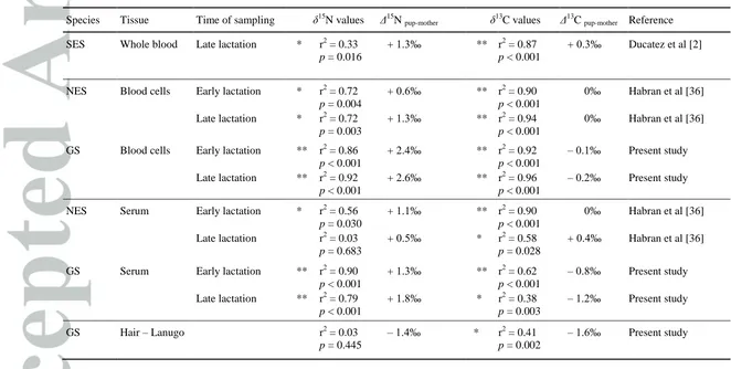
In GS, the δ15N values in blood cells were significantly higher in pups than in mothers (Δ15N = + 2.4‰ in T1
and + 2.6‰ in T2, for both p < 0.001, paired t test, Tables 1 and 5, Figure 1A). The δ15N values in serum were significantly higher in pups than in mothers (Δ15N = + 1.3‰ in T1 and + 1.8‰ in T2, for both p < 0.001, paired
t test, Tables 1 and 5). The δ15N value in lanugo was lower than that in maternal hair (Δ15N = 1.4‰, p < 0.001, paired t test, Tables 1 and 5, Figure 2A).
The δ13C values differed significantly between mother and pups in blood cells (Δ13C = 0.08‰, p = 0.015 in T1
and Δ13C = 0.2‰, p < 0.001 in T2, paired t test, Tables 1 and 5, Figure 1B), in serum (Δ13C = 0.7‰ in T1 and 1.2‰ in T2, for both p < 0.001, paired t test, Tables 1 and 5) and in lanugo and maternal hair (Δ13C = 1.5‰, p
< 0.001, paired t test, Tables 1 and 5, Figure 2B).
3.4. Changes of δ13C and δ15N values during fasting
Grey seal females. Stable isotope ratios did not differ between T1 and T2 in blood cells (Δ15N = 0.05‰, p = 0.244, Δ13C = 0.02‰, p = 0.361, paired t test) but were significantly different in maternal serum (Δ15N = 0.2‰,
p < 0.001, Δ13C = 0.3‰, p < 0.001, paired t test) and in milk (Δ15N = -2.7‰, p < 0.001, paired t test; Δ13C =
+1.1‰, p < 0.001, paired t test, Table 1).
Grey seal pups. Stable isotope ratios in blood cells and in serum did not differ significantly between T1, T2, T3 and T4 (For both δ13C and δ15N values: F3,66 = 6.3, p > 0.05; F3,66 = 4.0, p > 0.05; ANOVA with repeated
measures).
Northern elephant seals. δ15N and δ13C values in blood cells did not vary throughout the post-weaning fast (F3,84 = 1.00, p = 0.154 for δ15N and δ13C, ANOVA with repeated measures, Table 2). The δ15N values in serum remained stable at the beginning of the fast (F3,84 = 11.1, p > 0.05, ANOVA with repeated measures) and then
increased at week 7 (F3,84 = 11.1, p < 0.001, ANOVA with repeated measures). The δ13C values in serum
decreased at the beginning of the fast (F3,84 = 11.1, p < 0.001, ANOVA with repeated measures), then remained
stable (F3,84 = 11.1, p > 0.05, ANOVA with repeated measures). The δ15N and δ13C values in new hair were
4. Discussion
The aim of this study was to elucidate changes in isotopic composition during fasting in phocids. In that goal,
we investigated carbon and nitrogen (δ13C and δ15N) isotopic dynamics in blood cells, serum, milk and hair of
GS and NES during lactation and/or post-weaning fast.
We demonstrated that changes in the δ13C and δ15N values during lactation and post-weaning fast were similar
between the grey seal and the northern elephant seal. Both species showed a nitrogen isotope fractionation
between mother and offspring, but the amplitude of this fractionation was tissue-, time-, and species-specific.
Specifically, in contrast to the generally described enrichment of δ15N values in pups relative to maternal tissues
[22,23,26,40,41], the δ15N values in the lanugo were lower than those in maternal hair (15.1‰ in pups vs 16.5‰ in
mothers. The two species displayed a similar time-trend profile in the stable isotope ratios during fasting
associated with the lactation and the post-weaning period (Figure 3). The main results of this study is that isotope ratios in blood cells from pups strongly reflected those of their mothers (Figure 1), confirming the idea of using pups as proxies to investigate maternal foraging strategies, habits and places.
4.1. Fractionation between mothers and their pups
In the present study, the stable isotope ratios measured in the blood cells of the pups reflected those of their
mothers in a predictable way (r = 0.96, Figure 1A). Both phocid species showed a 15N fractionation between maternal and pup blood cells (from 0.6‰ to 2.6‰, Table 3). This fractionation is even more important in milk, consistent with the fact that pups do not feed on their mother’s blood but on milk: a Δ15N of 1.7‰ was
calculated between milk and pups (serum and blood cells) in grey seal pup at the end of their lactation period. A
Δ15N of 1.0‰ has previously been described between milk and serum in elephant seal pups at the end of their
lactation period [39]. Whole milk is often 15N-depleted compared with other maternal tissues 1. In grey seal our
data suggest higher or similar δ15N values in milk compared with blood cells and serum at the beginning and
end of lactation (Table 2). Ducatez et al 2 showed similar results in whole blood of southern elephant seals (Table 3). No fractionation was observed for 13C in NES and GS (Table 3).
4.2. Fasting and stable isotope ratios
Lactation: The δ15N and δ13C values in female were similar between the beginning and the end of the lactation (~ 2 weeks) for both the GS and the NES. Lactating females catabolize their tissues (blubber, muscle) to
previous meta-analysis showed large variations in the δ13C and δ15N values of various consumer species in
relation to starvation 15. During starvation, the N and C uptake is near zero, but N and C loss by excretion and
respiration remains, although at a low rate [43]. Fasting causes an average increase in the δ15N values of
organisms of 0.5‰, depending on the tissue type [44] and fasting duration [45]. δ15N values are often seen to
increase with fasting once an organism begins to catabolize tissues [46,47]. During fasting, an organism first
catabolizes its lipid reserves, before switching to catabolize proteins [47,48]. However, this enrichment in 15N
occurs when fasting or starvation is severe enough to cause protein, rather than lipid catabolism [45,47]. In grey
seals and elephant seals, fasting is associated with an very intense lactation process and the maternal mass and
fat content strongly influence the maternal investment in pinnipeds [49]. This rate of lipid energy output in
phocids requires extensive lipid mobilization, mainly from blubber [50]. The organs that will become 15
N-enriched during a fast are those that maintain significant synthesis which might not be the case for lactating
females [51].
Post-weaning fast: Stable isotope ratios in NES weaned pups (present study) are similar to those observed in suckling pups at the end of their lactation period [39]. The δ15N and δ13C values in blood cells of both species did
not vary significantly during this period. In contrast, NES serum was progressively enriched in 15N during the
fast (Δ15N = +0.9‰), while it was slightly depleted in 13C (Δ13C = -0.4‰). Δ15N values increase with fasting
time [45] which was longer in NES (9 weeks) than in GS (less than 2 weeks). These changes in the serum were
expected since catabolism of protein from lean tissues (e.g., muscle) during periods of nutritional stress may
cause an increase of δ15N values and a decrease of δ13C value because of the production of energy compounds
(e.g. body proteins synthesis) [23,33,46]. Changes were detectable in serum as it is a short-term integrator of diet
[52,53]. In contrast, blood cells integrate a period of several months in large mammals [54,55] as the half-life is
estimated to be 35 days in mammal blood [38], and thus buffer the short-term fasting effect found in tissues with
high turnover rates.
4.3. Special insight on hair tissue
In contrast to the enrichment in 15N of pup tissues relative to maternal tissues usually described [22,23,26,40,57], the
lanugo of GS pups showed δ15N values lower than those of maternal hair (Δ15N = 1.4‰ ± 0.7 and ± 0.5,
This highlights again that the isotopic fractionation between offspring and mother is tissue-specific and no
generalizations can be made across multiple tissues. Moreover, the relationship between δ15N values in lanugo
and maternal hair in the GS (Figure 2) was weaker than in RBC (Figure 1). Various studies have already used pup hair as a proxy to investigate the foraging ecology of adult females 3,54, but the key difference between these
studies and the present one is the hair physiology of the different mammal species. Dalerum et al, for example,
collected hair from meerkat pups and their mothers but the hair of mother and pups is synthesised during the
same period and is thus comparable 26. Porras-Peters analysed fur from suckling California sea lion pups,
assuming that they would accurately record differences in the foraging patterns in their mothers 54. However, the
growth histories and shedding phenologies of hair of sea lions (otarids) and GS/NES are different, with the NES
and GS undergoing moults during a different period from when the pup’s lanugo is synthesised. Our study
showed that using pup hair as a proxy to investigate the foraging ecology of adult femaleswas not appropriate in
these two specific species (GS and NES), as the hair physiology is very different from one mammal to another.
In the present study, we observed a negative fractionation between maternal hair and lanugo and no correlation
between the δ15N values of the maternal hair and the lanugo. This may be explained by the fact that the active
hair growth of phocids begins at the annual moult for ~12 weeks 55, meaning that the isotopic composition of the
hair after the moult and thus before the lactation represents prior moult foraging activities. On the other hand,
the lanugo is synthesized during gestation and thus according to the post moult foraging activities, meaning that
maternal hair and lanugo represent different period of feeding. Lanugo may not be used as a proxy of the
foraging habit of the mother as shown by our results. Considerable caution should be taken before comparing
hair and lanugo in GS and NES, as this assumes that the adult female uses the same foraging habitat and
resources before and after the moult and that there is no change of baseline isotopic ratios during these two
periods 56.
The δ15N and δ13C values observed in the lanugo of NES (15.9 ± 0.9‰ and −18.3 ± 0.3‰, respectively) were
similar to those reported previously in the lanugo of NES suckling pups from Año Nuevo 3, with values of 15.6
± 1.0‰ and −17.6 ± 0.4‰, respectively. In the present study the δ15N values in NES pup hair differed between
lanugo and the new hair of weaned pups (Δ1.9‰ ± 0.9 and ± 0.6, respectively; Table 2). Lanugo is produced during gestation and nutrients required for its growth were directly transferred from the maternal blood. In
contrast, the new hair of weaned pups is probably produced at the end of lactation and at the beginning of the
post-weaning fast; the protein required was thus obtained from the milk or from mobilization of pup tissue
lactation/fast), a likely different composition of hair might also influence the isotopic composition. Indeed, the
lanugo is composed of long, thin, woolly hair whereas the new hair of weaned pups looks like adult hair, which
is short, dense, and thick. Consequently, it is essential to clearly define the hair type of pups (lanugo or new
hair) in ecological studies using stable isotope analysis 3,54.
Although hair is a metabolically inert tissue, it would also be interesting to confirm the absence of changes in
isotopic composition through time, i.e. over the whole year, between new hair collected in a season and moult
hair in the following season (thus the same hair collected from a same animal). These changes might be due to
potential alteration, depigmentation, or exogenous deposit on hair surface. Once the methodology is validated,
monitoring of annual fluctuations in the isotopic signature of hair could be performed.
Conclusion
Many marine mammals, such as phocids, experience seasonal cycles in food intake and energy demands that
may impact the physiological processes governing isotopic fractionation during metabolism and tissue synthesis
57. In the light of our findings, isotopic consequences of lactation, fasting, and growth seem limited in NES and
GS, especially in medium-term integrator tissues of feeding activity such as blood cells. In addition, the pup
blood reflects the isotopic composition of maternal blood, supplemented by a subtle mother-to-pup fractionation
factor. Our results suggest that pup blood cells are mostly relevant for exploring the ecology of adult mammal
populations.
Acknowledgements
The authors are grateful to R. Biondo for his technical assistance. The authors would also like to thank M. Tift
(UC, San Diego) C. Champagne and M. Fowler for their assistance with sample collection and the park rangers
of Año Nuevo (University of California Natural Reserve system) for their cooperation in this study. We would
additionally like to acknowledge the Clairol Corporation for providing hair dye that was used as a marking
solution. The authors thank S. Moss, P. Reimann, C. Morris, W. Paterson, A. Hall, and N. Hanson for their
assistance with sample collection on the Isle of May. All capture and handling procedures were performed under
UK Home Office project licence #60/3303 and conformed to the Animals (Scientific Procedures) Act 1986.
by FRFC grant #2.4502.07 (F.R.S.-FNRS). Research was conducted under National Marine Fisheries Service
marine mammal research permit #786-1463. All animal handling procedures were approved by the Institutional
Animal Care and Utilization Committee of the University of California, Santa Cruz.
This paper is MARE publication XXX.
References
1. Cherel Y, Hobson KA, Guinet C. Milk isotopic values demonstrate that nursing fur seal pups are a full trophic level higher than their mothers. Rapid Commun Mass Spectrom. 2015;29(16):1485-1490. doi:10.1002/rcm.7243
2. Ducatez S, Dalloyau S, Richard P, Guinet C, Cherel Y. Stable isotopes document winter trophic ecology and maternal investment of adult female southern elephant seals (Mirounga leonina) breeding at the Kerguelen Islands. Mar Biol. 2008;155(4):413-420. doi:10.1007/s00227-008-1039-3
3. Aurioles D, Koch PL, Le Boeuf BJ. Differences in foraging location of Mexican and California elephant seals: Evidence from stable isotopes in pups. Mar Mammal Sci. 2006;22:326-338.
4. Costa D, Ortiz C. Blood chemistry homeostasis during prolonged fasting in the northern elephant seal.
Am J Physiol. 1982;242(5):R591-5.
5. Reiter J, Stinson NL, Le Boeuf BJ. Northern elephant seal development: The transition from weaning to nutritional independence. Behav Ecol Sociobiol. 1978;3(4):337-367. doi:10.1007/BF00303199
6. Hall A, Thompson D. Gray Seal (Halichoerus grypus). Encycl Mar Mamm. 2009;35:412-413. doi:10.1578/AM.35.3.2009.412
7. Hindell MA. Elephant Seals. In: Encyclopedia of Marine Mammals. Elsevier; 2018:303-307. doi:10.1016/B978-0-12-804327-1.00115-1
8. Champagne CD, Crocker DE, Fowler MA, Houser DS. Fasting Physiology of the Pinnipeds: The Challenges of Fasting While Maintaining High Energy Expenditure and Nutrient Delivery for Lactation. In: McCue MD, ed. Comparative Physiology of Fasting, Starvation, and Food Limitation. Springer Berlin Heidelberg; 2012:309-336. doi:10.1007/978-3-642-29056-5_19
9. Rosen D, Hindle A. Fasting. In: Castellini MA, Mellish J-A, eds. Marine Mammal Physiology. Boca Raton, FL, USA: CRC Press; 2016:169-191.
10. Crocker DE, Costa DP. Pinniped physiology. In: Perrin WE, Würisg B, Thewissen JGM, eds.
Encyclopedia of Marine Mammals. San Diego: Academic Press; 2002:935-937.
11. Fedak MA, Anderson SS. The energetics of lactation: accurate measurements from a large wild mammal, the grey seal (Halichoerus grypus). J Zool. 1982;198:473–479.
12. Noren SR, Boness DJ, Iverson SJ, McMillan J, Bowen WD. Body Condition at Weaning Affects the Duration of the Postweaning Fast in Gray Seal Pups (Halichoerus grypus). Physiol Biochem Zool. 2008;81(3):269-277. doi:10.1086/528777
13. Perrin WF, Würsig B, Thewissen JGM. Encyclopedia of Marine Mammals (Second Edition). London: Academic Press; 2009. doi:http://dx.doi.org/10.1016/B978-0-12-373553-9.00153-X
14. Boness DJ, Bowen WD. The Evolution of Maternal Care in Pinnipeds. Bioscience. 1996;46:645-654. doi:10.2307/1312894
an insight from meta-analysis of fasting experiments. R Soc Open Sci. 2017;4(8):170633. doi:10.1098/rsos.170633
16. Newsome SDDSD, Clementz MTTMT, Koch PLLPL. Using stable isotope biogeochemistry to study marine mammal ecology. Mar Mammal Sci. 2010;26(3):509-572.
17. DeNiro MJMJMJMJMJ, Epstein S. Influence of diet on the distribution of carbon isotopes in animals.
Geochim Cosmochim Acta. 1978;42:495-506. doi:10.1016/0016-7037(78)90199-0
18. DeNiro MJ, Epstein S. Influence of diet on the distribution of nitrogen isotopes in animals. Geochim Cosmochim Acta. 1981;45:341-351.
19. Louis C, Dirtu AC, Stas M, et al. Mobilisation of lipophilic pollutants from blubber in northern elephant seal pups (Mirounga angustirostris) during the post-weaning fast. Environ Res. 2014.
doi:10.1016/j.envres.2014.04.016
20. Smith RJ, Hobson KA, Koopman NH, Lavigne DM. Distinguishing between populations of fresh- and salt-water harbour seals ( Phoca vitulina ) using stable isotope ratios and fatty acid profiles. Can J Fish Aquac Sci. 1996;53:272-279.
21. Minagawa M, Wada E. Stepwise enrichment of 15N along food chains: further evidence and the relation between d15 N and animal age. Geochim Cosmochim Acta. 1984;48:1135-1140.
22. Nelson DE, Angerbjörn A, Lidén K, Turk I. Stable isotopes and the metabolism of the European cave bear. Oecologia. 1998;116:177-181.
23. Polischuk SC, Hobson KA, Ramsay MA. Use of stable-carbon and -nitrogen isotopes to assess weaning and fasting in female polar bears and their cubs. Can J Zool. 2001;79:499-511.
24. Sare DTJ, Millar JS, Longstaffe FJ. Nitrogen- and carbon-isotope fractionation between mothers and offspring in red-backed voles (Clethrionomys gapperi). Can J Zool. 2005;83:712-716.
25. Newsome SD, Koch PL, Etnier MA, Aurioles-Gamboa D. Using carbon and nitrogen isotope values to investigate maternal strategies in northeast pacific otariids. Mar Mammal Sci. 2006;22(556-572).
26. Dalerum F, Bennett NC, Clutton-Brock TH. Longitudinal differences in 15N between mothers and offspring during and after weaning in a small cooperative mammal, the meerkat (Suricata suricatta).
Rapid Commun Mass Spectrom. 2007;21:1889-1892.
27. Tieszen LLL, Boutton TWW, Tesdahl KGG, Slade NA a. Fractionation and turnover of stable carbon isotopes in animal tissues: Implications for d13C analysis of diet. Oecologia. 1983;57(1):32-37. doi:10.1007/bf00379558
28. Kelly JF. Stable isotopes of carbon and nitrogen in the study of avian and mammalian trophic ecology.
Can J Zool. 2000;78(1):1-27.
29. Fuller BT, Fuller JL, Sage NE, Harris DA, OʼConnell TC, Hedges REM. Nitrogen balance and d15N: Why youʼre not what you eat during nutritional stress. Rapid Commun Mass Spectrom. 2005;18:2481– 2728.
30. Cherel Y, Hobson KA, Bailleul F, Groscolas R. Nutrition, physiology, and stable isotopes: new information from fasting and molting penguins. Ecology. 2005;86:2881-2888.
31. Kurle CM. Stable-isotope ratios of blood components from captive northern fur seals (Callorhinus ursinus) and their diet:applications for studying the foraging ecology of wild otariids. Can J Zool. 2002;80:902-909.
32. Habran S, Pomeroy PP, Debier C, Das K. Changes in trace elements during lactation in a marine top predator, the grey seal. Aquat Toxicol. 2013;126:455-466. doi:10.1016/j.aquatox.2012.08.011
34. Chatt A, Katz S. Hair Analysis. Applications in the Biomedical and Environmental Sciences. New York: VCH Publisher; 1988.
35. Lehninger A, Nelson D, Cox M. Lehninger Principles of Biochemistry.; 2008.
36. Habran S, Debier C, Crocker DE, et al. Assessment of gestation, lactation and fasting on stable isotope ratios in northern elephant seals (Mirounga angustirostris). Mar Mammal Sci. 2010;4(4):26-880. doi:10.1111/j.1748-7692.2010.00372.x
37. Hobson KA, Sease JL. Stable isotope analyses of tooth annuli reveal temporal dietary records: An example using Steller sea lions. Mar Mammal Sci. 1998;14:116-129.
38. Jenkins SGG, Partridge ST, Stephenson TR, et al. Nitrogen and carbon isotope fractionation between mothers, neonates, and nursing offspring. Oecologia. 2001;129:336-341.
39. Plotnikoff NP. Prolyl-Leucyl-Glycine Amide (PLG) and Thyrotropin-Releasing Hormone (TRH): DOPA Potentiation and Biogenic Amine Studies. In: Progress in Brain Research. Vol 42. ; 1975:11-23. doi:10.1016/S0079-6123(08)63637-7
40. Lauff RF, Wood CM. Respiratory gas exchange, nitrogenous waste excretion, and fuel usage during starvation in juvenile rainbow trout, Oncorhynchus mykiss. J Comp Physiol B. 1996;165(7):542-551. doi:10.1007/BF00387515
41. Hertz E, Trudel M, Cox MK, Mazumder A. Effects of fasting and nutritional restriction on the isotopic ratios of nitrogen and carbon: A meta-analysis. Ecol Evol. 2015;5:4829-4839. doi:10.1002/ece3.1738
42. Martínez del Rio CM, Wolf B. Mass balance models for animal isotopic ecology. In: T.Wang, M.A.Starck, eds. Physiological and Ecological Adaptations to Feeding in Vertebrates. Science Publishers, Enfield, New Hampshire; 2005:141-174.
43. Hobson KA, Alisauska RT, Clark RG. Stable-nitrogen isotope enrichment in avian tissues due to fasting and nutritional stress: Implications for isotopic analyses of diet. Condor. 1993;95:388.
44. Hatch KA. The Use and Application of Stable Isotope Analysis to the Study of Starvation, Fasting, and Nutritional Stress in Animals BT - Comparative Physiology of Fasting, Starvation, and Food
Limitation. In: McCue MD, ed. Berlin, Heidelberg: Springer Berlin Heidelberg; 2012:337-364. doi:10.1007/978-3-642-29056-5_20
45. Doucett RR, Booth RK, Power G, McKinley RS. Effects of the spawning migration on the nutritional status of anadromous Atlantic salmon (Salmo salar): insights from stable-isotope analysis. Can J Fish Aquat Sci. 1999;56(11):2172-2180. doi:10.1139/f99-147
46. Crocker DE, McDonald BI. Chapter 10: Post-partum. In: Castellini MA, Mellish J.-A., eds. Marine Mammal Physiology. Requisite for Ocean Living. Boca Raton, FL: CRC Press; 2016:219-241.
47. Iverson SJ, Oftedal OT, Bowen WD, Boness DJ, Sampugna J. Prenatal and postnatal transfer of fatty acids from mother to pup in the hooded seal. J Comp Physiol B. 1995;165:1-12.
doi:10.1007/BF00264680
48. Wolf N, Carleton SA, Martínez del Rio C. Ten years of experimental animal isotopes ecology. Funct Ecol. 2009;23(1):17-26. doi:10.1111/j.1365-2435.2008.01529.x
49. Hobson KA, Clark RG. Turnover of 13C in cellular and plasma fractions of blood: implications for nondestructive sampling in avian dietary studies. Auk. 1993;110:638-641.
50. Hobson KA, Clark RG. Assessing avian diets using stable isotopes I: turnover of 13C in tissues.
Condor. 1992;94:181-188. doi:10.2307/1368807
51. Hilderbrand GV, Farley SD, Robbins CT, Hanley TA, Titus K, Servheen C. Use of stable isotopes to determine diets of living and extinct bears. Can J Zool. 1996;74:2080-2088.
53. Jenkins SG, Partridge ST, Stephenson TR, Farley SD, Robbins CT. Nitrogen and carbon isotope fractionation between mothers, neonates, and nursing offspring. Oecologia. 2001;129:336-341.
54. Porras-Peters H, Aurioles-Gamboa D, Cruz-Escalona VH, Koch PL. Trophic level and overlap of sea lions (Zalophus californianus) in the Gulf of California, Mexico. Mar Mammal Sci. 2008;24:554-576.
55. K. Ling J. The Skin and Hair of the Southern Elephant Seal, Mirounga Leonina (Linn.). IV. Annual Cycle of Pelage Follicle Activity and Moult. Vol 60.; 2012. doi:10.1071/ZO12049
56. Rita D, Drago M, Galimberti F, Cardona L. Temporal consistency of individual trophic specialization in southern elephant seals Mirounga leonina. Mar Ecol Prog Ser. 2017;585:229-242.
doi:10.3354/meps12411
Table.1. Mean (± SD) δ15N and δ13C values (‰) in blood cells, serum, milk, and hair of grey seals
(Halichoerus grypus). Mother-pup pairs (n = 21) were repeatedly captured during lactation and post-weaning fast (T1: early lactation, T2: late lactation, T3: early post-weaning fast, T4: middle post-weaning fast). Different letters (A, B for mothers and a, b, c, d for pups) indicate significant difference in values between T1, T2, T3, and T4 for each tissue (ANOVA with repeated measures, paired t-test and Scheffé post-hoc test).
Blood cells Serum Milk Hair
n ∂15N ∂13C ∂15N ∂13C ∂15N ∂13C ∂15N ∂13C
Mother T1 21 14.1 ±
0.6 A
-17.1 ± 0.4 A
15.1 ± 0.7 A
-17.4 ± 0.5 A
17.7 ± 1.0 A
-23.6 ± 0.5 A
T2 21 14.1 ±
0.6 A
-17.1 ± 0.4 A
15.0 ± 0.7 B
-17.1 ± 0.4 B
15.0 ± 0.7 B
-22.6 ± 0.6 B
16.5 ± 0.5
-15.3 ± 0.6
Pups T1 21 16.4 ±
0.6 a
-17.2 ± 0.5 a
16.4 ± 0.6 a
-18.2 ± 0.4 a
T2 21 16.7 ±
0.6 a
-17.3 ± 0.4 a
16.7 ± 0.6 a
-18.3 ± 0.4 a
15.1 ± 0.7
-16.8 ± 0.4
T3 16 17.0 ±
0.7 a
-17.2 ± 0.4 a
17.0 ± 0.7 a
-18.3 ± 0.4 a
T4 12 16.8 ±
0.5 a
-17.1 ± 0.4 a
16.8 ± 0.5 a
-18.6 ± 0.3 a
Note: For δ15N values in milk: n=21 in T1, n=20 in T2; for δ13C values in milk: n=18 in T1; n=14 in T2. For
δ15
Table 2. Mean (± SD) δ15N and δ13C values (‰) and C:N ratios in blood cells, serum, and hair of northern elephant seal (Mirounga angustirostris) at different stages of the post-weaning fast (weeks 1, 4, 7, and 9). Different letters indicate significant difference in values between weeks 1, 4, 7, and 9 for each tissue (ANOVA with repeated measures, paired t-test and Scheffé post-hoc test).
Blood cells Serum Hair
n ∂15N ∂13C ∂15N ∂13C ∂15N ∂13C
Week 1 22 16.2 ± 0.7
a
-19.3 ± 0.1 a 15.1 ± 0.7 A -17.4 ± 0.5 A
15.9 ± 0.9 a -18.3 ± 0.3 a
Week 4 22 16.3 ± 0.6
a
-19.2 ± 0.2 a 15.0 ± 0.7 B -17.1 ± 0.4 B
17.8 ± 0.7 b -18.1 ± 0.3 b
Week 7 22 16.2 ± 0.6
a
-19.2 ± 0.2 a 17.0 ± 0.7 a -18.3 ± 0.4 a
Week 9 14 16.3 ± 0.5
a
-19.2 ± 0.1 a 16.8 ± 0.5 a -18.6 ± 0.3 a
Table 3. Relationships of stable isotope ratios between maternal and pup tissues (whole blood, blood cells, serum, and hair). The mother-to-pup isotopic fractionation (ΔX
pup-mother) according to the tissue and the time of sampling (T1 vs T2) is also given. GS: grey seal, NES: northern elephant seal, SES: southern elephant seal. * significant (p < 0.05), ** highly significant (p < 0.001)
Species Tissue Time of sampling δ15N values Δ15N pup-mother δ13C values Δ13C pup-mother Reference
SES Whole blood Late lactation * r2 = 0.33
p = 0.016
+ 1.3‰ ** r2 = 0.87
p < 0.001
+ 0.3‰ Ducatez et al [2]
NES Blood cells Early lactation * r2 = 0.72
p = 0.004
+ 0.6‰ ** r2 = 0.90
p < 0.001
0‰ Habran et al [36]
Late lactation * r2 = 0.72
p = 0.003
+ 1.3‰ ** r2 = 0.94
p < 0.001
0‰ Habran et al [36]
GS Blood cells Early lactation ** r2 = 0.86
p < 0.001
+ 2.4‰ ** r2 = 0.92
p < 0.001
– 0.1‰ Present study
Late lactation ** r2 = 0.92
p < 0.001
+ 2.6‰ ** r2 = 0.96
p < 0.001
– 0.2‰ Present study
NES Serum Early lactation * r2 = 0.56
p = 0.030
+ 1.1‰ ** r2 = 0.90
p < 0.001
0‰ Habran et al [36]
Late lactation r2 = 0.03
p = 0.683
+ 0.5‰ * r2 = 0.58
p = 0.028
+ 0.4‰ Habran et al [36]
GS Serum Early lactation ** r2 = 0.90
p < 0.001
+ 1.3‰ ** r2 = 0.62
p < 0.001
– 0.8‰ Present study
Late lactation ** r2 = 0.79
p < 0.001
+ 1.8‰ * r2 = 0.38
p = 0.003
– 1.2‰ Present study
GS Hair – Lanugo r2 = 0.03
p = 0.445
– 1.4‰ * r2 = 0.41
p = 0.002