Title Page
Title:
Pharmacogenomics of drug-induced liver injury (DILI): Molecular biology to clinical applications.
Authors:
Kalaiyarasi Kaliyaperumal1, Jane I. Grove2,3, Robin M. Delahay2,3, William J.H. Griffiths4, Adam Duckworth5, Guruprasad P. Aithal2,3.
Affiliations:
1 Department of Gastroenterology and Hepatology, Tan Tock Seng Hospital, 11 Jalan Tan Tock Seng, Singapore 308433
2 NIHR Nottingham Biomedical Research Centre, Nottingham University Hospitals NHS Trust and University of Nottingham, Nottingham, UK.
3 Nottingham Digestive Diseases Centre, University of Nottingham, Nottingham, UK. 4 The Liver Unit, Cambridge University Hospitals NHS Foundation Trust, Cambridge, UK
5 Department of Pathology, Cambridge University Hospitals NHS Foundation Trust, Cambridge, UK
Corresponding Author: Prof Guruprasad P. Aithal,
Nottingham Digestive Diseases Centre, Queens Medical Centre,
Nottingham NG7 2UH
Tel: 0115 823 1149 Fax: 0115 970 9012
E-mail: Guru.Aithal@nottingham.ac.uk Author contributions:
KK, WJHG, AD, GPA identified patients and completed clinical analysis, data acquisition, evaluation and diagnosis of the patients forming the basis of this
manuscript. RMD generated and interpreted the three-dimensional protein structural model described. JIG and GPA reviewed literature, interpreted findings in context of published results and drafted the article with critical revision by all authors.
Key Words:
Drug-induced liver injury (DILI) Amoxicillin-clavulanic acid HLA-DRB1*15:02
diagnostic genotyping
Word count: 3458 Figures: 2
Tables: 2 Key Points:
1) Candidate gene and genome-wide association studies (GWAS) in well characterized cases have delineated molecular mechanisms underlying development of DILI.
2) Distinct physicochemical properties of the peptide-binding grooves of HLA molecules determine the specificity of drug-antigens presented and resultant activation of drug-specific T cells.
3) Carriage of single nucleotide polymorphisms (SNPs) in particular genes and Human Leucocyte Antigen (HLA) alleles can be utilized as tests to support or refute the diagnosis of DILI.
4) Different HLA alleles associated with DILI and autoimmune hepatitis (AIH) can be used to distinguish these two conditions with similar manifestations.
5) Evidence does not support treatment of DILI with corticosteroids routinely. 6) In cases of drug-induced AIH treated with corticosteroid therapy, withdrawal of
immunosuppressive therapy does not lead to relapse of liver injury.
Abbreviations:
AIH: autoimmune hepatitis; ALT: alanine transaminase; ANA antinuclear antibodies; ASM: anti-smooth muscle antibodies; CIOMS: Council for International
genome-wide association study; HLA: Human leukocyte antigen; H&E: Haemotoxylin and Eosin; LKM anti-liver-kidney microsomal-1 antibodies; MELD: Model for Endstage Liver Disease; MHC: Major histocompatibility complex; NAC: N-acetyl cysteine; SNP: single nucleotide polymorphism; TB: tuberculosis.
Conflicts of Interest:
Guru Aithal declares associations with SHIRE, AGIOS and GLAXO SMITH KLINE, and advises Medicines and Healthcare products Regulatory Agency (MHRA), all outside the submitted work. There are no other conflicts of interest to report.
Financial Disclosure:
GPA, JIG and RMD were supported by NIHR Nottingham Digestive Diseases Biomedical Research Unit and NIHR Nottingham Biomedical Research Centre. The funders had no role in study design, in the collection, analysis and interpretation of data; in the writing of the report and in the decision to submit the article for
publication.
Summary
A number of drug-specific and host-related factors contribute to the development of drug-induced liver injury (DILI). Investigations focused on genetic susceptibility to DILI have advanced our understanding of the pathogenesis of this rare, yet
Human Leucocyte Antigen (HLA) alleles that are associated with DILI secondary to compounds with dissimilar chemical structures, highlighting the role of adaptive immune responses in the development of liver damage. These risk alleles, such as HLA-DRB1*15:02 illustrated by the example presented in the clinical vignette,
determine the physicochemical properties of the peptide-binding grooves of the HLA molecules and increase the likelihood of DILI in a susceptible individual by altering the nature or the magnitude of immune-mediated liver injury. Associations of HLA alleles with DILI secondary to specific drugs can be translated into genetic tests, and when performed selectively, can improve the accuracy of diagnosis of DILI as well as assist in identifying the correct causal agent when the event could be attributed to more than one drug.
Clinical vignette
A 21-year old woman was admitted with a 2-week history of painless jaundice, fatigue and anorexia having been previously fit and well. One month prior to
presentation, the patient had taken a 5-day course of amoxicillin-clavulanic acid for an infected skin cyst. She was otherwise on the oral contraceptive pill only and reported minimal alcohol intake. On examination, she was deeply jaundiced, but, alert and oriented with no asterixis. She had no stigmata of chronic liver disease, but hepatomegaly extending 3 cm from below the right subcostal margin was evident. Investigations showed a white cell count of 13.4 x10⁹/L (normal 3.6-9.3), hemoglobin was 11.8 g/dL (normal 11-15), platelet count of 356 x109/L (normal 170-420), sodium was 138 mmol/L (normal 134-144), potassium was 3.5 mmol/L (normal 3.5-5.0), creatinine 32 µmol/L (normal 40-75), albumin 30 g/L (normal 35-48), alanine
30-130), bilirubin 384 µmol/L (normal 7-31) and prothrombin time 27.2 s (normal 11.7-14). Screening for hepatitis A, B, C, E, Epstein-Barr virus, cytomegalovirus and autoimmune hepatitis was negative. Tests for anti-smooth muscle (ASM), antinuclear (ANA), and anti-liver-kidney microsomal-1 (LKM) antibodies were negative;
immunoglobins levels and caeruloplasmin levels were normal. Liver ultrasonography demonstrated a liver of normal contour with no biliary dilatation, a normal spleen size and patent vessels. Liver biopsy revealed severe portal interface hepatitis with
lobular inflammation and scant plasma cells.
Her clinical condition deteriorated in the following days with prothrombin time and bilirubin rising to 56.6 s and 470 µmol/L, respectively. At follow-up after 11 days her ALT was 1931 IU/L. She developed grade 2 hepatic encephalopathy 14 days after presentation, and was listed for a super-urgent liver transplant. HLA typing was performed as a part of preparatory investigations and showed the patient carried the HLA haplotype HLA-DRB1*15:02-DQB1*06:01. Following orthotopic transplantation of a deceased donor graft her explant histology revealed severe ongoing hepatitis with multi-acinar necrosis (Fig. 1 A and B).
This case raised a number of important questions about the diagnosis of DILI and tools available for clinicians to make best decisions for patient care:
How is the diagnosis of DILI made?
What are the risk factors including genetic risk factors? What are the mechanisms of liver injury?
This grand round will try to explore these questions and describe the
pathophysiology, diagnostic and prognostic biomarkers, clinical management and discuss areas of uncertainty.
Clinical-molecular pathological correlation
The HLA DRB1*15:01-DQB1*06:02 haplotype common in European populations is a well-established risk factor for increased susceptibility to DILI on exposure to amoxicillin-clavulanic acid [1]. The patient described in the clinical vignette was of South Asian (Indian) origin and carried the HLA haplotype HLA-DRB1*15:02-DQB1*06:01. HLA-DRB1*15:02 is present in only 0.7% of the Caucasian population while its prevalence is 8% in Taiwan and Japan [2, 3] and 13%-18% among Asians [4, 5].
HLA-DRB1*15:02, derived from the solved structure of HLA-DRB1*15:01 ([8], Fig. 2B and D), shows broadly equivalent electrostatic potential of pocket P1 for both alleles ([9, 10], Fig 2E and F, [6]). This suggests that most DRB P1 pockets have an overall neutral charge which is also minimally affected by Val86Gly variation. Therefore, DRB1*15:02-DQB1*06:01 would be predicted to have similar association with amoxicillin-clavulanic acid DILI in Asian populations as described for DRB1*15:01-DQB1*06:02 in Caucasians [1].
Members of the DRB1*15 family of alleles, have been associated with immune-allergic reactions following exposure to a variety of potential toxins and xenobiotic agents including DILI due to nitrofurantoin and halothane [11, 12]. DRB1 is a HLA class II gene but genes from the closely related HLA class I family,
especially B, are also relevant to adverse drug reactions. Association of HLA-B*57:01 with serious cutaneous hypersensitivity secondary to abacavir, an anti-retroviral agent is the most well investigated example of a genetic risk factor for an adverse drug reaction with a negative predictive value of 100% and positive
predictive value of 48% [13]. Therefore, HLA-B*57:01 genotyping prior to abacavir prescription has been mandated by the Food and Drug Administration as well as the European Medicine Agency.
How is the diagnosis of DILI made?
manifestations to DILI inaccurately means that the correct underlying diagnosis such as autoimmune hepatitis or biliary obstruction is missed, with long-term adverse consequences to the patient.
Clinicians’ awareness of the association of a particular drug with a pattern of manifestation is an important first step in the process of diagnosis. However, barring a few exceptions such as ‘acute fatty liver’-associated drugs such as valproate, stavudine, zalcitabine or didanosine, the vast majority of DILI do not have sufficiently distinct ‘signature’ patterns that are consistently recognisable. Considering the fact that over 350 drugs have been convincingly linked to DILI [14] , many clinicians access dedicated websites such as LiverTox [15] as an up to date source of
information. A systematic evaluation of the circumstantial evidence that supports the diagnosis of DILI and exclusion of alternative aetiology that could lead to similar pattern of liver injury has been termed ‘causality assessment’. One such method involves the use of the ‘Council for International Organizations of Medical Sciences (CIOMS) Scale’ which includes a weighted scoring of an event according to distinct domains related to the temporal relationship between exposure to a particular drug and the liver injury, exclusion of alternative, non-drug related aetiologies, exposure to other medications that could explain DILI, risk factors for the adverse hepatic
reaction, evidence in the literature regarding DILI from drug in question and
secondary care hospital setting demonstrated that only 12% of drug-related liver enzyme elevations of more than 3 times upper limit of normal were identified by the clinical team in charge of the care for the patient [19].
Over the past decade, candidate gene studies, initially, and GWAS, more recently, have identified several genetic factors associated with DILI (Table 1) (reviewed by [20-22]). While associations with single nucleotide polymorphismas (SNPs) in genes involved in drug metabolism and excretion have been drug specific, some HLA alleles have been associated with DILI from a number of drugs of varying structure. There are over 15 currently used drugs where HLA genotype or haplotype increases the susceptibility to DILI and, as demonstrated by high relative risks, some of these associations are strong [23-26]. Rarity of occurrence of DILI in relationship with a given drug means that many of these HLA alleles have a negative predictive value of >95%. Consequently, genetic tests have been used to exclude the
diagnosis of DILI or identify the right aetiological agent when more than one potential medication could have caused DILI [27, 28].In addition to the above, HLA genotyping in combination with other serological markers could strengthen the diagnosis of DILI.
There are substantial overlaps between DILI and autoimmune hepatitis (AIH), so that 9% of DILI cases are indistinguishable from AIH [29]. On the other hand, 9% of AIH occurrences are considered drug-induced, highlighting the challenge in
establishing the diagnosis of both clinical scenarios [30]. In routine clinical practice, a combination of clinical features, serological, histological parameters as well as
genetic tests are considered in reaching the diagnosis of idiopathic AIH as none of the individual features are pathognomonic [31]. In a recent nationwide cohort
in particular has been included within the original International AIH Group criteria [33]. Therefore, when a patient suspected to have DILI also tests positive for liver specific auto-antibodies (ANA, anti-SMA, anti-LKM) or has raised immunoglobulins [34], carriage of specific HLA alleles in the patient would support the diagnosis of AIH (DRB1*03:01 and *04:01) or DILI (risk allele for specific drug in question).
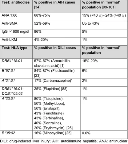
Table 2 lists diagnostic tests useful for diagnosis of DILI and the frequencies for specific HLA genes of interest in the reference population and in cases of DILI due to common drugs. These data provide an indication of the positive yield of the genetic tests in cases of DILI compared to their frequency in the reference
population. For example, if applied in clinical practice, genotyping for
HLA-DRB1*15:01 when amoxicillin-clavulanic acid DILI is suspected, HLA-B*57:01 in suspected flucloxacillin-induced DILI and HLA-B*35:02 in a possible minocycline DILI case would have similar performance characteristics to anti-nuclear antibody (ANA), immunoglobulin G estimation and anti-liver kidney microsomal (LKM)
antibody test, respectively in a case of suspected AIH. Considering the importance of clinical decision-making, such as permanent withdrawal of an effective medication in a patient and/or initiation of long-term immunosuppressive regimen, incorporating genetic tests into diagnostic armamentarium is justified and would increase the accuracy and confidence in the diagnosis.
What are the risk factors including genetic risk factors?
A number of drug-related and host factors increase an individual’s
daily dose and reports of liver failure, liver transplantation, as well as death caused by DILI [35]. In addition, compounds with >50% hepatic metabolism are more likely to be associated with ALT >3 times the upper limit of normal, liver failure and fatal DILI [36]. Suggestion that lipophilicity of a compound, independently of other
properties, may increase its hepatotoxic potential has recently been refuted [37, 38]. Drugs with dual potency as mitochondrial and bile salt export pump inhibitors are associated with severe human DILI [39].
Age may be a risk factor increasing susceptibility to DILI due to anti-tuberculosis drugs and flucloxacillin [40, 41]. Chronic hepatitis B and C are also associated with increased risk of DILI from anti-HIV and anti-tuberculosis therapies [40, 42, 43]. In different ethnic groups, significantly different medications underlie DILI [44]. Trimethoprim/sulfamethoxazole, methyldopa and phenytoin are more often the cause of DILI among African-Americans, while amoxicillin-clavulanic acid is a causative agent in a higher proportion of Caucasians. In addition, severe
cutaneous reactions, rates of hospitalisation, liver transplantation or liver-related deaths are more frequent among African–Americans, compared to Caucasians, after controlling for selected covariates [44] . As discussed previously, accumulating evidence over the past decade points consistently towards genetic factors being associated with the individual’s risk of developing liver injury on exposure to medications.
What are the mechanisms of liver injury?
predominantly undergo hepatic metabolism and biliary excretion are more frequently associated with severe DILI suggesting that formation and clearance of reactive metabolites, steps that are common for many compounds, are likely upstream events mediating liver injury. Consistent with this, SNPs in genes coding for drug-metabolizing enzymes and transporters involved in the excretion of drug metabolites have been associated with increased susceptibility to DILI [21]. An increasing
number of drugs are shown to be ‘haptens’, generating sensitizing molecules that are directly protein reactive and form covalent adducts in vivo [45-47]. The
observation that the formation of reactive metabolites and covalent adducts are associated with exposure to the drug, even in the absence of clinically significant liver injury, indicates that, while upstream events are essential steps in the
pathogenesis, they are not sufficient on their own to lead to severe DILI. Rather, a subclinical event may release drug-altered peptide to be taken up by antigen presenting cells or stimulate production of ‘danger signals’, hence triggering a pathogenic adaptive immune response [48].
In the past 8 years, GWASs have identified several HLA alleles and
manifesting as DILI. While HLA class I molecules can be associated with cytotoxic CD8+ T cell-mediated hepatocyte injury, HLA class II molecules invoke interaction between antigen presenting cells and CD4+ T cells, leading to inflammation and liver injury (Fig. 2G). With regards to flucloxacillin and amoxicillin-clavulanic acid-induced DILI, experimental evidence supports the ‘hapten mechanism’ where MHC class II and I restricted CD4+ and CD8+ clones are activated by a drug-derived antigen [49, 50].
Considering that the structure of drugs are often designed to facilitate binding to specific receptors, some compounds or their metabolites may bind directly to HLA molecules or T cell receptors. Such ‘pharmacological interaction’ (p-i concept) may trigger activation and proliferation of T cells leading to immune-mediated liver injury [51]. More recently, some drugs have been shown to occupy the peptide-binding groove of HLA molecules, hence altering the repertoire of peptides (altered peptide repertoire model) that can bind to a particular HLA protein [52, 53]. This permits interaction between self-peptides and HLA molecules leading to immune-mediated DILI.
Prognostic markers
A number of serum/ plasma biomarkers are being investigated with a view to develop tests that can assist in identifying patients with DILI who are likely to
progress to acute liver failure or develop chronicity (reviewed in [54]). These include: organ-specific markers such as microRNA-122 associated with hepatocellular injury [55]; organelle-specific markers such as glutamate dehydrogenase, reflecting
mitochondrial injury; mechanistic biomarkers such as cytokeratin-18
necrosis, respectively; macrophage colony-stimulating factor receptor 1 associated with liver inflammation, and osteopontin associated with inflammatory cell activation and with liver regeneration due to activation of hepatic stem cells [54]. None of these have been validated in a well-characterized cohort of patients with idiosyncratic DILI and hence, they are not yet ready for clinical application.
Areas of uncertainty
While we apply the knowledge gained with regards to molecular mechanisms underlying the development of DILI in clinical practice, there is a compelling case of need for a technique that is capable of reflecting host genetic factors at the same time, recapitulating initial essential steps involving the metabolism and clearance of drugs as well as key events related to immune-mediated liver injury. Such a
methodology could bring about a step change in investigations related to hepatotoxicity both in experimental and clinical settings.
of the adaptive immune system in DILI pathogenesis and, hence, the application of this methodology in investigations related to hepatotoxicity from a wide range of drugs is uncertain.
Management
Early recognition of DILI and prompt withdrawal of the causal medication is crucial to minimise injury and its progression. In addition, measures such as clear documentation in the medical notes, information provided to the general practitioner, referring clinician or pharmacist and the patient to avoid inadvertent re-exposure to the causal medication are important. Although re-exposure does not always lead to recurrence of DILI, in 11% to 51% of cases when liver injury manifests (called positive rechallenge), [58] the consequences could be serious including mortality in 2% to 13% of cases [59, 60]. So re-introduction of medication that has caused DILI in an individual should only be considered in discussion with the patient and under circumstances where benefits clearly outweigh the risks [58]. For example, when providing potentially life-saving cancer chemotherapy or when alternative
medications to treat underlying conditions are not accessible or affordable as in cases of anti-tuberculosis treatment [61].
Currently, treatment of DILI is limited to management of the patient’s symptoms such as itching [62]. There is not yet sufficient evidence to support the treatment of DILI with corticosteroids or ursodeoxycholic acid although these have been used in clinical practice and have been thought to enhance recovery [63]. In a cohort of patients with acute liver failure, there was no statistically significant
subgroup of patients with a Model for Endstage Liver Disease (MELD) score of more than 40 [64]. However, initiation of corticosteroid treatment is justified in certain cases where drug-induced AIH is indistinguishable from the acute presentation of idiopathic AIH. Nevertheless, once the liver injury has resolved, as evidenced by normalization of liver enzymes, immunosuppression could be completely withdrawn with regular and close monitoring of patients. Long-term follow-up studies
demonstrate that drug-induced AIH does not recur over a median follow up of 4 years [65] with idiopathic AIH relapses in 63% of cases in 1 year and 75% of cases in 5 years [66]. Therefore, this is a reasonable approach considering that treatment of relapse of AIH is identical to that of its initial presentation.
Potential Therapies
With regards treatment of severe liver disease, administration of N-acetyl cysteine (NAC) significantly improved transplant-free survival in early stage non-acetaminophen acute liver failure [67]. Transplant-free survival for DILI patients specifically was 58% (95% CI=33-83%) for those receiving NAC compared to 27% (95% CI=8-46%) receiving placebo. High-volume plasma exchange has also been shown to significantly improve outcome in acute liver failure in a prospective randomised multicentre trial of 182 patients [68]. Finally, emergency liver transplantation is an established procedure for refractory fulminant hepatic failure as in the case presented here.
hepatitis [69], and nor-ursodeoxycholic acid, for its properties of reducing expression of MHC class II molecules on macrophages and inhibiting the proliferation of CD4+ T lymphocytes [70]. International consortia that have collaborated successfully in the last decade are well-placed to coordinate randomised clinical trials of treatments for DILI in the near future.
Acknowledgements
The views expressed are those of the authors and not necessarily those of the NHS, the NIHR or the Department of Health.
Figure Legends:
Fig. 1. Liver explant histology in the DILI patient described. (A) Reticulin stained section x10 magnification. There is panacinar cell loss with occasional multiacinar collapse of the reticulin framework and regenerative-appearing islands of residual parenchyma.(B)H&E stained section x10 magnification. There is extensive bridging parenchymal collapse with haemorrhage into cell plates and residual islands of regenerative-appearing hepatocytes. The porto-septal areas contain a mild chronic inflammatory infiltrate, predominantly lymphocytic, and with florid ductular reaction adjacent to areas of hepatocyte loss. The findings are in keeping with a severe ongoing acute hepatitis with massive cell necrosis.
Fig 2. Structure and electrostatic properties of the peptide binding groove of heterodimeric HLA-DRB1 alleles and their role in antigen presentation and stimulation of an immune response during DILI. (A) Structure of the
HLA-DRB1*15:02 peptide binding groove highlighting the overall neutral charge of pocket P1 [8-10]. Potentials are graded from -10 kT/e, shown in blue, to +10 kT/e in red, with neutral potentials (0 kT/e) coloured white. (G) Suggested role of HLA-DRB1 in presentation of drug-derived antigens by antigen presenting cells in DILI. Circulating drug or drug metabolite arising from hepatic metabolism, ‘hapten’, may be taken up by antigen presenting cells and processed as an antigen. The subsequent
Table 1. Genetic susceptibility loci for DILI identified in GWAS and candidate gene studies. Association described: HLA allele Drug studied:
Study type & cohort population OR
A*02:01 rs2523822 TRNAI25
Amoxi-clav GWAS: 201 cases 532 P controls (European) [24]
2.3
A*30:02 Amoxi-clav CGS: 75 cases 885 P controls (European) [1] 6.7 A*33 Tiopronin CGS: 14 cases, 472 T controls (Japanese)
[71]
NR
A*33:01 Multiple GWAS: 862 cases (21 terbinafine; 7 fenofibrate; 5 ticlopidine cases) 10588 P controls (European) [26]
40.5; 58.7; 163.1 A*33:03 Ticlopidine CGS: 22 cases 85 T controls (Japanese) [72] 13
B*08 Clometacin CGS: 30 cases (European) [73]
B*18:01 Amoxi-clav CGS: 75 cases, 885 P control (European) [1] 2.9 B*35:02 Minocycline GWAS: 25 cases, 10588 P controls
(European) [25]
29.6
B*57:01
rs2395029 HCP5
Flucloxacillin CGS: 51 cases, 282 P controls (European)
[23]
45
B*57:01 Pazopanib CGS: 429 cases, 1761 T controls [74] 2.0
B*57:02 Efavirenz +
Anti-TB
CGS: 46 cases, 46 controls (African) [75] 8.1
B*57:03 Efavirenz + Anti-TB
CGS: 46 cases, 46 controls (African) 26.8
B*58:01 Nevirapine CGS: 57 cases, 111 T controls (South African) [76]
DQA1*01:02 protective
Anti-TB CGS: 56 cases, 209 T controls (Indian) [77] 4
DRB1*15:01- DRB5*0101-DQB1*06:02; DQB1*06:02 rs9274407
Amoxi-clav GWAS: 201 cases, 532 P controls (European) [24];
CGS: (European) 35 cases, 300 P controls [78]; 22 cases 134 P controls [79]; 40 cases, 140 P controls [80]; 75 cases, 885 P controls [1]
3.1
DRB1*01 Nevirapine CGS: 21 cases 133 T control (Caucasian) [81] NR DRB1*01:02 Nevirapine CGS: 57 cases 111 t controls (South African)
[76]
NR DRB1*07
protectivea
Amoxi-clav CGS: 40 cases, 140 P controls (European) [80]
0.18 DRB1*07: Ximelagatran GWAS: 74 cases, 130 T controls (European)
[82]
4.4 DRB1*07:01
(linkage with DQA1*02:01)
Lapatinib GWAS: 37 cases, 286 T controls [83]; CGS: 37 cases 1071 T controls. (European) [84];
GWAS: 34 cases, 810 T controls [85]
NR
DRB1*13 protectivea
DRB1*15:01 Lumiracoxib GWAS 41 cases, 176 T controls (International) [87]
5
DRB1*16:01-DQB1*05:02
Flupirtine GWAS: 614 cases (6 flupirtine) 10588 P controls (European) [88]
18.7 DQB1*0201 Anti-TB CGS: 56 cases, 209 T controls (Indian) [77];
GWAS: 59 cases, 111 T controls, 109 P controls (Indian): association not confirmed [89] 1.9 Association described: drug metabolism loci Drug studied:
Study type & cohort population OR
ABCC2 rs717620
Diclofenac CGS: 24 cases, 48 T controls (European) [90] 5
CYP2B6 *6 Efavirenz CGS: 41 cases, 160 T controls (South African) [91]
NR CYP2B6
rs7254579
Ticlopidine CGS: 22 cases, 92 T controls (Japanese) [92] NR NAT2 slow
acetylator alleles
Isoniazid CGS: 26 cases, 101 P controls
(European/Asian) [93];
GWAS:24 cases - association not confirmed [94]
4.25
UGT1A6/1A9 Tolcapone CGS: 135 cases, 234 T controls (European) [95]; CGS: 2 cases (European) [96]
NR
UGT2B7*2 Diclofenac CGS: 24 cases, 48 T controls (European) [90];
GWAS:34 cases - association partly confirmed [94]
8.5
Various other associations:
Drug studied:
Study type & cohort population OR
ALG10B rs6582630
Flucloxacillin GWAS: 51 cases, 282 P controls (European) [23]
2.8 C9orf82
(CAAP1) rs10812428
Flucloxacillin GWAS: 51 cases, 282 P controls (European) [23]
2.9
ERN1
rs199650082
Efavirenz GWAS: 21 cases, 234 T controls [97] (African) 18.2
FAM65B intron
rs10946737
Rifapicin GWAS: 48 cases, 354 T controls [98]; CGS: 27 cases, 217 T controls (African) [98];
3.4 lincRNA
rs4842407
Efavirenz + anti-TB
GWAS: 42 cases, 292 T controls [97] (African) 5.4
MCTP2 rs4984390
Flucloxacillin GWAS: 51 cases, 282 P controls (European) [23]
3.3 OR5H2
rs1497546
Flucloxacillin GWAS: 51 cases, 282 P controls (European) [23] 6.6 PPARG rs17036170 Multiple (Diclofenac)
GWAS: 783 cases (30 diclofenac) 3001 P controls (European) [94]
11.3 ST6GAL1
rs10937275
Flucloxacillin GWAS: 51 cases, 282 P controls (European) [23]
Table 2. Summary of tests utilized for diagnosis of DILI and distinction from AIH and prevalence of variant alleles.
Test: antibodies % positive in AIH cases [34]
% positive in ‘normal’ population [99-101]
ANA 1:60 68%-75% 15% (<40 ♀) - 24% (>40 ♀)
Anti-SMA 52%-59% Up to 43%
IgG >1600 mg/dl 86% 5%
Anti-LKM 4%-20% 1%
Test: HLA type % positive in DILI cases % positive in ‘normal’ population
DRB1*15:01 57%-67% (Amoxicillin-clavulanic acid) [1]
15%-20%
B*57:01 84%-87% (Flucloxacillin) [23]
6%
A*31:01 17% (Carbamazepine)* 2%
DRB1*16:01-DQB1*05:02
25% (Flupirtine) [88] 1%
A*33:01 80% (Ticlopidine), 50% (Methyldopa), 50% (Enalapril), 43% (Fenofibrate), 43% (Terbinafine), 40% (Sertraline),
20% (Erythrmycin), [26]
1%
B*35:02 16% (Minocycline) [25] 0.6%
References
[1] Stephens C, Lopez-Nevot MA, Ruiz-Cabello F, Ulzurrun E, Soriano G, Romero-Gomez M, et al. HLA alleles influence the clinical signature of amoxicillin-clavulanate hepatotoxicity. PloS one 2013;8:e68111.
[2] Chen P, Lin JJ, Lu CS, Ong CT, Hsieh PF, Yang CC, et al. Carbamazepine-induced toxic effects and HLA-B*1502 screening in Taiwan. The New England journal of medicine 2011;364:1126-1133.
[3] Valdes AM, Styrkarsdottir U, Doherty M, Morris DL, Mangino M, Tamm A, et al. Large scale replication study of the association between HLA class II/BTNL2 variants and osteoarthritis of the knee in European-descent populations. PloS one 2011;6:e23371.
[4] Kelly MA, Alvi NS, Croft NJ, Mijovic CH, Bottazzo GF, Barnett AH. Genetic and immunological characteristics of Type I diabetes mellitus in an Indo-Aryan population. Diabetologia 2000;43:450-456.
[5] Walker DG, Williams HR, Bancil AS, Rai P, Pantelidis P, Chambers J, et al. Ethnicity differences in genetic susceptibility to ulcerative colitis: a comparison of Indian asians and white northern Europeans. Inflamm Bowel Dis 2013;19:2888-2894. [6] Hov JR, Kosmoliaptsis V, Traherne JA, Olsson M, Boberg KM, Bergquist A, et al. Electrostatic modifications of the human leukocyte antigen-DR P9 peptide-binding pocket and susceptibility to primary sclerosing cholangitis. Hepatology 2011;53:1967-1976.
[7] DeLano WL. The PyMol Molecular Graphics System http://www.pymol.org DeLano Scientific LLC, Palo Alto, California, USA. 2008.
[8] Webb B, Sali A. Comparative Protein Structure Modeling Using MODELLER. Curr Protoc Bioinformatics 2014;47:5 6 1-32.
[9] Baker NA, Sept D, Joseph S, Holst MJ, McCammon JA. Electrostatics of nanosystems: application to microtubules and the ribosome. Proceedings of the National Academy of Sciences of the United States of America 2001;98:10037-10041. [10] PDB2PQR. http://nbcr-222.ucsd.edu/pdb2pqr_2.0.0/.
[11] Stricker BH, Blok AP, Claas FH, Van Parys GE, Desmet VJ. Hepatic injury associated with the use of nitrofurans: a clinicopathological study of 52 reported cases. Hepatology 1988;8:599-606.
[12] Otsuka S, Yamamoto M, Kasuya S, Ohtomo H, Yamamoto Y, Yoshida TO, et al. HLA antigens in patients with unexplained hepatitis following halothane anesthesia. Acta Anaesthesiol Scand 1985;29:497-501.
[13] Mallal S, Phillips E, Carosi G, Molina JM, Workman C, Tomazic J, et al. HLA-B*5701 screening for hypersensitivity to abacavir. The New England journal of medicine 2008;358:568-579.
[14] Bjornsson ES, Hoofnagle JH. Categorization of drugs implicated in causing liver injury: Critical assessment based on published case reports. Hepatology 2016;63:590-603.
[15] http://livertox.nih.gov.
[16] Aithal GP, Rawlins MD, Day CP. Clinical diagnostic scale: a useful tool in the evaluation of suspected hepatotoxic adverse drug reactions. Journal of hepatology 2000;33:949-952.
[18] Chalasani NP, Hayashi PH, Bonkovsky HL, Navarro VJ, Lee WM, Fontana RJ, et al. ACG Clinical Guideline: the diagnosis and management of idiosyncratic drug-induced liver injury. The American journal of gastroenterology 2014;109:950-966; quiz 967.
[19] M'Kada H, Perazzo H, Munteanu M, Ngo Y, Ramanujam N, Fautrel B, et al. Real time identification of drug-induced liver injury (DILI) through daily screening of ALT results: a prospective pilot cohort study. PloS one 2012;7:e42418.
[20] Aithal GP, Grove JI. Genome-Wide Association Studies in Drug-Induced Liver Injury: Step Change in Understanding the Pathogenesis. Seminars in liver disease 2015;35:421-431.
[21] Daly AK. Are Polymorphisms in Genes Relevant to Drug Disposition Predictors of Susceptibility to Drug-Induced Liver Injury? Pharm Res 2017;34:1564-1569.
[22] Clare KE, Miller MH, Dillon JF. Genetic Factors Influencing Drug-Induced Liver Injury: Do They Have a Role in Prevention and Diagnosis? Current hepatology reports 2017;16:258-264.
[23] Daly AK, Donaldson PT, Bhatnagar P, Shen Y, Pe'er I, Floratos A, et al. HLA-B*5701 genotype is a major determinant of drug-induced liver injury due to flucloxacillin. Nature genetics 2009;41:816-819.
[24] Lucena MI, Molokhia M, Shen Y, Urban TJ, Aithal GP, Andrade RJ, et al. Susceptibility to amoxicillin-clavulanate-induced liver injury is influenced by multiple HLA class I and II alleles. Gastroenterology 2011;141:338-347.
[25] Urban TJ, Nicoletti P, Chalasani N, Serrano J, Stolz A, Daly AK, et al. Minocycline hepatotoxicity: Clinical characterization and identification of HLA-B *35:02 as a risk factor. Journal of hepatology 2017;67:137-144.
[26] Nicoletti P, Aithal GP, Bjornsson ES, Andrade RJ, Sawle A, Arrese M, et al. Association of Liver Injury From Specific Drugs, or Groups of Drugs, With Polymorphisms in HLA and Other Genes in a Genome-Wide Association Study. Gastroenterology 2017;152:1078-1089.
[27] El Sherrif Y, Potts JR, Howard MR, Barnardo A, Cairns S, Knisely AS, et al. Hepatotoxicity from anabolic androgenic steroids marketed as dietary supplements: contribution from ATP8B1/ABCB11 mutations? Liver international : official journal of the International Association for the Study of the Liver 2013;33:1266-1270.
[28] Invernizzi P. Drug-induced liver injury: is it time for genetics to change our clinical practice? Journal of hepatology 2010;53:993-994.
[29] Licata A, Maida M, Cabibi D, Butera G, Macaluso FS, Alessi N, et al. Clinical features and outcomes of patients with drug-induced autoimmune hepatitis: a retrospective cohort study. Digestive and liver disease : official journal of the Italian Society of Gastroenterology and the Italian Association for the Study of the Liver 2014;46:1116-1120.
[30] Bjornsson E, Talwalkar J, Treeprasertsuk S, Kamath PS, Takahashi N, Sanderson S, et al. Drug-induced autoimmune hepatitis: clinical characteristics and prognosis. Hepatology 2010;51:2040-2048.
[31] EASL Clinical Practice Guidelines: Autoimmune hepatitis. Journal of hepatology;63:971-1004.
[33] Alvarez F, Berg PA, Bianchi FB, Bianchi L, Burroughs AK, Cancado EL, et al. International Autoimmune Hepatitis Group Report: review of criteria for diagnosis of autoimmune hepatitis. Journal of hepatology 1999;31:929-938.
[34] Donaghy L, Barry FJ, Hunter JG, Stableforth W, Murray IA, Palmer J, et al. Clinical and laboratory features and natural history of seronegative hepatitis in a nontransplant centre. European journal of gastroenterology & hepatology 2013;25:1159-1164.
[35] Lammert C, Einarsson S, Saha C, Niklasson A, Bjornsson E, Chalasani N. Relationship between daily dose of oral medications and idiosyncratic drug-induced liver injury: search for signals. Hepatology 2008;47:2003-2009.
[36] Lammert C, Bjornsson E, Niklasson A, Chalasani N. Oral medications with significant hepatic metabolism at higher risk for hepatic adverse events. Hepatology 2010;51:615-620.
[37] Chen M, Borlak J, Tong W. High lipophilicity and high daily dose of oral medications are associated with significant risk for drug-induced liver injury. Hepatology 2013;58:388-396.
[38] Weng Z, Wang K, Li H, Shi Q. A comprehensive study of the association between drug hepatotoxicity and daily dose, liver metabolism, and lipophilicity using 975 oral medications. Oncotarget 2015;6:17031-17038.
[39] Aleo MD, Luo Y, Swiss R, Bonin PD, Potter DM, Will Y. Human drug-induced liver injury severity is highly associated with dual inhibition of liver mitochondrial function and bile salt export pump. Hepatology 2014;60:1015-1022.
[40] Ramappa V, Aithal GP. Hepatotoxicity Related to Anti-tuberculosis Drugs: Mechanisms and Management. J Clin Exp Hepatol 2013;3:37-49.
[41] Wing K, Bhaskaran K, Pealing L, Root A, Smeeth L, van Staa TP, et al. Quantification of the risk of liver injury associated with flucloxacillin: a UK population-based cohort study. The Journal of antimicrobial chemotherapy 2017;72:2636-2646. [42] Nunez M. Hepatotoxicity of antiretrovirals: incidence, mechanisms and management. Journal of hepatology 2006;44:S132-139.
[43] Wang NT, Huang YS, Lin MH, Huang B, Perng CL, Lin HC. Chronic hepatitis B infection and risk of antituberculosis drug-induced liver injury: Systematic review and meta-analysis. J Chin Med Assoc 2016;79:368-374.
[44] Chalasani N, Reddy KRK, Fontana RJ, Barnhart H, Gu J, Hayashi PH, et al. Idiosyncratic Drug Induced Liver Injury in African-Americans Is Associated With Greater Morbidity and Mortality Compared to Caucasians. The American journal of gastroenterology 2017;112:1382-1388.
[45] Park BK, Laverty H, Srivastava A, Antoine DJ, Naisbitt D, Williams DP. Drug bioactivation and protein adduct formation in the pathogenesis of drug-induced toxicity. Chemico-biological interactions 2011;192:30-36.
[46] Aithal GP, Ramsay L, Daly AK, Sonhit N, Leathart JBS, Alexander G, et al. Hepatic adducts, circulating antibodies, and cytokine polymorphisms in patients with diclofenac hepatotoxicity. Hepatology 2004;39:1430-1440.
[47] Hammond TG, Meng X, Jenkins RE, Maggs JL, Castelazo AS, Regan SL, et al. Mass spectrometric characterization of circulating covalent protein adducts derived from a drug acyl glucuronide metabolite: multiple albumin adductions in diclofenac patients. The Journal of pharmacology and experimental therapeutics 2014;350:387-402.
[49] Monshi MM, Faulkner L, Gibson A, Jenkins RE, Farrell J, Earnshaw CJ, et al. Human leukocyte antigen (HLA)-B*57:01-restricted activation of drug-specific T cells provides the immunological basis for flucloxacillin-induced liver injury. Hepatology 2013;57:727-739.
[50] Kim SH, Saide K, Farrell J, Faulkner L, Tailor A, Ogese M, et al. Characterization of amoxicillin- and clavulanic acid-specific T cells in patients with amoxicillin-clavulanate-induced liver injury. Hepatology 2015;62:887-899.
[51] Pichler WJ. Consequences of drug binding to immune receptors: Immune stimulation following pharmacological interaction with immune receptors (T-cell receptor for antigen or human leukocyte antigen) with altered peptide-human leukocyte antigen or peptide. Dermatol Sin 2013;31:181-190.
[52] Ostrov DA, Grant BJ, Pompeu YA, Sidney J, Harndahl M, Southwood S, et al. Drug hypersensitivity caused by alteration of the MHC-presented self-peptide repertoire. Proceedings of the National Academy of Sciences of the United States of America 2012;109:9959-9964.
[53] Illing PT, Vivian JP, Dudek NL, Kostenko L, Chen Z, Bharadwaj M, et al. Immune self-reactivity triggered by drug-modified HLA-peptide repertoire. Nature 2012;486:554-558.
[54] Kullak-Ublick GA, Andrade RJ, Merz M, End P, Benesic A, Gerbes AL, et al. Drug-induced liver injury: recent advances in diagnosis and risk assessment. Gut 2017;66:1154-1164.
[55] McGill MR, Jaeschke H. MicroRNAs as Signaling Mediators and Biomarkers of Drug- and Chemical-Induced Liver Injury. J Clin Med 2015;4:1063-1078.
[56] Benesic A, Leitl A, Gerbes AL. Monocyte-derived hepatocyte-like cells for causality assessment of idiosyncratic drug-induced liver injury. Gut 2016;65:1555-1563.
[57] Benesic A, Rahm NL, Ernst S, Gerbes AL. Human monocyte-derived cells with individual hepatocyte characteristics: a novel tool for personalized in vitro studies. Lab Invest 2012;92:926-936.
[58] Hunt CM, Papay JI, Stanulovic V, Regev A. Drug rechallenge following drug-induced liver injury. Hepatology 2017;66:646-654.
[59] Papay JI, Clines D, Rafi R, Yuen N, Britt SD, Walsh JS, et al. Drug-induced liver injury following positive drug rechallenge. Regul Toxicol Pharmacol 2009;54:84-90.
[60] Andrade RJ, Robles M, Lucena MI. Rechallenge in drug-induced liver injury: the attractive hazard. Expert opinion on drug safety 2009;8:709-714.
[61] Sharma SK, Singla R, Sarda P, Mohan A, Makharia G, Jayaswal A, et al. Safety of 3 different reintroduction regimens of antituberculosis drugs after development of antituberculosis treatment-induced hepatotoxicity. Clinical infectious diseases : an official publication of the Infectious Diseases Society of America 2010;50:833-839. [62] Beuers U, Kremer AE, Bolier R, Elferink RP. Pruritus in cholestasis: facts and fiction. Hepatology 2014;60:399-407.
[63] Wree A, Dechene A, Herzer K, Hilgard P, Syn WK, Gerken G, et al. Steroid and ursodesoxycholic Acid combination therapy in severe drug-induced liver injury. Digestion 2011;84:54-59.
[64] Karkhanis J, Verna EC, Chang MS, Stravitz RT, Schilsky M, Lee WM, et al. Steroid use in acute liver failure. Hepatology 2014;59:612-621.
: the official clinical practice journal of the American Gastroenterological Association 2017;15:1635-1636.
[66] Czaja AJ. Difficult treatment decisions in autoimmune hepatitis. World journal of gastroenterology : WJG 2010;16:934-947.
[67] Lee WM, Hynan LS, Rossaro L, Fontana RJ, Stravitz RT, Larson AM, et al. Intravenous N-acetylcysteine improves transplant-free survival in early stage non-acetaminophen acute liver failure. Gastroenterology 2009;137:856-864, 864 e851. [68] Larsen FS, Schmidt LE, Bernsmeier C, Rasmussen A, Isoniemi H, Patel VC, et al. High-volume plasma exchange in patients with acute liver failure: An open randomised controlled trial. Journal of hepatology 2016;64:69-78.
[69] Manns MP, Woynarowski M, Kreisel W, Lurie Y, Rust C, Zuckerman E, et al. Budesonide induces remission more effectively than prednisone in a controlled trial of patients with autoimmune hepatitis. Gastroenterology 2010;139:1198-1206.
[70] Sombetzki M, Fuchs CD, Fickert P, Osterreicher CH, Mueller M, Claudel T, et al. 24-nor-ursodeoxycholic acid ameliorates inflammatory response and liver fibrosis in a murine model of hepatic schistosomiasis. Journal of hepatology 2015;62:871-878. [71] Kurosaki M, Takagi H, Mori M. HLA-A33/B44/DR6 is highly related to intrahepatic cholestasis induced by tiopronin. Digestive diseases and sciences 2000;45:1103-1108.
[72] Hirata K, Takagi H, Yamamoto M, Matsumoto T, Nishiya T, Mori K, et al. Ticlopidine-induced hepatotoxicity is associated with specific human leukocyte antigen genomic subtypes in Japanese patients: a preliminary case-control study. The pharmacogenomics journal 2008;8:29-33.
[73] Pariente EA, Hamoud A, Goldfain D, Latrive JP, Gislon J, Cassan P, et al. [Hepatitis caused by clometacin (Duperan). Retrospective study of 30 cases. A model of autoimmune drug-induced hepatitis?]. Gastroenterologie clinique et biologique 1989;13:769-774.
[74] Xu C-F, Johnson T, Wang X, Carpenter C, Graves AP, Warren L, et al. HLA-B*57:01 Confers Susceptibility to Pazopanib-Associated Liver Injury in Patients with Cancer. Clinical Cancer Research 2016;22:1371-1377.
[75] Petros Z, Kishikawa J, Makonnen E, Yimer G, Habtewold A, Aklillu E. HLA-B(*)57 Allele Is Associated with Concomitant Anti-tuberculosis and Antiretroviral Drugs Induced Liver Toxicity in Ethiopians. Frontiers in Pharmacology 2017;8:90.
[76] Phillips E, Bartlett JA, Sanne I, Lederman MM, Hinkle J, Rousseau F, et al. Associations between HLA-DRB1*0102, HLA-B*5801, and hepatotoxicity during initiation of nevirapine-containing regimens in South Africa. Journal of acquired immune deficiency syndromes 2013;62:e55-57.
[77] Sharma SK, Balamurugan A, Saha PK, Pandey RM, Mehra NK. Evaluation of clinical and immunogenetic risk factors for the development of hepatotoxicity during antituberculosis treatment. American journal of respiratory and critical care medicine 2002;166:916-919.
[78] Hautekeete ML, Horsmans Y, Van Waeyenberge C, Demanet C, Henrion J, Verbist L, et al. HLA association of amoxicillin-clavulanate--induced hepatitis. Gastroenterology 1999;117:1181-1186.
[79] O'Donohue J, Oien KA, Donaldson P, Underhill J, Clare M, MacSween RN, et al. Co-amoxiclav jaundice: clinical and histological features and HLA class II association. Gut 2000;47:717-720.
[81] Yuan J, Guo S, Hall D, Cammett AM, Jayadev S, Distel M, et al. Toxicogenomics of nevirapine-associated cutaneous and hepatic adverse events among populations of African, Asian, and European descent. Aids 2011;25:1271-1280.
[82] Kindmark A, Jawaid A, Harbron CG, Barratt BJ, Bengtsson OF, Andersson TB, et al. Genome-wide pharmacogenetic investigation of a hepatic adverse event without clinical signs of immunopathology suggests an underlying immune pathogenesis. The pharmacogenomics journal 2008;8:186-195.
[83] Spraggs CF, Budde LR, Briley LP, Bing N, Cox CJ, King KS, et al. HLA-DQA1*02:01 is a major risk factor for lapatinib-induced hepatotoxicity in women with advanced breast cancer. Journal of clinical oncology : official journal of the American Society of Clinical Oncology 2011;29:667-673.
[84] Schaid DJ, Spraggs CF, McDonnell SK, Parham LR, Cox CJ, Ejlertsen B, et al. Prospective Validation of HLA-DRB1*07:01 Allele Carriage As a Predictive Risk Factor for Lapatinib-Induced Liver Injury. Journal of clinical oncology : official journal of the American Society of Clinical Oncology 2014;32:2296-2303.
[85] Parham LR, Briley LP, Li L, Shen J, Newcombe PJ, King KS, et al. Comprehensive genome-wide evaluation of lapatinib-induced liver injury yields a single genetic signal centered on known risk allele HLA-DRB1[ast]07:01. The pharmacogenomics journal 2015.
[86] Aithal GP. Hepatotoxicity related to antirheumatic drugs. Nature reviews Rheumatology 2011;7:139-150.
[87] Singer JB, Lewitzky S, Leroy E, Yang F, Zhao X, Klickstein L, et al. A genome-wide study identifies HLA alleles associated with lumiracoxib-related liver injury. Nature genetics 2010;42:711-714.
[88] Nicoletti P, Werk AN, Sawle A, Shen Y, Urban TJ, Coulthard SA, et al. HLA-DRB1*16: 01-DQB1*05: 02 is a novel genetic risk factor for flupirtine-induced liver injury. Pharmacogenetics and genomics 2016;26:218-224.
[89] Nicoletti PD, H.; Goel, A.; Eapen, C.E.; Venkatesan, R.; Grove, J.I.; Daly, A.K.; Aithal, G.P. Genome-wide association study (GWAS) to identify genetic risk factors that increase susceptibility to anti-tuberculosis drug-induced liver injury (ATDILI). Hepatology 2017;66:1-148.
[90] Daly AK, Aithal GP, Leathart JB, Swainsbury RA, Dang TS, Day CP. Genetic susceptibility to diclofenac-induced hepatotoxicity: contribution of UGT2B7, CYP2C8, and ABCC2 genotypes. Gastroenterology 2007;132:272-281.
[91] Yimer G, Ueda N, Habtewold A, Amogne W, Suda A, Riedel KD, et al. Pharmacogenetic & pharmacokinetic biomarker for efavirenz based ARV and rifampicin based anti-TB drug induced liver injury in TB-HIV infected patients. PloS one 2011;6:e27810.
[92] Ariyoshi N, Iga Y, Hirata K, Sato Y, Miura G, Ishii I, et al. Enhanced susceptibility of HLA-mediated ticlopidine-induced idiosyncratic hepatotoxicity by CYP2B6 polymorphism in Japanese. Drug metabolism and pharmacokinetics 2010;25:298-306.
[93] Ng CS, Hasnat A, Al Maruf A, Ahmed MU, Pirmohamed M, Day CP, et al. N-acetyltransferase 2 (NAT2) genotype as a risk factor for development of drug-induced liver injury relating to antituberculosis drug treatment in a mixed-ethnicity patient group. European journal of clinical pharmacology 2014;70:1079-1086.
[95] Acuna G, Foernzler D, Leong D, Rabbia M, Smit R, Dorflinger E, et al. Pharmacogenetic analysis of adverse drug effect reveals genetic variant for susceptibility to liver toxicity. The pharmacogenomics journal 2002;2:327-334.
[96] Martignoni E, Cosentino M, Ferrari M, Porta G, Mattarucchi E, Marino F, et al. Two patients with COMT inhibitor-induced hepatic dysfunction and UGT1A9 genetic polymorphism. Neurology 2005;65:1820-1822.
[97] Petros Z, Lee MT, Takahashi A, Zhang Y, Yimer G, Habtewold A, et al. Genome-Wide Association and Replication Study of Hepatotoxicity Induced by Antiretrovirals Alone or with Concomitant Anti-Tuberculosis Drugs. OMICS 2017;21:207-216.
[98] Petros Z, Lee MM, Takahashi A, Zhang Y, Yimer G, Habtewold A, et al. Genome-wide association and replication study of anti-tuberculosis drugs-induced liver toxicity. BMC genomics 2016;17:755.
[99] Craig WY, Ledue TB, Johnson AM, Ritchie RF. The distribution of antinuclear antibody titers in "normal" children and adults. J Rheumatol 1999;26:914-919.
[100] Zeman MV, Hirschfield GM. Autoantibodies and liver disease: uses and abuses. Canadian journal of gastroenterology = Journal canadien de gastroenterologie 2010;24:225-231.
[image:29.595.300.500.481.645.2][101] Gonzalez-Quintela A, Alende R, Gude F, Campos J, Rey J, Meijide LM, et al. Serum levels of immunoglobulins (IgG, IgA, IgM) in a general adult population and their relationship with alcohol consumption, smoking and common metabolic abnormalities. Clinical and experimental immunology 2008;151:42-50.
Fig. 1.
Fig. 2.