metal-organic papers
m1228
Wilkinson and Harrison (C4H8N3O)[H2AsO4] doi:10.1107/S1600536805016144 Acta Cryst.(2005). E61, m1228–m1230 Acta Crystallographica Section E
Structure Reports Online
ISSN 1600-5368
Creatininium dihydrogenarsenate
Hazel S. Wilkinson and William T. A. Harrison*
Department of Chemistry, University of Aberdeen, Meston Walk, Aberdeen AB24 3UE, Scotland
Correspondence e-mail: w.harrison@abdn.ac.uk
Key indicators
Single-crystal X-ray study
T= 293 K
Mean(C–C) = 0.003 A˚
Rfactor = 0.027
wRfactor = 0.069
Data-to-parameter ratio = 26.9
For details of how these key indicators were automatically derived from the article, see http://journals.iucr.org/e.
#2005 International Union of Crystallography Printed in Great Britain – all rights reserved
The title compound, (C4H8N3O)[H2AsO4], contains a network
of creatininium cations and dihydrogenarsenate anions [mean As—O = 1.681 (2) A˚ ]. The crystal packing involves
anion-to-anion O—H O, cation-to-anion N—H O and
cation-to-cation N—H O hydrogen bonds, resulting in a chain
structure.
Comment
The title compound, (I) (Fig. 1), was prepared as part of our ongoing structural studies of hydrogen-bonding interactions in protonated-amine (di)hydrogen arsenate molecular salts (Wilkinson & Harrison, 2004; Todd & Harrison, 2005). The [H2AsO4]
dihydrogenarsenate group in (I) shows a normal tetrahedral geometry [mean As—O = 1.681 (2) A˚ ], with the protonated As1—O3 and As1—O4 vertices showing their usual lengthening relative to the unprotonated As1—O1 and As1—O2 bonds, which have formal partial double-bond character (Table 1).
The creatininium cation is approximately planar [r.m.s. deviation for the non-H atoms = 0.031 A˚ ; maximum deviation from the mean plane = 0.0597 (16) A˚ for N2]. The three C1— N bond distances (Table 1) are distinctly different, with C1— N1 much longer than the other two. This configuration perhaps indicates that the canonical form of the molecule, with a formal double bond in the C1—N1 position and a formal positive charge on N1, is of less importance than the forms that place the double bond in the C1—N2 and C1—N3 posi-tions and the positive charge on the respective N atoms. However, the rather short C2—N1 bond length suggests that
some conjugation involving the C2 O5 group may also be
significant. In the structure of creatininium dipicolinate monohydrate (Moghimiet al., 2004), the creatininium cation is constrained to be planar by mirror symmetry and an almost identical pattern of C—N bond lengths is observed.
As well as electrostatic attractions, the component species in (I) interact by means of a network of cation-to-anion N—
H O, anion-to-anion O—H O and cation-to-cation N—
H O hydrogen bonds (Table 2). The [H2AsO4]
units are linked into polymeric chains (Fig. 2) propagating along [100]
by way of inversion-generated pairs of O3—H1 O2i and
O4—H2 O1ii bonds (see Table 2 for symmetry codes). In graph-set notation (Bernstein et al., 1995), each
inter-tetra-hedral linking motif corresponds to an R2
2(8) loop. The
As1 As1i and As1 As1ii separations are 4.0608 (3) and 3.9286 (3) A˚ , respectively.
The organic species interacts with the dihydrogenarsenate chains by way of two N—H O hydrogen bonds (Fig. 1), such that both sides of each [100] chain are decorated by the creatininium cations. The third creatininium N—H group is involved in a cation-to-cation N—H O bond (Fig. 2) that appears to reinforce the chains. Overall, a chain structure along theaaxis arises for (I), as shown in Fig. 3. Atoms O1 and O2 accept two hydrogen bonds each (bond angle sums about these atoms are 358.3 and 359.3, respectively). APLATON (Spek, 2003) analysis of (I) flagged the possible presence of
two short C—H O interactions (Table 2), although their
structural significance is not clear.
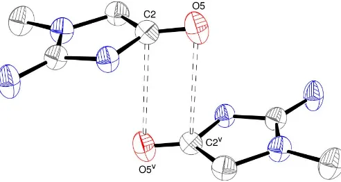
PLATON also flagged a short C2 C2v [symmetry code: (v) x, 1 y, z] intermolecular contact of 3.158 (3) A˚ ,
compared with a van der Waals radius sum of 3.40 A˚ (Bondi, 1964). This close contact might arise as part of a carbonyl– carbonyl interaction, as described by Allenet al.(1998). In the notation of these workers, the situation in (I) corresponds to a ‘sheared antiparallel’ or motif II interaction (Fig. 4), generated
by a centre of symmetry. Here, the O5 C2 O5v and
C2 O5 C2v interaction angles are 100.50 (14) and
79.50 (13), respectively, compared with the nominal values of 290for a perfect rectangle of the four constituent atoms. This is slightly more distorted than the mean O C O and
C O C angles of 96.5 (4) and 83.5 (4) based on 553
contributors, as cited by Allen et al. (1998). The C2 O5v separation of 3.147 (3) A˚ in (I) is slightly less than the C O van der Waals separation of 3.22 A˚ .
The dihydrogenarsenate chain motif in (I) replicates that seen int-butylammonium dihydrogenarsenate (Wilkinson &
Harrison, 2004). However, a different cation-to-anion
hydrogen-bonding scheme leads to a layered structure in this phase. The intrachain As As separations of 4.2662 (3) and 4.3002 (4) A˚ in the t-butylammonium compound are signifi-cantly larger than those seen in (I).
Experimental
A 0.5M aqueous creatine solution (10 ml) was added to a 0.5M
aqueous H3AsO4solution (20 ml) to result in a clear solution. A mass
of block-like crystals of (I) grew as the water evaporated over the course of a few days. The creatine transformed to creatinine under the low-pH conditions of the reaction.
metal-organic papers
Acta Cryst.(2005). E61, m1228–m1230 Wilkinson and Harrison (C
[image:2.610.322.510.71.376.2]4H8N3O)[H2AsO4]
m1229
Figure 2
Detail of a hydrogen-bonded chain in (I). Hydrogen bonds are indicated by dashed lines. [Symmetry codes as in Table 2; additionally, (vi)x1,y,
z.] Figure 1
[image:2.610.45.299.71.227.2]View of the asymmetric unit of (I), showing 50% probability displace-ment ellipsoids, with hydrogen bonds indicated by dashed lines.
Figure 3
[image:2.610.46.295.267.470.2]Crystal data
(C4H8N3O)[H2AsO4] Mr= 255.07
Monoclinic, P21=n a= 7.3576 (3) A˚
b= 10.4263 (5) A˚
c= 11.9471 (5) A˚ = 102.908 (1)
V= 893.33 (7) A˚3
Z= 4
Dx= 1.897 Mg m
3
MoKradiation Cell parameters from 5020
reflections = 2.6–32.2 = 3.80 mm1
T= 293 (2) K Chunk, colourless 0.490.330.24 mm
Data collection
Bruker SMART 1000 CCD diffractometer
!scans
Absorption correction: multi-scan (SADABS; Bruker, 1999)
Tmin= 0.223,Tmax= 0.401
11192 measured reflections
3202 independent reflections 2403 reflections withI> 2(I)
Rint= 0.029 max= 32.5
h=11!11
k=15!15
l=17!18
Refinement
Refinement onF2 R[F2> 2(F2)] = 0.027
wR(F2) = 0.069 S= 1.00 3202 reflections 119 parameters
H-atom parameters constrained
w= 1/[2
(Fo2) + (0.0381P)2] whereP= (Fo2+ 2Fc2)/3
(/)max= 0.002
max= 0.59 e A˚
3
min=0.52 e A˚
3
Table 1
Selected interatomic distances (A˚ ).
As1—O1 1.6512 (13)
As1—O2 1.6563 (12)
As1—O4 1.7013 (15)
As1—O3 1.7134 (13)
C1—N2 1.305 (2)
C1—N3 1.321 (2)
C1—N1 1.374 (2)
[image:3.610.47.293.71.202.2] [image:3.610.313.565.95.176.2]C2—N1 1.367 (2)
Table 2
Hydrogen-bond geometry (A˚ ,).
D—H A D—H H A D A D—H A
O3—H1 O2i
0.85 1.77 2.618 (2) 177
O4—H2 O1ii
0.82 1.79 2.598 (2) 169
N1—H3 O2 0.86 1.89 2.703 (2) 158
N2—H4 O5iii
0.86 2.16 2.983 (2) 161
N2—H5 O1 0.86 1.89 2.747 (2) 172
C3—H6 O1iv
0.97 2.46 3.334 (2) 149
C3—H7 O2v
0.97 2.46 3.384 (2) 159
Symmetry codes: (i) x;yþ2;z; (ii) xþ1;yþ2;z; (iii) xþ1;y;z; (iv)
xþ1 2;y
1 2;zþ
1
2; (v)x;yþ1;z.
The hydroxy H atoms were found in difference maps and refined as riding on their carrier O atoms in their as-found relative positions. The H atoms bonded to C and N atoms were placed in idealized positions (C—H = 0.96–0.97 A˚ and N—H = 0.86 A˚) and refined as riding, allowing for free rotation of the –CH3group. The constraint Uiso(H) = 1.2Ueq(carrier) or 1.5Ueq(methyl carrier) was applied.
Data collection:SMART(Bruker, 1999); cell refinement:SAINT
(Bruker, 1999); data reduction:SAINT; program(s) used to solve structure:SHELXS97(Sheldrick, 1997); program(s) used to refine structure: SHELXL97 (Sheldrick, 1997); molecular graphics:
ORTEP-3(Farrugia, 1997); software used to prepare material for publication:SHELXL97.
HSW thanks the Carnegie Trust for the Universities of Scotland for an undergraduate vacation studentship.
References
Allen, F. H., Baalham, C. A., Lommerse, J. P. M. & Raithby, P. R. (1998).Acta Cryst.B54, 320–329.
Bernstein, J., Davis, R. E., Shimoni, L. & Chang, N.-L. (1995).Angew. Chem. Int. Ed. Engl.34, 1555–1573.
Bondi, A. (1964).J. Phys. Chem.68, 441–451.
Bruker (1999). SMART (Version 5.624), SAINT (Version 6.02A) and
SADABS. Bruker AXS Inc., Madison, Wisconsin, USA. Farrugia, L. J. (1997).J. Appl. Cryst.30, 565.
Moghimi, A., Sharif, M. A. & Aghabozorg, H. (2004).Acta Cryst.E60, o1790– o1792.
Sheldrick, G. M. (1997). SHELXS97 and SHELXL97. University of Go¨ttingen, Germany.
Spek, A. L. (2003).J. Appl. Cryst.36, 7–13.
Todd, M. J. & Harrison, W. T. A. (2005).Acta Cryst.E61, m1024–m1026. Wilkinson, H. S. & Harrison, W. T. A. (2004). Acta Cryst.E60, m1359–
m1361.
metal-organic papers
m1230
Wilkinson and Harrison (C4H8N3O)[H2AsO4] Acta Cryst.(2005). E61, m1228–m1230
Figure 4
supporting information
sup-1
Acta Cryst. (2005). E61, m1228–m1230
supporting information
Acta Cryst. (2005). E61, m1228–m1230 [https://doi.org/10.1107/S1600536805016144]
Creatininium dihydrogenarsenate
Hazel S. Wilkinson and William T. A. Harrison
Creatininium dihydrogenarsenate
Crystal data
(C4H8N3O)[H2AsO4] Mr = 255.07 Monoclinic, P21/n
Hall symbol: -P 2yn
a = 7.3576 (3) Å
b = 10.4263 (5) Å
c = 11.9471 (5) Å
β = 102.908 (1)°
V = 893.33 (7) Å3 Z = 4
F(000) = 512
Dx = 1.897 Mg m−3
Mo Kα radiation, λ = 0.71073 Å Cell parameters from 5020 reflections
θ = 2.6–32.2°
µ = 3.80 mm−1 T = 293 K Chunk, colourless 0.49 × 0.33 × 0.24 mm
Data collection
Bruker SMART 1000 CCD diffractometer
Radiation source: fine-focus sealed tube Graphite monochromator
ω scans
Absorption correction: multi-scan (SADABS; Bruker, 1999)
Tmin = 0.223, Tmax = 0.401
11192 measured reflections 3202 independent reflections 2403 reflections with I > 2σ(I)
Rint = 0.029
θmax = 32.5°, θmin = 2.6° h = −11→11
k = −15→15
l = −17→18
Refinement
Refinement on F2
Least-squares matrix: full
R[F2 > 2σ(F2)] = 0.027 wR(F2) = 0.069 S = 1.00 3202 reflections 119 parameters 0 restraints
Primary atom site location: structure-invariant direct methods
Secondary atom site location: difference Fourier map
Hydrogen site location: inferred from neighbouring sites
H-atom parameters constrained
w = 1/[σ2(F
o2) + (0.0381P)2]
where P = (Fo2 + 2Fc2)/3
(Δ/σ)max = 0.002
Δρmax = 0.59 e Å−3
Δρmin = −0.52 e Å−3
Special details
supporting information
sup-2
Acta Cryst. (2005). E61, m1228–m1230
Refinement. Refinement of F2 against ALL reflections. The weighted R-factor wR and goodness of fit S are based on F2,
conventional R-factors R are based on F, with F set to zero for negative F2. The threshold expression of F2 > σ(F2) is used
only for calculating R-factors(gt) etc. and is not relevant to the choice of reflections for refinement. R-factors based on F2
are statistically about twice as large as those based on F, and R- factors based on ALL data will be even larger.
Fractional atomic coordinates and isotropic or equivalent isotropic displacement parameters (Å2)
x y z Uiso*/Ueq
As1 0.26213 (2) 0.928771 (15) 0.019997 (18) 0.03047 (7) O1 0.46360 (18) 0.87172 (12) 0.09239 (13) 0.0373 (3) O2 0.08787 (18) 0.82749 (11) 0.01759 (13) 0.0398 (3) O3 0.2283 (2) 1.06885 (12) 0.08753 (15) 0.0438 (3) H1 0.1274 1.1029 0.0514 0.053* O4 0.2634 (2) 0.96492 (14) −0.11868 (13) 0.0444 (3) H2 0.3451 1.0179 −0.1198 0.053* C1 0.3742 (2) 0.54747 (15) 0.13831 (17) 0.0311 (4) C2 0.0711 (3) 0.51202 (17) 0.13441 (17) 0.0338 (4) C3 0.1748 (3) 0.38752 (17) 0.16057 (18) 0.0355 (4) H6 0.1640 0.3537 0.2344 0.043* H7 0.1294 0.3240 0.1016 0.043* C4 0.5201 (3) 0.33417 (18) 0.1848 (2) 0.0446 (5) H8 0.4793 0.2528 0.1506 0.067* H9 0.5636 0.3239 0.2662 0.067* H10 0.6196 0.3664 0.1527 0.067* N1 0.2003 (2) 0.60209 (13) 0.12221 (14) 0.0316 (3) H3 0.1761 0.6817 0.1067 0.038* N2 0.5212 (2) 0.61337 (15) 0.12952 (17) 0.0430 (4) H4 0.6280 0.5763 0.1395 0.052* H5 0.5112 0.6939 0.1138 0.052* N3 0.3657 (2) 0.42406 (13) 0.16181 (15) 0.0345 (3) O5 −0.0931 (2) 0.53087 (16) 0.12300 (15) 0.0489 (4)
Atomic displacement parameters (Å2)
U11 U22 U33 U12 U13 U23
supporting information
sup-3
Acta Cryst. (2005). E61, m1228–m1230
Geometric parameters (Å, º)
As1—O1 1.6512 (13) C2—C3 1.503 (3) As1—O2 1.6563 (12) C3—N3 1.452 (2) As1—O4 1.7013 (15) C3—H6 0.9700 As1—O3 1.7134 (13) C3—H7 0.9700 O3—H1 0.8488 C4—N3 1.451 (2) O4—H2 0.8185 C4—H8 0.9600 C1—N2 1.305 (2) C4—H9 0.9600 C1—N3 1.321 (2) C4—H10 0.9600 C1—N1 1.374 (2) N1—H3 0.8600 C2—O5 1.202 (2) N2—H4 0.8600 C2—N1 1.367 (2) N2—H5 0.8600
O1—As1—O2 112.36 (6) C2—C3—H7 111.2 O1—As1—O4 112.96 (7) H6—C3—H7 109.1 O2—As1—O4 107.28 (7) N3—C4—H8 109.5 O1—As1—O3 105.56 (7) N3—C4—H9 109.5 O2—As1—O3 111.00 (7) H8—C4—H9 109.5 O4—As1—O3 107.62 (8) N3—C4—H10 109.5 As1—O3—H1 108.6 H8—C4—H10 109.5 As1—O4—H2 109.2 H9—C4—H10 109.5 N2—C1—N3 127.51 (17) C2—N1—C1 110.36 (14) N2—C1—N1 122.20 (16) C2—N1—H3 124.8 N3—C1—N1 110.29 (15) C1—N1—H3 124.8 O5—C2—N1 125.52 (18) C1—N2—H4 120.0 O5—C2—C3 128.01 (18) C1—N2—H5 120.0 N1—C2—C3 106.45 (15) H4—N2—H5 120.0 N3—C3—C2 102.73 (14) C1—N3—C4 126.66 (16) N3—C3—H6 111.2 C1—N3—C3 110.16 (15) C2—C3—H6 111.2 C4—N3—C3 123.18 (15) N3—C3—H7 111.2
O5—C2—C3—N3 179.0 (2) N2—C1—N3—C4 0.9 (3) N1—C2—C3—N3 0.8 (2) N1—C1—N3—C4 −179.61 (18) O5—C2—N1—C1 −178.5 (2) N2—C1—N3—C3 −178.4 (2) C3—C2—N1—C1 −0.2 (2) N1—C1—N3—C3 1.0 (2) N2—C1—N1—C2 179.00 (19) C2—C3—N3—C1 −1.1 (2) N3—C1—N1—C2 −0.5 (2) C2—C3—N3—C4 179.53 (18)
Hydrogen-bond geometry (Å, º)
D—H···A D—H H···A D···A D—H···A
O3—H1···O2i 0.85 1.77 2.618 (2) 177
O4—H2···O1ii 0.82 1.79 2.598 (2) 169
N1—H3···O2 0.86 1.89 2.703 (2) 158 N2—H4···O5iii 0.86 2.16 2.983 (2) 161
supporting information
sup-4
Acta Cryst. (2005). E61, m1228–m1230
C3—H6···O1iv 0.97 2.46 3.334 (2) 149
C3—H7···O2v 0.97 2.46 3.384 (2) 159