organic papers
o2556
Anithaet al. C6H6NO2+C6H2N3O7 doi:10.1107/S160053680502221X Acta Cryst.(2005). E61, o2556–o2558
Acta Crystallographica Section E
Structure Reports
Online
ISSN 1600-5368
Nicotinium picrate
K. Anitha, S. Athimoolam and R. K. Rajaram*
Department of Physics, Madurai Kamaraj University, Madurai 625 021, India
Correspondence e-mail: [email protected]
Key indicators
Single-crystal X-ray study T= 293 K
Mean(C–C) = 0.004 A˚ Rfactor = 0.045 wRfactor = 0.136
Data-to-parameter ratio = 11.0
For details of how these key indicators were automatically derived from the article, see http://journals.iucr.org/e.
#2005 International Union of Crystallography Printed in Great Britain – all rights reserved
The title compound, C6H6NO2 +
C6H2N3O7
, is the picrate salt of the nicotinium cation. In the picrate anion, theorthonitro groups are twisted out of the plane of the ring, whereas the
para nitro group lies approximately in the ring plane. Hydrogen bonds from the nicotinate cation link two different picrate anions, forming a straight chain along thebaxis. The picrate anions are stacked in columns along [010].
Comment
Nicotinic acid (3-pyridine carboxylic acid) is a B vitamin known as niacin. The crystal structures of nicotinic acid (Wright & King, 1953; Kutoglu & Scheringer, 1983), nicotin-amide (Wright & King, 1954), isonicotinic acid hydrazide (Bhatet al., 1974), 1-methyl nicotinamide iodide, chloride and picrate (Freeman & Bugg, 1974), isonicotinic acid (Takusa-gawa & Shimada, 1976), nicotinoylglycine (Krishnaswamy et al., 1987), 6-aminonicotinic acid hydrochloride (Giantsidis & Turnbull, 2000), and dinicotinamidium squarate (Bulutet al., 2003) have been reported. The structure of the title compound, (I), is reported here.
In (I) (Fig. 1), the H atom of the hydroxy group of picric acid has been transferred to the N atom of nicotinic acid, leading to the formation of a molecular complex. The bond lengths and bond angles in the pyridine ring of the nicotinium cation are comparable to the average values of 1.38 A˚ for the C—C bonds and 1.33 A˚ for the C—N bonds found in dinico-tinamidium squarate (Bulutet al., 2003) and nicotinoylglycine (Krishnaswamy et al., 1987). The C11—C12 bond length is similar to that observed in dinicotinamidium squarate (Bulut
et al., 2003), while atom O1B is identified as the hydroxy O atom by comparison of the C11—O1A and C11—O1B
distances. A comparison of equivalent bond distances invol-ving the pyridine ring atoms of (I) with the values found in nicotinic acid (Wright & King, 1953) shows that the positive charge is localized on the pyridine N atom and does not have much effect on the ring structure. These distances also
compare well with those for 6-aminonicotinic acid hydro-chloride (Giantsidis & Turnbull, 2000) and dinicotinamidium squarate (Bulutet al., 2003). The nicotinium cation is planar, and the planes of the nicotinium cation and the ring of the picrate anion are inclined to one another at 62.9 (1). In the
picrate anion, removal of the phenol H atom leads to a shortening of the C1—O1 bond, while the C1—C2 and C1— C6 distances increase as observed previously (Anitha et al., 2004; Muthamizhchelvan et al., 2005). The torsion angles involving theortho-related nitro groups in the picrate anion (C1—C6—N3—O6,O7 and C1—C2—N1—O2,O3) are 144.0 (3), 139.6 (3), 131.5 (3) and 132.1 (3), respectively
(Table 1). It has been found that in most picrates theortho -related nitro groups, which are commonly involved in hydrogen-bonding interactions, are more likely to be rotated out of the molecular plane than the para nitro substituent (Anithaet al., 2004; Kaiet al., 1994; Smithet al., 2004; Gartland
et al., 1974). Here, even though one of theorthonitro groups is not involved in hydrogen bonding, it is still twisted from the plane of the ring, while theparanitro group lies approximately in the ring plane. It is also found that the twisting of these nitro groups is independent of C—N bond distances (Soriano-Garciaet al., 1978; Srikrishnanet al., 1980). The nitro O atoms not involved in hydrogen bonding have largeUeqvalues. In the
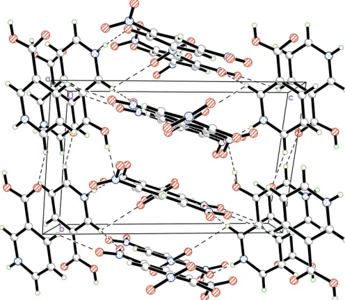
crystal structure, the cations and anions are linked by strong N11—H111 O1 and O1B—H11B O1 hydrogen bonds (Table 2). The structure is also stabilized by C—H O hydrogen bonding. These hydrogen bonds link the layers of cations with the layers of anions, with each nicotinium cation linking two different picrate anions to form a straight chain along the b axis (Fig. 2). The picrate anions are stacked in columns along [010].
Experimental
The title compound was crystallized from a nicotonic acid and picric acid mixture in the stoichiometric ratio of 1:1 at room temperature by slow evaporation.
Crystal data
C6H6NO2+C6H2N3O7
Mr= 352.22
Triclinic,P1
a= 8.063 (3) A˚
b= 8.080 (3) A˚
c= 12.030 (5) A˚
= 93.27 (3)
= 95.87 (4)
= 113.46 (3) V= 711.1 (5) A˚3
Z= 2
Dx= 1.645 Mg m
3
Dm= 1.639 Mg m
3
Dmmeasured by flotation in CHBr3
and CCl4
MoKradiation Cell parameters from 25
reflections
= 10.2–14.3
= 0.14 mm1
T= 293 (2) K Block, yellow 0.190.170.15 mm
Data collection
Nonius MACH3 four-circle diffractometer
!–2scans
Absorption correction: scan (Northet al., 1968)
Tmin= 0.973,Tmax= 0.978
3091 measured reflections 2509 independent reflections 1765 reflections withI> 2(I)
Rint= 0.012 max= 25
h=1!9
k=9!9
l=14!14 3 standard reflections
frequency: 60 min intensity decay: none
Refinement
Refinement onF2 R[F2> 2(F2)] = 0.045
wR(F2) = 0.136
S= 1.02 2509 reflections 228 parameters
H-atom parameters constrained
w= 1/[2
(Fo2) + (0.0601P)2
+ 0.5668P]
whereP= (Fo2+ 2Fc2)/3
(/)max< 0.001 max= 0.50 e A˚
3 min=0.26 e A˚
3
Extinction correction:SHELXTL/
PC
Extinction coefficient: 0.029 (5)
Table 1
Selected geometric parameters (A˚ ,).
O1A—C11 1.196 (3)
O1B—C11 1.308 (3)
N11—C15 1.332 (4)
N11—C16 1.332 (3)
O1—C1 1.279 (3)
C1—C6 1.423 (3)
C1—C2 1.431 (3)
C15—N11—C16 122.7 (2)
O1A—C11—O1B 125.6 (3)
O1A—C11—C12 122.2 (2)
O1B—C11—C12 112.3 (2)
N11—C15—C14 119.8 (3)
N11—C16—C12 119.7 (2)
O1—C1—C6 123.0 (2)
O1—C1—C2 124.4 (2)
C6—C1—C2 112.5 (2)
O2—N1—C2—C3 144.0 (3)
O3—N1—C2—C1 139.6 (3)
O4—N2—C4—C5 170.2 (3)
O5—N2—C4—C3 171.0 (3)
O7—N3—C6—C5 131.5 (3)
[image:2.610.313.565.599.648.2]O6—N3—C6—C1 132.1 (3)
Table 2
Hydrogen-bond geometry (A˚ ,).
D—H A D—H H A D A D—H A
N11—H11 O1 0.86 1.85 2.711 (3) 174
O1B—H11B O1i
0.82 1.75 2.563 (3) 170
C16—H16 O7 0.93 2.55 2.962 (4) 107
Symmetry code: (i)x;y1;z.
All H atoms were placed in geometrically calculated positions, with C—H distances of 0.93 A˚ , an N—H distance of 0.86 A˚ and an O—H distance of 0.82 A˚ , and allowed to ride on the carrier atom with
Uiso(H) equal to 1.2Ueq(C,N) and 1.5Ueq(O).
Data collection: CAD-4 EXPRESS (Enraf–Nonius, 1994); cell refinement:CAD-4 EXPRESS; data reduction:XCAD4(Harms & Wocadlo, 1995); program(s) used to solve structure:SHELXTL/PC
organic papers
Acta Cryst.(2005). E61, o2556–o2558 Anithaet al. C
6H6NO2+C6H2N3O7
o2557
Figure 1
(Bruker, 2000); program(s) used to refine structure:SHELXTL/PC; molecular graphics:SHELXTL/PC; software used to prepare mate-rial for publication:SHELXTL/PC.
The authors thank the Department of Science and Tech-nology, Government of India, for establishing a single-crystal
diffractometer facility at the School of Physics, Madurai Kamaraj University, Madurai, through the FIST programme.
References
Anitha, K., Sridhar, B. & Rajaram, R. K. (2004).Acta Cryst.E60, o1530– o1532.
Bhat, T. N., Singh, T. P. & Vijayan, M. (1974).Acta Cryst.B30, 2921–2922. Bruker (2000). SHELXTL/PC. Version 6.10. Bruker AXS Inc., Madison,
Wisconsin, USA.
Bulut, A., Yesilel, O. Z., Dege, N., Icbudak, H., Olmaz, H. & Bujukgungor, O. (2003).Acta Cryst.C59, o727–o729.
Enraf–Nonius (1994). CAD-4 EXPRESS. Version 5.1/1.2. Enraf–Nonius, Delft, The Netherlands.
Freeman, G. R. & Bugg, C. E. (1974).Acta Cryst.B30, 431–443.
Gartland, G. L., Freeman, G. R. & Bugg, C. E. (1974).Acta Cryst.B30, 1841– 1849.
Giantsidis, J. & Turnbull, M. M. (2000).Acta Cryst.C56, 334–335.
Harms, K. & Wocadlo, S. (1995).XCAD4.University of Marburg, Germany. Kai, T., Goto, M., Furuhata, K. & Takayanagi, H. (1994).Anal. Sci.10, 359–
360.
Krishnaswamy, S., Pattabhi, V. & Guru Row, T. N. (1987).Acta Cryst.C43, 728– 729.
Kutoglu, A. & Scheringer, C. (1983).Acta Cryst.C39, 232–234.
Muthamizhchelvan, C., Saminathan, K., Fraanje, J., Pescher, R. & Sivakumar, K. (2005).Acta Cryst.E61, o1153–o1155.
North, A. C. T., Phillips, D. C. & Mathews, F. S. (1968).Acta Cryst.A24, 351– 359.
Smith, G., Wermuth, U. D. & Healy, P. C. (2004).Acta Cryst.E60, o1800–o1803. Soriano-Garcia, M., Srikrishnan, T. & Parthasarathy, R. (1978).Acta Cryst.
A34, s114.
Srikrishnan, T., Soriano-Garcia, M. & Parthasarathy, R. (1980).Z. Kristallogr.
151, 317–323.
Takusagawa, F. & Shimada, A. (1976).Acta Cryst.B32, 1925–1927. Wright, W. B. & King, G. S. D. (1953).Acta Cryst.6, 305–310. Wright, W. B. & King, G. S. D. (1954).Acta Cryst.7, 283–288.
organic papers
o2558
Anithaet al. C [image:3.610.46.294.73.289.2]6H6NO2+C6H2N3O7 Acta Cryst.(2005). E61, o2556–o2558
Figure 2
supporting information
sup-1 Acta Cryst. (2005). E61, o2556–o2558
supporting information
Acta Cryst. (2005). E61, o2556–o2558 [https://doi.org/10.1107/S160053680502221X]
Nicotinium picrate
K. Anitha, S. Athimoolam and R. K. Rajaram
Nicotinium picrate
Crystal data
C6H6NO2+·C6H2N3O7−
Mr = 352.22 Triclinic, P1 Hall symbol: -P 1 a = 8.063 (3) Å b = 8.080 (3) Å c = 12.030 (5) Å α = 93.27 (3)° β = 95.87 (4)° γ = 113.46 (3)° V = 711.1 (5) Å3
Z = 2
F(000) = 360 Dx = 1.645 Mg m−3
Dm = 1.639 Mg m−3
Dm measured by flotation; CHBr3 & CCl4
Mo Kα radiation, λ = 0.71073 Å Cell parameters from 25 reflections θ = 10.2–14.3°
µ = 0.15 mm−1
T = 293 K Needle, yellow 0.19 × 0.17 × 0.15 mm
Data collection
Nonius MACH3 sealed-tube diffractometer
Radiation source: fine-focus sealed tube Graphite monochromator
ω–2θ scans
Absorption correction: ψ scan (North et al., 1968)
Tmin = 0.973, Tmax = 0.978
3091 measured reflections
2509 independent reflections 1765 reflections with I > 2σ(I) Rint = 0.012
θmax = 25°, θmin = 2.8°
h = −1→9 k = −9→9 l = −14→14
3 standard reflections every 60 min intensity decay: none
Refinement
Refinement on F2
Least-squares matrix: full R[F2 > 2σ(F2)] = 0.045
wR(F2) = 0.136
S = 1.02 2509 reflections 228 parameters 0 restraints
Primary atom site location: structure-invariant direct methods
Secondary atom site location: difference Fourier map
Hydrogen site location: inferred from neighbouring sites
H-atom parameters constrained w = 1/[σ2(F
o2) + (0.0601P)2 + 0.5668P]
where P = (Fo2 + 2Fc2)/3
(Δ/σ)max < 0.001
Δρmax = 0.50 e Å−3
Δρmin = −0.26 e Å−3
Extinction correction: SHELXTL/PC, Fc*=kFc[1+0.001xFc2λ3/sin(2θ)]-1/4
supporting information
sup-2 Acta Cryst. (2005). E61, o2556–o2558
Special details
Geometry. All e.s.d.'s (except the e.s.d. in the dihedral angle between two l.s. planes) are estimated using the full covariance matrix. The cell e.s.d.'s are taken into account individually in the estimation of e.s.d.'s in distances, angles and torsion angles; correlations between e.s.d.'s in cell parameters are only used when they are defined by crystal symmetry. An approximate (isotropic) treatment of cell e.s.d.'s is used for estimating e.s.d.'s involving l.s. planes.
Refinement. Refinement of F2 against ALL reflections. The weighted R-factor wR and goodness of fit S are based on F2,
conventional R-factors R are based on F, with F set to zero for negative F2. The threshold expression of F2 > σ(F2) is used
only for calculating R-factors(gt) etc. and is not relevant to the choice of reflections for refinement. R-factors based on F2
are statistically about twice as large as those based on F, and R- factors based on ALL data will be even larger.
Fractional atomic coordinates and isotropic or equivalent isotropic displacement parameters (Å2)
x y z Uiso*/Ueq
O1A 0.6967 (4) −0.3057 (3) 0.94419 (19) 0.0712 (7)
O1B 0.5023 (3) −0.3033 (3) 0.79944 (17) 0.0587 (6)
H11B 0.4910 −0.4076 0.7837 0.088*
N11 0.6202 (3) 0.2269 (3) 0.87177 (18) 0.0421 (6)
H11 0.5668 0.2776 0.8287 0.051*
C11 0.6248 (4) −0.2291 (4) 0.8880 (2) 0.0396 (6)
C12 0.6672 (3) −0.0319 (3) 0.91400 (19) 0.0339 (6)
C13 0.7897 (4) 0.0683 (4) 1.0069 (2) 0.0391 (6)
H13 0.8490 0.0142 1.0529 0.047*
C14 0.8236 (4) 0.2488 (4) 1.0312 (2) 0.0448 (7)
H14 0.9041 0.3169 1.0941 0.054*
C15 0.7367 (4) 0.3266 (4) 0.9609 (2) 0.0463 (7)
H15 0.7592 0.4485 0.9755 0.056*
C16 0.5828 (4) 0.0519 (4) 0.8465 (2) 0.0384 (6)
H16 0.5002 −0.0133 0.7837 0.046*
O1 0.4312 (3) 0.3651 (2) 0.73443 (15) 0.0466 (5)
O2 0.1092 (5) 0.3296 (5) 0.8122 (2) 0.1135 (13)
O3 −0.0094 (4) 0.4605 (4) 0.7055 (2) 0.0866 (9)
O4 −0.1795 (3) 0.1255 (4) 0.3199 (2) 0.0717 (7)
O5 0.0448 (4) 0.1230 (5) 0.24188 (19) 0.0938 (10)
O6 0.6432 (3) 0.3136 (4) 0.4727 (2) 0.0802 (8)
O7 0.5993 (3) 0.1686 (4) 0.61744 (19) 0.0703 (7)
N1 0.0753 (4) 0.3677 (4) 0.7216 (2) 0.0549 (7)
N2 −0.0215 (3) 0.1476 (4) 0.3238 (2) 0.0517 (6)
N3 0.5514 (3) 0.2449 (3) 0.54566 (19) 0.0414 (5)
C1 0.3226 (3) 0.3086 (3) 0.64204 (19) 0.0326 (6)
C2 0.1439 (3) 0.3077 (4) 0.6254 (2) 0.0367 (6)
C3 0.0333 (3) 0.2593 (4) 0.5240 (2) 0.0392 (6)
H3 −0.0812 0.2626 0.5177 0.047*
C4 0.0953 (3) 0.2055 (4) 0.4315 (2) 0.0374 (6)
C5 0.2661 (4) 0.2040 (3) 0.4391 (2) 0.0368 (6)
H5 0.3085 0.1718 0.3758 0.044*
supporting information
sup-3 Acta Cryst. (2005). E61, o2556–o2558
Atomic displacement parameters (Å2)
U11 U22 U33 U12 U13 U23
O1A 0.108 (2) 0.0492 (12) 0.0591 (13) 0.0441 (13) −0.0265 (13) −0.0010 (10)
O1B 0.0718 (15) 0.0482 (12) 0.0531 (12) 0.0315 (11) −0.0225 (11) −0.0160 (10)
N11 0.0485 (13) 0.0431 (13) 0.0404 (12) 0.0251 (11) −0.0002 (10) 0.0105 (10)
C11 0.0505 (16) 0.0425 (14) 0.0302 (13) 0.0248 (13) 0.0008 (12) 0.0018 (11)
C12 0.0384 (14) 0.0403 (14) 0.0265 (12) 0.0202 (12) 0.0017 (10) 0.0031 (10)
C13 0.0450 (15) 0.0445 (15) 0.0313 (13) 0.0239 (13) −0.0036 (11) 0.0024 (11)
C14 0.0501 (17) 0.0427 (15) 0.0380 (14) 0.0188 (13) −0.0058 (12) −0.0030 (12)
C15 0.0547 (17) 0.0362 (14) 0.0486 (16) 0.0189 (13) 0.0080 (14) 0.0045 (12)
C16 0.0398 (15) 0.0445 (15) 0.0319 (13) 0.0198 (12) −0.0019 (11) 0.0030 (11)
O1 0.0588 (12) 0.0456 (11) 0.0363 (10) 0.0302 (10) −0.0189 (9) −0.0114 (8)
O2 0.165 (3) 0.178 (4) 0.0506 (16) 0.118 (3) 0.0379 (18) 0.0215 (19)
O3 0.0907 (19) 0.103 (2) 0.0904 (19) 0.0675 (18) 0.0152 (15) −0.0139 (16)
O4 0.0431 (13) 0.0940 (18) 0.0681 (15) 0.0256 (12) −0.0209 (11) −0.0015 (13)
O5 0.0771 (18) 0.173 (3) 0.0332 (12) 0.061 (2) −0.0124 (12) −0.0163 (15)
O6 0.0556 (14) 0.126 (2) 0.0836 (17) 0.0523 (15) 0.0308 (13) 0.0499 (17)
O7 0.0622 (14) 0.111 (2) 0.0645 (14) 0.0587 (15) 0.0103 (11) 0.0338 (14)
N1 0.0548 (16) 0.0732 (18) 0.0442 (15) 0.0350 (14) 0.0071 (12) −0.0026 (13)
N2 0.0462 (15) 0.0632 (16) 0.0403 (14) 0.0214 (13) −0.0122 (11) 0.0002 (12)
N3 0.0353 (12) 0.0534 (14) 0.0399 (12) 0.0231 (11) 0.0014 (10) 0.0065 (11)
C1 0.0379 (13) 0.0299 (12) 0.0296 (12) 0.0161 (11) −0.0041 (10) −0.0012 (10)
C2 0.0384 (14) 0.0425 (14) 0.0339 (13) 0.0214 (12) 0.0062 (11) 0.0022 (11)
C3 0.0292 (13) 0.0448 (15) 0.0453 (15) 0.0182 (12) −0.0009 (11) 0.0043 (12)
C4 0.0342 (14) 0.0442 (15) 0.0304 (13) 0.0152 (12) −0.0065 (11) 0.0020 (11)
C5 0.0398 (14) 0.0455 (15) 0.0275 (12) 0.0210 (12) 0.0020 (10) 0.0010 (11)
C6 0.0280 (12) 0.0364 (13) 0.0348 (13) 0.0165 (11) 0.0004 (10) 0.0036 (10)
Geometric parameters (Å, º)
O1A—C11 1.196 (3) O3—N1 1.210 (3)
O1B—C11 1.308 (3) O4—N2 1.209 (3)
O1B—H11B 0.8200 O5—N2 1.211 (3)
N11—C15 1.332 (4) O6—N3 1.220 (3)
N11—C16 1.332 (3) O7—N3 1.207 (3)
N11—H11 0.8600 N1—C2 1.469 (3)
C11—C12 1.497 (4) N2—C4 1.454 (3)
C12—C16 1.377 (3) N3—C6 1.458 (3)
C12—C13 1.384 (4) C1—C6 1.423 (3)
C13—C14 1.380 (4) C1—C2 1.431 (3)
C13—H13 0.9300 C2—C3 1.372 (4)
C14—C15 1.375 (4) C3—C4 1.382 (4)
C14—H14 0.9300 C3—H3 0.9300
C15—H15 0.9300 C4—C5 1.377 (4)
C16—H16 0.9300 C5—C6 1.369 (3)
O1—C1 1.279 (3) C5—H5 0.9300
supporting information
sup-4 Acta Cryst. (2005). E61, o2556–o2558
C11—O1B—H11B 109.5 O4—N2—O5 123.0 (3)
C15—N11—C16 122.7 (2) O4—N2—C4 119.0 (3)
C15—N11—H11 118.6 O5—N2—C4 118.0 (2)
C16—N11—H11 118.6 O7—N3—O6 123.2 (2)
O1A—C11—O1B 125.6 (3) O7—N3—C6 119.4 (2)
O1A—C11—C12 122.2 (2) O6—N3—C6 117.3 (2)
O1B—C11—C12 112.3 (2) O1—C1—C6 123.0 (2)
C16—C12—C13 119.0 (2) O1—C1—C2 124.4 (2)
C16—C12—C11 120.4 (2) C6—C1—C2 112.5 (2)
C13—C12—C11 120.6 (2) C3—C2—C1 124.1 (2)
C14—C13—C12 119.8 (2) C3—C2—N1 117.3 (2)
C14—C13—H13 120.1 C1—C2—N1 118.5 (2)
C12—C13—H13 120.1 C2—C3—C4 118.7 (2)
C15—C14—C13 119.0 (3) C2—C3—H3 120.6
C15—C14—H14 120.5 C4—C3—H3 120.6
C13—C14—H14 120.5 C5—C4—C3 121.2 (2)
N11—C15—C14 119.8 (3) C5—C4—N2 118.8 (2)
N11—C15—H15 120.1 C3—C4—N2 119.9 (2)
C14—C15—H15 120.1 C6—C5—C4 118.7 (2)
N11—C16—C12 119.7 (2) C6—C5—H5 120.7
N11—C16—H16 120.2 C4—C5—H5 120.7
C12—C16—H16 120.2 C5—C6—C1 124.7 (2)
O2—N1—O3 122.1 (3) C5—C6—N3 116.6 (2)
O2—N1—C2 119.6 (3) C1—C6—N3 118.7 (2)
O3—N1—C2 118.2 (3)
O1A—C11—C12—C16 179.4 (3) C1—C2—C3—C4 0.8 (4)
O1B—C11—C12—C16 −1.1 (4) N1—C2—C3—C4 178.8 (2)
O1A—C11—C12—C13 −1.5 (4) C2—C3—C4—C5 −1.5 (4)
O1B—C11—C12—C13 178.0 (3) C2—C3—C4—N2 177.9 (2)
C16—C12—C13—C14 0.7 (4) O4—N2—C4—C5 170.2 (3)
C11—C12—C13—C14 −178.4 (3) O5—N2—C4—C5 −9.5 (4)
C12—C13—C14—C15 −1.1 (4) O4—N2—C4—C3 −9.2 (4)
C16—N11—C15—C14 −0.1 (4) O5—N2—C4—C3 171.0 (3)
C13—C14—C15—N11 0.8 (4) C3—C4—C5—C6 2.3 (4)
C15—N11—C16—C12 −0.3 (4) N2—C4—C5—C6 −177.1 (2)
C13—C12—C16—N11 −0.1 (4) C4—C5—C6—C1 −2.6 (4)
C11—C12—C16—N11 179.1 (2) C4—C5—C6—N3 179.3 (2)
O1—C1—C2—C3 175.1 (2) O1—C1—C6—C5 −174.2 (2)
C6—C1—C2—C3 −0.9 (4) C2—C1—C6—C5 1.8 (4)
O1—C1—C2—N1 −2.9 (4) O1—C1—C6—N3 3.9 (4)
C6—C1—C2—N1 −178.9 (2) C2—C1—C6—N3 179.9 (2)
O2—N1—C2—C3 144.0 (3) O7—N3—C6—C5 −131.5 (3)
O3—N1—C2—C3 −38.5 (4) O6—N3—C6—C5 46.1 (4)
O2—N1—C2—C1 −37.9 (5) O7—N3—C6—C1 50.2 (3)
supporting information
sup-5 Acta Cryst. (2005). E61, o2556–o2558
Hydrogen-bond geometry (Å, º)
D—H···A D—H H···A D···A D—H···A
N11—H11···O1 0.86 1.85 2.711 (3) 174
O1B—H11B···O1i 0.82 1.75 2.563 (3) 170
C16—H16···O7 0.93 2.55 2.962 (4) 107