organic papers
o860
Kedziorek Mariuszet al. C11H9NO DOI: 10.1107/S1600536803008158 Acta Cryst.(2003). E59, o860±o861 Acta Crystallographica Section EStructure Reports
Online ISSN 1600-5368
(E)-Azulene-1-carboxaldehyde oxime
Kedziorek Mariusz,a² Klaus
Hafnerb and Hans J. Lindnerb*
aInstitute of Organic Chemistry, Polish Academy
of Sciences, ul. Kasprzaka 44, 01-224 Warszawa 42, Poland, andbInstitut fuÈr Organische Chemie, Darmstadt University of Technology, Petersenstrasse 22, D-64287 Darmstadt, Germany
² Research fellow at the Institut fuÈr Organische Chemie, Darmstadt University of Technology, July 1±September 1, 2002.
Correspondence e-mail:
Key indicators
Single-crystal X-ray study
T= 293 K
Mean(C±C) = 0.004 AÊ
Rfactor = 0.052
wRfactor = 0.149
Data-to-parameter ratio = 14.9
For details of how these key indicators were automatically derived from the article, see http://journals.iucr.org/e.
#2003 International Union of Crystallography Printed in Great Britain ± all rights reserved
In the title compound, C11H9NO, the azulene moiety is planar with a delocalized 10-electron perimeter. In the crystal structure, the molecules are connected by hydrogen bonds to form centrosymmetric dimers.
Comment
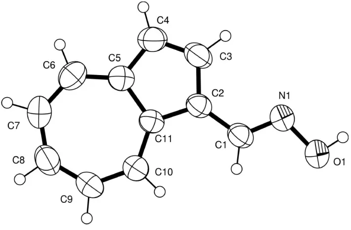
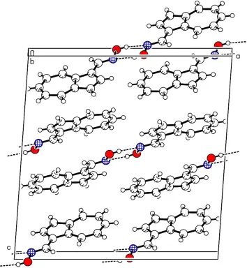
Azulene-1-carboxaldehyde oxime was ®rst obtained by Hafner & Bernhard (1959) as a cystalline derivative of azulene-1-carbaldehyde. To determine the con®guration of the oxime, the synthesis was optimized. The (E )-azulene-1-carboxaldehyde oxime, (I), could be separated from the isomer (Z)-azulene-1-carboxaldehyde and crystallized. No isomerization could be observed in solution in the absence of acids. The (E)-azulene-1-carboxaldehyde oxime shows the expected molecular geometry (see Fig. 1), viz. a planar azulene moiety with a delocalized 10-electron perimeter [mean CÐC distance 1.392 (4) AÊ] and a central bond length of 1.489 (4) AÊ. The crystal packing is determined by inter-molecular hydrogen bonds and-stacking, as shown in Fig. 2. Hydrogen-bonded centrosymmetric dimers are stacked along thecaxis.
Experimental
To a mixture of hydroxylammonium chloride (460 mg, 6.6 mmol) and potassium acetate (668 mg, 6.8 mmol) in 40 ml ethanol azulene-1-carbaldehyde (1.0 g, 6.5 mmol) was added and the mixture was
Received 8 April 2003 Accepted 9 April 2003 Online 23 May 2003
Figure 1
tography with silica gel (hexane/ethyl acetate 4:1) yielded the oxime isomers. Dark green crystals of (I) were obtained from a toluene solution by evaporation. (E)-Azulene-1-carboxaldehyde oxime, (I), m.p. 393±394 K;1H NMR (500 MHz, [d
6]DMSO):= 10.94 (s, 1H,
H11O), 8.92 (d, 1H, H8), 8.72 (s, 1H, H11), 8.42 (d, 1H, H4), 8.13 (d, 1H, H2), 7.74 (approx.t, 1H, H6), 7.43 (d, 1H, H3), 7.32 (approx.q, 2H, H5, H7);J2,3= 4.0,J4,5= 9.3,J7,8= 9.8,J5,6=J6,7= 9.8 Hz.13C
NMR (125.75 MHz, [d6]DMSO):= 144.9 (C11), 143.2 (C10), 139.6
(C6), 138.2 (C4), 136.4 (C2), 136.3 (C9), 136.1 (C8), 125.4 (C5), 125.1 (C7), 121.8 (C1), 119.2 (C3).
Crystal data C11H9NO Mr= 171.19
Monoclinic,C2=c a= 16.931 (3) AÊ b= 6.174 (2) AÊ c= 17.028 (5) AÊ
= 94.00 (2)
V= 1775.6 (8) AÊ3 Z= 8
Dx= 1.281 Mg mÿ3
MoKradiation Cell parameters from 5481
re¯ections
= 3.5±26.4
= 0.08 mmÿ1 T= 293 (2) K Needle, dark green 0.520.280.14 mm Data collection
Oxford Diffraction Excalibur diffractometer with Sapphire CCD detector
!androtation scans Absorption correction: none 5481 measured re¯ections 1803 independent re¯ections
815 re¯ections withI> 2(I) Rint= 0.066
max= 26.4 h=ÿ21!21 k=ÿ7!5 l=ÿ21!21
Re®nement Re®nement onF2 R[F2> 2(F2)] = 0.052 wR(F2) = 0.149 S= 0.98 1803 re¯ections 121 parameters
H atoms treated by a mixture of independent and constrained re®nement
w= 1/[2(F
o2) + (0.0524P)2]
whereP= (Fo2+ 2Fc2)/3
(/)max< 0.001
max= 0.13 e AÊÿ3
min=ÿ0.14 e AÊÿ3
Table 1
Hydrogen-bonding geometry (AÊ,).
DÐH A DÐH H A D A DÐH A
O11ÐH11O N11i 0.840 (17) 2.043 (19) 2.841 (3) 158 (3)
Symmetry code: (i) 2ÿx;ÿy;1ÿz.
The position of the hydroyxl H atom was found in a difference Fourier map and re®ned. The other H atoms were treated as riding atoms.
Data collection: CrysAlis CCD (Oxford Diffraction, 2001); cell re®nement: CrysAlis CCD; data reduction:CrysAlis RED (Oxford Diffraction, 2001); program(s) used to solve structure:SHELXS97 (Sheldrick, 1997); program(s) used to re®ne structure:SHELXL97 (Sheldrick, 1997); molecular graphics: PLATON(Spek, 1999) and
ORTEPIII (Johnson & Burnett, 1998); software used to prepare material for publication: SHELXL97, CIF and IUCr SHELXL97 template.
We thank H. Fuess, Fachgebiet Strukturforschung, TU Darmstadt, for diffractometer time, S. Foro for technical assistance, and the Fonds der Chemischen Industrie for partial funding. MK thanks the Dr. Otto RoÈhm-GedaÈchtnisstiftung, Darmstadt, for a research fellowship.
References
Hafner, K. & Bernhard, C. (1959).Liebigs Ann. Chem.625, 108±123. Johnson, C. K. & Burnett, M. N. (1998).ORTEPIII. Version 1.0.2. Oak Ridge
National Laboratory, Tennessee, USA.
Oxford Diffraction (2001). CrysAlis CCD and CrysAlis RED. Oxford Diffraction Limited, Oxford, England.
Sheldrick, G. M. (1997). SHELXS97 and SHELXL97. University of GoÈttingen, Germany.
Spek, A. L. (1999).PLATON.Version of October 1999. Utrecht University, The Netherlands.
Figure 2
supporting information
sup-1
Acta Cryst. (2003). E59, o860–o861supporting information
Acta Cryst. (2003). E59, o860–o861 [doi:10.1107/S1600536803008158]
(
E
)-Azulene-1-carboxaldehyde oxime
Kedziorek Mariusz, Klaus Hafner and Hans J. Lindner
S1. Comment
Azulene-1-carboxaldehyde oxime was first obtained by Hafner (Hafner & Bernhard, 1959) as cystalline derivative of azulene-1-carbaldehyde. To determine the configuration of the oxime, the synthesis was optimized. The (E )-azulene-1-carboxaldehyde oxime, (I), could be separated from the isomer (Z)-azulene-1-carboxaldehyde and crystallized. No isomerization could be observed in solution in the absence of acids. The (E)-azulene-1-carboxaldehyde oxime shows the expected molecular geometry (see Fig. 1), viz. a planar azulene moiety with a delocalized 10π-electron perimeter [mean C —C distance 1.392 (4) Å] and a central bond length of 1.489 (4) Å. The crystal packing is determined by intermolecular hydrogen bonds and π-stacking as shown in Fig. 2. Hydrogen bonded centrosymmetric dimers are stacked along the z
axis.
S2. Experimental
To a mixture of hydroxylammoniumchloride (460 mg, 6.6 mmol) and potassium acetate (668 mg, 6.8 mmol) in 40 ml e thanol azulene-1-carbaldehyde (1.0 g, 6.5 mmol) was added and heated to 323 K. After 1.5 h, the solvent was evaporated. Chromatography with silica gel (hexane/ethyl acetate 4:1) yielded the isomer oximes. Dark green crystals of (I) were obtained from a toluene solution by evaporation. (E)-Azulene-1-carboxaldehyde oxime, (I), m.p. 393–394 K; 1H NMR
(500 MHz, [D6]DMSO): δ = 10.94 (s, 1H, H11O), 8.92 (d, 1H, H8), 8.72 (s, 1H, H11), 8.42 (d, 1H, H4), 8.13 (d, 1H, H2),
7.74 (app.t, 1H, H6), 7.43 (d, 1H, H3), 7.32 (app. q, 2H, H5,H7); J2,3 = 4.0, J4,5 = 9.3, J7,8 = 9.8, J5,6 = J6,7=9.8 Hz. 13C
NMR (125.75 MHz, [D6]DMSO): δ = 144.9 (C11), 143.2 (C10), 139.6 (C6), 138.2 (C4), 136.4 (C2), 136.3 (C9), 136.1
(C8), 125.4 (C5), 125.1 (C7), 121.8 (C1), 119.2 (C3).
S3. Refinement
Figure 1
supporting information
[image:5.610.129.483.72.456.2]sup-3
Acta Cryst. (2003). E59, o860–o861Figure 2
A packing plot of (I), viewed along the b axis.
E-azulenecarboxaldehyde oxime
Crystal data
C11H9NO
Mr = 171.19
Monoclinic, C2/c a = 16.931 (3) Å
b = 6.174 (2) Å
c = 17.028 (5) Å
β = 94.00 (2)°
V = 1775.6 (8) Å3
Z = 8
F(000) = 720
Dx = 1.281 Mg m−3
Melting point = 120–121 K Mo Kα radiation, λ = 0.71073 Å Cell parameters from 5481 reflections
θ = 3.5–26.4°
µ = 0.08 mm−1
T = 293 K
Oxford Diffraction Excalibur (TM) single-crystal X-ray
diffractometer with Sapphire CCD detector Radiation source: fine-focus sealed tube Graphite monochromator
Rotation method data acquisition using ω and θ
scans
5481 measured reflections
1803 independent reflections 815 reflections with I > 2σ(I)
Rint = 0.066
θmax = 26.4°, θmin = 3.5°
h = −21→21
k = −7→5
l = −21→21
Refinement
Refinement on F2
Least-squares matrix: full
R[F2 > 2σ(F2)] = 0.052
wR(F2) = 0.149
S = 0.98 1803 reflections 121 parameters 1 restraint
Primary atom site location: structure-invariant direct methods
Secondary atom site location: difference Fourier map
Hydrogen site location: inferred from neighbouring sites
H atoms treated by a mixture of independent and constrained refinement
w = 1/[σ2(F
o2) + (0.0524P)2]
where P = (Fo2 + 2Fc2)/3
(Δ/σ)max < 0.001
Δρmax = 0.13 e Å−3
Δρmin = −0.14 e Å−3
Special details
Geometry. All e.s.d.'s (except the e.s.d. in the dihedral angle between two l.s. planes) are estimated using the full covariance matrix. The cell e.s.d.'s are taken into account individually in the estimation of e.s.d.'s in distances, angles and torsion angles; correlations between e.s.d.'s in cell parameters are only used when they are defined by crystal symmetry. An approximate (isotropic) treatment of cell e.s.d.'s is used for estimating e.s.d.'s involving l.s. planes.
Mean-plane data from final SHELXL refinement run:
Least-squares planes (x,y,z in crystal coordinates) and deviations from them (* indicates atom used to define plane) 2.9924 (0.0090) x + 2.8321 (0.0046) y + 14.5824 (0.0084) z = 10.5556 (0.0085)
* −0.0070 (0.0022) C1 * −0.0130 (0.0024) C2 * 0.0078 (0.0025) C3 * 0.0068 (0.0024) C4 * −0.0034 (0.0025) C5 * −0.0134 (0.0026) C6 * 0.0017 (0.0024) C7 * 0.0095 (0.0023) C8 * 0.0092 (0.0023) C9 * 0.0018 (0.0024) C10 − 0.0039 (0.0036) C11 − 0.0506 (0.0036) N11 − 0.0125 (0.0047) O11
Rms deviation of fitted atoms = 0.0084
Refinement. Refinement of F2 against ALL reflections. The weighted R-factor wR and goodness of fit S are based on F2,
conventional R-factors R are based on F, with F set to zero for negative F2. The threshold expression of F2 > σ(F2) is used
only for calculating R-factors(gt) etc. and is not relevant to the choice of reflections for refinement. R-factors based on F2
are statistically about twice as large as those based on F, and R- factors based on ALL data will be even larger.
Fractional atomic coordinates and isotropic or equivalent isotropic displacement parameters (Å2)
x y z Uiso*/Ueq
C1 0.81777 (15) −0.2035 (4) 0.59510 (16) 0.0523 (8) C2 0.86254 (17) −0.3869 (5) 0.62111 (19) 0.0678 (9)
H2 0.9158 −0.4081 0.6132 0.081*
C3 0.81635 (18) −0.5287 (5) 0.65957 (19) 0.0684 (9)
H3 0.8335 −0.6588 0.6824 0.082*
C4 0.67554 (19) −0.5457 (5) 0.69167 (17) 0.0639 (9)
supporting information
sup-5
Acta Cryst. (2003). E59, o860–o861C6 0.56442 (18) −0.2881 (6) 0.66306 (19) 0.0718 (9)
H6 0.5107 −0.2719 0.6696 0.086*
C7 0.59873 (17) −0.1188 (5) 0.62419 (19) 0.0705 (9)
H7 0.5648 −0.0052 0.6095 0.085*
C8 0.67591 (16) −0.0935 (5) 0.60397 (16) 0.0584 (8)
H8 0.6865 0.0338 0.5775 0.070*
C9 0.73988 (15) −0.2334 (4) 0.61798 (15) 0.0473 (7) C10 0.73862 (16) −0.4461 (5) 0.65905 (16) 0.0531 (8) C11 0.84635 (16) −0.0184 (5) 0.55349 (16) 0.0566 (8)
H11 0.8120 0.0959 0.5409 0.068*
N11 0.91781 (13) −0.0081 (4) 0.53362 (14) 0.0616 (7) O11 0.93267 (11) 0.1898 (4) 0.49475 (14) 0.0763 (7) H11O 0.9787 (12) 0.168 (5) 0.4808 (19) 0.092*
Atomic displacement parameters (Å2)
U11 U22 U33 U12 U13 U23
C1 0.0514 (17) 0.0573 (18) 0.0485 (18) 0.0087 (15) 0.0053 (14) −0.0002 (15) C2 0.0526 (19) 0.076 (2) 0.075 (2) 0.0178 (17) 0.0090 (16) 0.003 (2) C3 0.072 (2) 0.061 (2) 0.073 (2) 0.0147 (18) 0.0120 (17) 0.0080 (18) C4 0.080 (2) 0.0551 (19) 0.057 (2) −0.0070 (17) 0.0051 (16) −0.0009 (16) C5 0.062 (2) 0.078 (2) 0.076 (2) −0.0146 (19) 0.0142 (17) 0.003 (2) C6 0.0479 (18) 0.089 (3) 0.079 (2) −0.0009 (19) 0.0069 (16) 0.003 (2) C7 0.0509 (19) 0.077 (2) 0.084 (2) 0.0124 (17) 0.0064 (17) 0.009 (2) C8 0.0533 (18) 0.0608 (18) 0.061 (2) 0.0034 (15) 0.0051 (14) 0.0022 (17) C9 0.0496 (17) 0.0505 (17) 0.0418 (17) 0.0070 (14) 0.0034 (13) −0.0044 (14) C10 0.0576 (19) 0.0542 (18) 0.0477 (19) 0.0038 (15) 0.0041 (14) −0.0025 (15) C11 0.0504 (18) 0.062 (2) 0.058 (2) 0.0106 (15) 0.0076 (15) −0.0015 (16) N11 0.0511 (15) 0.0637 (17) 0.0714 (19) 0.0034 (12) 0.0137 (12) 0.0062 (14) O11 0.0603 (14) 0.0758 (15) 0.0952 (18) 0.0054 (12) 0.0239 (12) 0.0206 (13)
Geometric parameters (Å, º)
C1—C9 1.413 (3) C6—C7 1.386 (4)
C1—C2 1.416 (3) C6—H6 0.9300
C1—C11 1.446 (3) C7—C8 1.383 (4)
C2—C3 1.371 (4) C7—H7 0.9300
C2—H2 0.9300 C8—C9 1.393 (3)
C3—C10 1.411 (3) C8—H8 0.9300
C3—H3 0.9300 C9—C10 1.489 (4)
C4—C10 1.382 (3) C11—N11 1.281 (3)
C4—C5 1.392 (4) C11—H11 0.9300
C4—H4 0.9300 N11—O11 1.420 (3)
C5—C6 1.374 (4) O11—H11O 0.840 (17)
C5—H5 0.9300
C9—C1—C2 107.4 (3) C8—C7—C6 129.7 (3)
C3—C2—C1 110.5 (3) C7—C8—C9 128.6 (3)
C3—C2—H2 124.8 C7—C8—H8 115.7
C1—C2—H2 124.8 C9—C8—H8 115.7
C2—C3—C10 109.2 (3) C8—C9—C1 127.0 (3)
C2—C3—H3 125.4 C8—C9—C10 126.2 (2)
C10—C3—H3 125.4 C1—C9—C10 106.8 (2)
C10—C4—C5 129.3 (3) C4—C10—C3 125.7 (3)
C10—C4—H4 115.4 C4—C10—C9 128.2 (3)
C5—C4—H4 115.4 C3—C10—C9 106.1 (2)
C6—C5—C4 128.0 (3) N11—C11—C1 121.6 (3)
C6—C5—H5 116.0 N11—C11—H11 119.2
C4—C5—H5 116.0 C1—C11—H11 119.2
C5—C6—C7 130.0 (3) C11—N11—O11 111.6 (2)
C5—C6—H6 115.0 N11—O11—H11O 101 (2)
C7—C6—H6 115.0
C9—C1—C2—C3 −0.4 (3) C11—C1—C9—C10 −179.9 (2) C11—C1—C2—C3 179.0 (3) C5—C4—C10—C3 −179.7 (3) C1—C2—C3—C10 1.1 (4) C5—C4—C10—C9 1.2 (5) C10—C4—C5—C6 −0.4 (6) C2—C3—C10—C4 179.4 (3) C4—C5—C6—C7 −1.0 (6) C2—C3—C10—C9 −1.4 (3) C5—C6—C7—C8 1.2 (6) C8—C9—C10—C4 −0.7 (4) C6—C7—C8—C9 −0.3 (5) C1—C9—C10—C4 −179.7 (3) C7—C8—C9—C1 178.8 (3) C8—C9—C10—C3 −179.9 (3) C7—C8—C9—C10 −0.1 (5) C1—C9—C10—C3 1.1 (3) C2—C1—C9—C8 −179.5 (3) C9—C1—C11—N11 −178.4 (3) C11—C1—C9—C8 1.1 (5) C2—C1—C11—N11 2.3 (5) C2—C1—C9—C10 −0.4 (3) C1—C11—N11—O11 −178.9 (2)
Hydrogen-bond geometry (Å, º)
D—H···A D—H H···A D···A D—H···A
O11—H11O···N11i 0.84 (2) 2.04 (2) 2.841 (3) 158 (3)