Impact of estrogens in males and androgens in
females
Stephen R. Hammes, Ellis R. Levin
J Clin Invest.
2019;
129(5)
:1818-1826.
https://doi.org/10.1172/JCI125755
.
Androgens and estrogens are known to be critical regulators of mammalian physiology and
development. While these two classes of steroids share similar structures (in general,
estrogens are derived from androgens via the enzyme aromatase), they subserve markedly
different functions via their specific receptors. In the past, estrogens such as estradiol were
thought to be most important in the regulation of female biology, while androgens such as
testosterone and dihydrotestosterone were believed to primarily modulate development and
physiology in males. However, the emergence of patients with deficiencies in androgen or
estrogen hormone synthesis or actions, as well as the development of animal models that
specifically target androgen- or estrogen-mediated signaling pathways, have revealed that
estrogens and androgens regulate critical biological and pathological processes in both
males and females. In fact, the concept of “male” and “female” hormones is an
oversimplification of a complex developmental and biological network of steroid actions that
directly impacts many organs. In this Review, we will discuss important roles of estrogens in
males and androgens in females.
Review
Find the latest version:
Introduction
The sex steroids testosterone and estradiol, the latter of which is derived from the former, play important developmental and func-tional roles in many organs of male and female humans, respec-tively (1). These roles include actions in the reproductive organs, brain, bones, heart and vasculature, and liver. However, substan-tial evidence now indicates that endogenous estrogens in men and androgens in women are not only integral to health, but can addi-tionally promote aspects of disease.
Ovaries are the primary source of estrogen in females, and lev-els fluctuate with phases of the menstrual cycle. Local estrogen pro-duction is also present in female tissues such as the bone and breast secondary to aromatization of androgens. In males, both testicu-lar Leydig and Sertoli cells synthesize estrogen that likely remains relatively local, while most estrogen in the blood comes from aro-matization of testosterone in peripheral organs. In both males and females, estrogens act upon binding either of the two isoforms of estrogen receptors (ERs), ERα (1) and ERβ (2), which then modulate nuclear transcriptional responses as well as extranuclear kinase and G protein signaling. This results from multiple cellular pools of ERs acting both in nuclear and extranuclear locations.
Similarly to estrogen, testosterone is synthesized by both males and females, albeit in different quantities. Testosterone production by Leydig cells in the testes of males is 7 to 8 times higher than that produced by the ovaries in females (3). Testoster-one is converted to dihydrotestosterTestoster-one (DHT), and both of these bind the androgen receptor (AR), although DHT has a greater affinity and cannot be aromatized to estrogen. Like estrogen, androgens act in both nuclear and extranuclear domains of many cells in multiple organs.
We propose that the crossover effects of androgens and estro-gens in females and males, respectively, are likely programmed through evolution to ensure the health of humans during their reproductive years, thereby promoting survival. This Review will examine physiological human estrogen actions in males and andro-gen actions in females, with a focus on steroid hormone actions in the bone, reproductive system, CNS, and metabolism. In addition, we will describe recent data implicating androgen in breast cancer and estrogen in prostate cancer growth and progression.
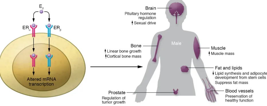
Estrogen action in men
Preface. 17-β-estradiol (E2) is the common form of serum and tissue estrogen in men and women. The CYP19A1 gene encodes the aromatase enzyme that converts testosterone to E2 in both sexes (4). Aromatase is expressed in many organs and cells; thus, local production and action of E2 in men is likely physiological-ly relevant (5). Strong support for important roles of E2 comes from studies in men with inactivating mutations of either ERα or aromatase (6, 7). E2 insensitivity was found in a 28-year-old man diagnosed with a homozygous ERα mutation that produced a trun-cated nonfunctional protein (6). The individual presented with continued linear growth and tall stature due in part to unfused epiphyses, despite normal serum testosterone. Significant osteo-porosis was noted, indicating that endogenous estrogen and ERα are important in men for normal bone growth and development. This individual was also overweight for his height and showed excess abdominal fat. Elevated endogenous estrogen levels in this individual failed to suppress the pituitary gonadotropins, luteiniz-ing and follicle-stimulatluteiniz-ing hormones (LH and FSH, respectively) in the absence of the functional ERα receptor. Thus, while direct action of male sex steroids at ARs in the brain may play some role in negative feedback that regulates LH and FSH, estrogen signal-ing via ERα is also required.
Similarly, men who are functionally deficient in aromatase activity, and therefore cannot make estrogens, have abnormalities
Androgens and estrogens are known to be critical regulators of mammalian physiology and development. While these two classes of steroids share similar structures (in general, estrogens are derived from androgens via the enzyme aromatase), they subserve markedly different functions via their specific receptors. In the past, estrogens such as estradiol were thought to be most important in the regulation of female biology, while androgens such as testosterone and dihydrotestosterone were believed to primarily modulate development and physiology in males. However, the emergence of patients with deficiencies in androgen or estrogen hormone synthesis or actions, as well as the development of animal models that specifically target androgen- or estrogen-mediated signaling pathways, have revealed that estrogens and androgens regulate critical biological and pathological processes in both males and females. In fact, the concept of “male” and “female” hormones is an oversimplification of a complex developmental and biological network of steroid actions that directly impacts many organs. In this Review, we will discuss important roles of estrogens in males and androgens in females.
Impact of estrogens in males and androgens in females
Stephen R. Hammes1 and Ellis R. Levin2,3
1Division of Endocrinology and Metabolism, Department of Medicine, University of Rochester School of Medicine, Rochester, New York, USA. 2Departments of Medicine and Biochemistry, UCI, Irvine,
California, USA. 3Division of Endocrinology, UCI and United States Department of Veterans Affairs Medical Center, Long Beach, California, USA.
Conflict of interest: The authors have declared that no conflict of interest exists.
Copyright: © 2019, American Society for Clinical Investigation.
with WT mice, while trabecular bone showed increased miner-alizing surface, likely due to reduced bone resorption (22). The cortical bone discrepancy may result from completeness of the ERβ deletion. Interestingly, the male K/G mice showed no bone abnormalities (20). Deletion of ERβ in osteoprogenitor cells also showed increased trabecular but not cortical bone mass in female mice (23). Overall, these results indicate that ERβ has no signifi-cant effect in male mice, but may restrain trabecular bone miner-alization that is dependent on ERα in female mice.
In summary, testosterone conversion to estrogen in human males is important for both normal cortical bone development and preservation of healthy bone metabolism during aging that likely reduces fractures.
Reproduction. Although only a few men with aromatase gene
mutation have been studied, these individuals consistently show oligospermia and at least one presented with infertility (24). These reproductive abnormalities may reflect the loss of estrogen production in testicular Leydig cells. Severely decreased sperm motility was also noted in the man with mutated ERα and in ERα genetically deleted male mice (6, 8), suggesting that signaling via ERα regulates spermatogenesis. These findings are supported by more recent mouse studies, wherein loss of membrane or nuclear ERα in the testes results in abnormal sperm production and func-tion, leading to infertility as the male mice advance in age (25). Mechanistically, loss of ERα results in excessive fluid accumula-tion in the epididymis, which may contribute to abnormal sperm morphology and function (26).
In contrast to male ERα KO mice, male mice with KO of ERβ retain relatively normal fertility in two different models (18, 19). Surprisingly, however, a small number of male humans with muta-tions in ERβ are associated with 46, XY disorders of sex develop-ment, showing markedly abnormal or absent gonads (27). These differences between mice and men with mutated or no ERβ high-light the importance of studying estrogen signaling in both mouse models and human patients.
Interestingly, a gain-of-function mutation of the aromatase gene CYP19A1, which causes increased levels of the estrogen estrone, is linked to familial gynecomastia in young males (28). This perturbation of the normal ratio of testosterone to estrogen in men underlies most forms of gynecomastia.
Additional studies suggest that estrogen contributes to libido and sexual performance in men. For example, 202 healthy men given an analogue of gonadotropin-releasing hormone (GnRH) to inhibit endogenous androgen production had loss of sexual drive and erectile function. These men then received testosterone replacement without or with an aromatase inhibitor (anastrozole) for 16 weeks. Although testosterone administration significant-ly improved these functions, addition of the aromatase inhibitor attenuated improvement in both libido and penile erections (29).
It is well recognized that nitric oxide (NO) formation in penile blood vessels is necessary for vasodilation and erection (30). Estrogen acting at both ERα and ERβ strongly stimulates several isoforms of the NO synthase enzyme to produce NO in endothe-lial and other vascular cells (31–33), which may explain the erec-tile dysfunction associated with loss of estradiol production from testosterone. Once NO production is impaired from penile arte-rial disease, such as in diabetes, perhaps estrogen can no longer of bone formation, glucose and lipid metabolism (trending toward
the metabolic syndrome), and reproductive tract development and function (ultimately impairing fertility) (8, 9), many of which improve with estradiol. These examples in human males confirm the importance of estrogen in normal male physiology and are supported by studies in ERα-deficient mice, where similar pheno-types are observed (6, 7, 10).
Bone development and function. Many studies have shown
important effects of estrogen for bone health in elderly men and for bone development in young men. In the latter, there is evidence that estrogen strongly contributes to the closure of the epiphyses, thus limiting linear growth (11). This role of E2 is consistent with periosteal bone expansion during puberty, which is also seen with E2 replacement in men with aromatase gene mutations (8, 9, 11).
In elderly hypogonadal men with elevated markers of bone resorption characteristic of enhanced osteoclast activity, testos-terone replacement is minimally effective in suppressing these markers (12). In contrast, estrogen replacement strongly sup-presses the increase of bone resorption markers. The authors of this study conclude that in men, estrogen accounted for approx-imately 70% of antiresorptive effects on bone, with testoster-one contributing about 30%. These findings are consistent with osteopenia/osteoporosis observed in men with either mutated aromatase or ERα genes (6, 7). Studies in elderly men treated with an aromatase inhibitor provide additional support for estrogen’s role in preserving bone (13).
Mechanistically, in mouse models, estrogen suppresses IL-6– dependent osteoclast differentiation, which then may attenuate bone loss. However, TNF-α likely is more important for mediating estrogen deficiency–related bone loss, since ovariectomy increas-es bone marrow production of TNF-α, accompanied by bone loss, while ovariectomy in TNF-α–deficient mice does not lead to bone loss (14). The roles of TNF-α and possibly IL-1β suppression in mediating the antiresorptive effects of estrogen were confirmed in studies in women (15). However, comparable studies have not been done in men. Other purported mediators of osteoclast devel-opment and/or resorption in females that are inhibited by estro-gen include activation of NF-κΒ and sclerostin (16). However, again, little has been validated in men.
While aromatase in bone cells facilitates the local estrogen synthesis needed for bone formation in normal men, different conclusions have been drawn from genetic mouse models of ERα deletion in osteoblast (bone-forming) precursor cells, suggesting little contribution by E2 and ERα in male mice. A potential expla-nation is that estrogen effects in humans are mainly on cortical bone that comprises approximately 80% of the human skeleton, whereas cortical bone is quite different in the mouse and may be regulated differently by estrogens (17). This might be clinically relevant to prevent osteoporosis-related fractures in long bones of both human sexes.
it appears that local estrogen signaling in adipocytes may play a major role in modulating its own production (44). Estrogen may also directly affect weight and fat formation by regulating ener-gy intake and output. In genetic mouse models, loss of ERα from specific hypothalamic regions results in excessive food intake and decreased energy expenditure (45). Furthermore, studies in ERα-deficient mice demonstrate that estrogen enhances insulin action in the liver, muscle, and fat of both males and females (46).
In addition to regulating insulin signaling, estrogens also modulate β cell function in the pancreas. ERα in pancreatic islets suppresses fatty acid synthesis via STAT3-mediated suppression of the fatty acid synthase gene in male rats, contributing to the prevention of β cell failure (47). In both sexes of aromatase-gene– deficient mice given streptozotocin (which causes β cell apoptosis), estrogen/ERα sustains insulin secretion by mitigating β cell death (48). Nuclear ERα in the CNS helps to maintain insulin sensitivity in female mice, while loss of nuclear ERα impairs the ability of glu-cose injected into the carotid artery to stimulate brain regulation of insulin secretion only in male mice (49). These findings indicate roles for ERα to positively regulate normal glucose homeostasis in both male and female mice. This is consistent with impaired glu-cose homeostasis in men with aromatase gene mutations (9, 11). However, both in ERα deletion models in mice and the aforemen-tioned male humans, there is no evidence of diabetes, indicating a moderate regulatory role for the sex steroid receptor.
Aromatase-deficient men and the ERα mutant male also showed indications of the metabolic syndrome, including hyper-tension. Estrogen administration reversed many of these disor-ders, including improvements in insulin resistance and glucose intolerance (24). Interestingly, ERβ-KO male and female mice (specifically C-ERβ-KO) become hypertensive with aging (32), suggesting that this ER isoform may also contribute to normaliza-tion of blood pressure. In mice, delenormaliza-tion of ERα confined to adipo-cytes resulted in increased markers of fibrosis and inflammation in the fat niche, as well as impaired overall glucose homeostasis, effects that were more pronounced in males (50).
In summary, various aspects of the metabolic syndrome are clearly improved in male mouse models of disease, but, while sugges-tive, whether this extends to men requires additional determination.
Prostate cancer. In the normal human prostate, both ERα and ERβ are expressed mainly in the stroma and epithelium, respec-tively (51). ERα is generally felt to be proproliferative in normal and malignant prostate, contributing to the development of premalig-nant lesions and cancer in rodent models. In contrast, ERβ main-tains epithelial differentiation while inhibiting proliferation caused by ERα, thereby promoting normal development and at least ini-tially acting as a suppressor of prostate cancer development. A syn-thetic estrogen, diethylstilbestrol (DES), was used to successfully treat prostate cancer in the 1960s and 1970s, suppressing andro-gen production through feedback upon the hypothalamic-pituitary axis (52). However, due to its prothrombotic effects, a high number of myocardial infarctions resulted in DES-treated patients. Nev-ertheless, these studies suggested targeting ER in prostate cancer could be therapeutically advantageous.
Clinical trials using an ERα agonist (53) or a selective ER mod-ulator (54) have not generated sufficiently impactful evidence for the treatment of prostate cancer to justify comprehensive promote vasodilation, as the sex steroid appears to prevent early
arterial disease in mouse models. Thus, estradiol in men func-tions both in the brain (libido) and the gonads (erection) to mod-ulate male reproduction.
CNS. Studies in animals and in humans have demonstrated
that estrogen’s actions in the CNS play critical roles in aggression and in sexual behavior in males, most likely due to local production of estradiol by aromatase. For example, treatment of macaques with aromatase inhibitors leads to decreased sexual motivation and ejaculatory actions (34). Human males with aromatase muta-tions have decreased libido and reduced sexual behavior, despite high testosterone levels, and estrogen treatment enhances libi-do and sexual activity (35). Similarly, as mentioned, testosterone replacement in the presence of an aromatase inhibitor in hypo-gonadal males leads to only a partial decrease in sexual function compared with testosterone replacement alone (29). Interestingly, aromatase expression is abundant in numerous brain nuclei of both females and males (36, 37), and local estradiol production in these regions appears to be critical in mediating aggressive and sexual behaviors. For example, mouse models of aromatase deficiency have shown that its actions in the hypothalamus and amygdala are important for male aggression (38, 39). Furthermore, male mice lacking AR expression in the CNS still exhibit male sexual and ter-ritorial actions (40), indicating that aromatization of androgens to estrogens, followed by estrogen actions on ERs, plays an essential role in what are commonly thought to be “male” behaviors.
Finally, estrogen may play a critical role in male brains beyond its actions in sexual and aggressive behavior. Local production of estradiol in the male cerebellum appears to be important for ves-tibular-ocular reflex adaptation (41), which coordinates eye and head movements to help stabilize vision. Estrogen also enhances spatial memory in females via hippocampus ERα, but through ERβ in the hippocampus of male mice (42). These studies indicate that estrogen production and actions in the CNS are diverse and that more estrogen-mediated processes will likely be discovered.
Fat and the metabolic syndrome. Men with aromatase mutations
often display low HDL cholesterol, high LDL cholesterol, increased triglycerides and visceral fat, and impaired glucose homeostasis (8, 24). These lipid abnormalities are reversed by treatment with estrogen (8). Aromatase-deficient men and the previously dis-cussed individual with an ERα mutation show reduced endothelial function and premature atherosclerosis, including plaque forma-tion. Estrogen replacement resolved these conditions in one indi-vidual (24). The hepatic steatosis reported in several of these men may be the result of elevated triglycerides (24).
the bone depends on its aromatase-mediated conversion to estra-diol. However, men still have bigger bones, suggesting that andro-gens may play an independent role in regulating bone size. Data from humans and mouse models suggest that androgens enhance bone size by maintaining cancellous (trabecular) bone mass (62). Rodent models also suggest that androgens enhance bone size by promoting periosteal bone apposition (63). However, in one aro-matase-deficient human male with high testosterone levels, quan-titative CT demonstrated decreased periosteal bone apposition that improved upon estradiol therapy (11); thus, the role of andro-gens versus estroandro-gens in human periosteal bone apposition is still not clearly defined. Notably, individuals with complete androgen insensitivity (e.g., XY individuals with inactivating mutations in ARs) have decreased bone size and density (including trabecu-lar bone) relative to both normal males and females, yet have no increase in fracture rate (64). Accordingly, in AR-KO male mice, cancellous/trabecular bone volumes are smaller and less dense, even though androgen and estradiol levels are normal (65). Simi-larly, in male mice, specific inactivation of the AR in bone-forming osteoblasts confers no change in cortical bone, but a significant reduction in trabecular bone volume (66), again suggesting that androgens maintain trabecular/cancellous bone mass. AR defi-ciency has less dramatic effects on the bones of female mice, but these mice still display reduced cortical bone mass and signifi-cantly altered cancellous bone architecture (66, 67). These obser-vations indicate that, in females, androgens may have estrogen- independent effects on bone metabolism and growth via the AR.
Reproduction. It is established that androgens are important
in female reproduction. For example, the most common cause of infertility in women is polycystic ovary syndrome (PCOS), which is characterized by androgen excess, oligomenorrhea or amenor-rhea, and polycystic ovaries. While two out of these three are suf-ficient for diagnosis, the majority of PCOS patients have androgen excess and ovulatory dysfunction, and androgens are thought to be a critical mediator of the reproductive dysfunction (68). The patho-physiology is unclear, but in part, it involves a powerful positive feedback loop whereby ovarian androgens increase the frequency of hypothalamic GnRH pulsations. This activity favors pituitary LH over FSH secretion, resulting in the loss of an estradiol and sub-sequent LH surge, leading to anovulation and even more ovarian androgen production (69, 70). In addition, excess androgens have direct effects on the ovary, perhaps promoting increased or unreg-ulated follicle growth, which then prevents normal selection of a single follicle for ovulation (71). In fact, KO of the AR in mouse fol-licular theca cells slightly protects cycling in hyperandrogenic mice (72), suggesting that AR signaling in theca cells may partially drive the abnormal reproductive phenotype seen in PCOS.
Whereas androgen excess clearly impairs female fertility, physiologic androgen levels play a positive role. For many years, work in vitro and in animal models suggested that androgens might play an important role in follicle growth, perhaps acting to enhance FSH-mediated signaling (73, 74). Proof of this concept came with the description of a female mouse in which the AR was specifically ablated in ovarian granulosa cells (75). These mice have decreased ovulation rates, smaller litter sizes, and premature ovarian failure, primarily due to the loss of AR-mediated signals that prevent follicle atresia and augment FSH-mediated follicle studies in this malignancy. This may reflect the complexity of the
nuclear ER working in conjunction with the nuclear AR in myriad ways, depending upon the stage of the tumor. In addition, there are various ERβ isoforms that have either tumor-suppressive or tumor-promoting functions (55). ERβ isoform switching has been observed in castration-resistant and metastatic prostate cancer in men, perhaps explaining the dichotomy of ERβ actions in various types of this malignancy. Interestingly, recent studies in humans show that a high expression of ERβ occurs in many prostate can-cers and correlates to a favorable prognosis (56), whereas high levels of estradiol or estrone are significantly associated with a shorter time to the development of castration-resistant prostate cancer, presumably through actions at ERα (57). In aromatase-KO mice, an ERβ agonist induces apoptosis of stromal, luminal, and epithelial cells within the prostate. Agonists for this receptor also induce apoptosis in stroma and epithelial progenitor cells using patient-derived, Gleason-7 xenograft tissues in mice. This process is mediated by TNF-α–mediated upregulation of caspase-8 (58). Realizing the importance of ER in the prostate, interventional par-adigms continue to be developed.
Androgen action in women
Preface. While estrogens are considered the dominant sex steroid
in women, in fact, serum androgen levels in women are higher than estrogen levels most of the time. The exception is during the preovulatory and midluteal phases of the menstrual cycle, when androgen and estrogen levels are similar. Therefore, it is reason-able to consider that androgens might have important physiologic effects in women. However, there are many difficulties relating androgen levels to physiological or disease processes, primarily due to unknowns about steroid metabolism and inefficiencies in accurately measuring testosterone levels. With regard to steroid metabolism, most androgen actions are likely mediated by intra-cellular conversion to DHT; thus, it is unclear whether serum tes-tosterone levels truly reflect active androgen levels. In addition, testosterone is readily converted to E2 by aromatase in most tis-sues; therefore, observations associated with high testosterone levels might really reflect estrogen actions.
Even more troubling is determining how to measure testos-terone. First, most testosterone immunoassays are inaccurate below approximately 100 ng/ml, which is where testosterone levels in women rest. In fact, even in hypogonadal men with tes-tosterone levels in the 100–200 ng/ml range, immunoassays are not accurate. Second, it is unclear which testosterone moiety should be measured. More than 98% of testosterone is bound to proteins, either tightly to sex hormone–binding globulin (SHBG) (about 66%) or weakly to other proteins such as albumin (about 33%) (59). Therefore, it is not clear which measurement (total tes-tosterone, free testes-tosterone, or bioavailable testosterone— non-SHBG-bound) is most meaningful. The answer is still not known; however, the Endocrine Society suggests that total testosterone measured by liquid chromatography/mass spectroscopy in a quali-fied laboratory is most useful, although other validated assays may be appropriate in some instances (60, 61).
Bone development and function. In humans, the role of
this decrease in testosterone has been implicated in postmeno-pausal decreases in libido, and possibly to hypoactive sexual desire disorder (HSDD), defined as a deficiency of sexual fantasies and desire for normal sexual activity that causes significant stress or interpersonal difficulty (89). In general, some studies suggest a correlation between serum testosterone levels and sexual desire (90, 91), but others do not (92, 93). These discrepancies may in part be related to the varying methods used to measure testoster-one; thus, to date, no clear evidence exists that definitively relates androgen levels to sexual desire. Several randomized placebo- controlled studies have suggested that androgen treatment (either testosterone or dehydroepiandrosterone [DHEA]) improves sexual desire and performance (94, 95) independently of androgen levels, especially in HSDD (96, 97). However, most of these studies are small, demonstrate large placebo effects, and are based on surveys rather than concrete quantitative measurements.
After careful examination of the available data, the recom-mendations of the Endocrine Society are against making a diag-nosis of androgen deficiency in women (98). Furthermore, the Society recommends against prescribing testosterone for women except those with HSDD. Notably, such replacement medications are not available in many countries, including the United States, and long-term safety data are not known. Care with monitoring for signs of androgen excess (e.g., hirsutism, acne, thinning of scalp hair) should be taken, and testosterone should be stopped if no significant improvement is seen.
Fat and the metabolic syndrome. As mentioned, women with
PCOS have androgen excess and reduced fertility. In addition, PCOS is closely linked with obesity, insulin resistance, and the metabolic syndrome. Studies suggest that obesity, insulin resis-tance, and subsequent hyperinsulinemia lead to androgen excess, perhaps in part via selective insulin sensitivity in the ovaries that results in excessive insulin-mediated androgen production (99, 100). However, androgen treatment of mice and rats can also promote weight gain and insulin resistance in female mice, ulti-growth (76). These findings in mice are consistent with
observa-tions in women with diminished ovarian reserve, in whom studies suggest that androgen pretreatment improves fertilization rates (77). The human studies are small and not always well controlled; thus, larger randomized placebo-controlled studies are necessary. However, the concept that a tightly balanced androgen milieu in the ovary is necessary for normal fertility is now well established.
Androgens may also play a role in the maintenance of pregnan-cy, especially with regard to parturition. Total testosterone levels reportedly increase slightly during pregnancy (78); however, due to the use of less accurate immunoassays, along with increased SHBG production during pregnancy, it is unclear whether free tes-tosterone levels change during pregnancy. In contrast, androgen levels are higher in pregnant women with PCOS, with a report-ed increase in the incidence of preterm labor (79). Treatment of female primates with the androgen androstenedione triggers premature uterine contractions in some but not all studies (80, 81). Animal models support a potential role for androgen in par-turition, as androgen actions through the AR promote collagenase expression in the cervix, leading to cervical remodeling critical for normal delivery (82, 83). In animals and in humans, AR levels are high in myometrial cells early in pregnancy but are reduced by par-turition (84, 85). Studies in rodents suggest that androgen signal-ing via the AR suppresses myometrial contractility by modulatsignal-ing calcium signaling in a transcription-independent fashion; thus, as myometrial AR levels drop at the end of gestation, suppression of myometrial contractions may be reduced, which, in combination with the aforementioned cervical remodeling, leads to parturition (85–87). More studies on androgen actions during pregnancy are needed; however, combined with the studies on ovarian function, androgens appear important for female reproduction from follicle development through parturition.
CNS. Like estradiol, testosterone levels in women decrease
[image:6.585.60.526.60.245.2]with age, although postmenopausal serum testosterone levels drop only 2-fold, while estradiol levels drop 10-fold (88). Nonetheless,
mately leading to hyperglycemia. This androgen-induced phe-nomenon is substantially attenuated in female mice with the AR knocked out specifically in β cells (101), suggesting a positive feedback loop whereby, in PCOS, insulin from β cells promotes ovarian androgen production, which in turn stimulates more insulin production by the β cell.
Breast cancer. The role of ARs in breast cancer has been
dis-cussed for years but has recently returned to the limelight. ARs are expressed in many different breast cell types, and androgens have long been considered “stop” signals during normal estrogen- and progesterone-mediated breast development, which might explain why men do not develop breasts unless testosterone levels are low. AR signaling, however, may only be a minor contributor to nor-mal breast development, as global AR null fenor-male mice have sub-tle to no changes in their mammary glands (65). With regard to breast cancer, up to 90% of tumors express ARs. In general, AR expression is considered a positive prognostic factor for breast cancer, especially when tumors are both ERα and AR positive (102–104). Accordingly, androgens were once used to treat breast cancer, with general efficacy in blocking cancer progression (105, 106). These observations are consistent with postulated antipro-liferative effects of androgens on normal breast development. In contrast, only 50% of ERα-negative breast cancers express ARs, and AR expression seems to have a neutral to possibly negative effect on prognosis (102, 107). Finally, approximately 30% of tri-ple-negative breast cancers (ERα, PR, and ERB2/Her2 negative) are AR positive, and AR expression again predicts a neutral to worse outcome (108–110). In vitro studies suggest that AR signal-ing suppresses growth in ERα-positive breast cancer cells but pro-motes growth in ERα-negative cells. Even in the presence of ERα, if AR is overexpressed, or if breast cancer cells become resistant to tamoxifen suppression, androgens become promoters of prolifera-tion (111, 112). These studies suggest a complex crosstalk between
AR and ERα signaling (113, 114) whereby the higher the ratio of AR to ER, either in terms of levels or activities, the more sensitive cells become to AR-mediated growth. Interestingly, antiandrogen treatment of ER+/AR+ breast cancer cell lines reduces
ER-mediat-ed proliferation both in vitro and in xenograft models (115), again indicating a complex crosstalk between AR and ER signaling. Fur-thermore, AR/HER2 crosstalk has been reported: antiandrogen treatment suppresses HER2 phosphorylation and activation in vitro, and combined antiandrogen/anti-HER2 suppresses xeno-graft growth more than either treatment individually (116, 117).
Clinically, a phase II clinical trial using the AR antagonist bicalutamide to treat ERα/progesterone-receptor–negative tumors demonstrates a 19% clinical benefit rate (progression-free survival) over 6 months (118). A second phase II clinical trial using the more potent anti-AR drug enzalutamide in AR+, triple-negative tumors
demonstrates a similar 25% clinical benefit rate at 16 weeks (119). Finally, a phase II trial using the CYP17 inhibitor abiraterone, which suppresses androgen production, in AR+, triple-negative breast
cancer demonstrates a similar 6-month clinical benefit rate of 20% (120). Thus, antiandrogen treatment holds promise for the treat-ment of triple-negative breast cancer, though randomized place-bo-controlled studies are needed to truly address their benefit.
Conclusions
[image:7.585.61.532.61.242.2]the physiologic response can vary tremendously depending on the ratio of ER/AR signaling, as well as the amount of steroid metab-olism occurring locally. Together, the examples presented here, as well as many others, suggest that traditional views whereby sex-ual dimorphisms between women and men are explained almost exclusively by the presence of estrogens in women and androgens in men must be modified to reflect the complexities of steroid hor-mone signaling and biology.
Address correspondence to: Stephen R. Hammes, Division of Endocrinology and Metabolism, Department of Medicine, Uni-versity of Rochester School of Medicine, 601 Elmwood Avenue, Rochester, New York 14642, USA. Phone: 585.275.2901; Email: stephen_hammes@urmc.rochester.edu. Or to: Ellis R. Levin, Medical Service (111-I), Long Beach Veterans Affairs Medical Center, 5901 East 7th Street, Long Beach, California 90822, USA. Phone: 562.826.8000, ext. 24147; Email: ellis.levin@va.gov.
1. Jensen EV, Desombre ER, Kawashima T, Suzuki T, Kyser K, Jungblut PW. Estrogen-bind-ing substances of target tissues. Science. 1967;158(3800):529–530.
2. Kuiper GG, Enmark E, Pelto-Huikko M, Nilsson S, Gustafsson JA. Cloning of a novel receptor expressed in rat prostate and ovary. Proc Natl
Acad Sci U S A. 1996;93(12):5925–5930.
3. Franchimont P. Regulation of gonadal androgen secretion. Horm Res. 1983;18(1–3):7–17. 4. Harada N. Cloning of a complete cDNA encoding
human aromatase: immunochemical identifica-tion and sequence analysis. Biochem Biophys Res
Commun. 1988;156(2):725–732.
5. Jones ME, Boon WC, McInnes K, Maffei L, Cara-ni C, Simpson ER. RecogCara-nizing rare disorders: aromatase deficiency. Nat Clin Pract Endocrinol
Metab. 2007;3(5):414–421.
6. Smith EP, et al. Estrogen resistance caused by a mutation in the estrogen-receptor gene in a man.
N Engl J Med. 1994;331(16):1056–1061.
7. Rochira V, et al. Hypothalamic-pituitary-gonadal axis in two men with aromatase deficiency: evi-dence that circulating estrogens are required at the hypothalamic level for the integrity of gonad-otropin negative feedback. Eur J Endocrinol. 2006;155(4):513–522.
8. Carani C, et al. Effect of testosterone and estra-diol in a man with aromatase deficiency. N Engl J
Med. 1997;337(2):91–95.
9. Miedlich SU, Karamooz N, Hammes SR. Aro-matase deficiency in a male patient - Case report and review of the literature. Bone. 2016;93:181–186.
10. Lindzey J, Wetsel WC, Couse JF, Stoker T, Cooper R, Korach KS. Effects of castration and chronic steroid treatments on hypothalamic gonado-tropin-releasing hormone content and pituitary gonadotropins in male wild-type and estrogen receptor-alpha knockout mice. Endocrinology. 1998;139(10):4092–4101.
11. Bouillon R, Bex M, Vanderschueren D, Boonen S. Estrogens are essential for male pubertal peri-osteal bone expansion. J Clin Endocrinol Metab. 2004;89(12):6025–6029.
12. Falahati-Nini A, Riggs BL, Atkinson EJ, O’Fallon WM, Eastell R, Khosla S. Relative contributions of testosterone and estrogen in regulating bone resorption and formation in normal elderly men.
J Clin Invest. 2000;106(12):1553–1560.
13. Leder BZ, Rohrer JL, Rubin SD, Gallo J, Longcope C. Effects of aromatase inhibition in elderly men with low or borderline-low serum testosterone lev-els. J Clin Endocrinol Metab. 2004;89(3):1174–1180. 14. Roggia C, et al. Up-regulation of
TNF-produc-ing T cells in the bone marrow: a key
mech-anism by which estrogen deficiency induces bone loss in vivo. Proc Natl Acad Sci U S A. 2001;98(24):13960–13965.
15. Charatcharoenwitthaya N, Khosla S, Atkinson EJ, McCready LK, Riggs BL. Effect of blockade of
TNF-α and interleukin-1 action on bone
resorp-tion in early postmenopausal women. J Bone
Miner Res. 2007;22(5):724–729.
16. Eghbali-Fatourechi G, Khosla S, Sanyal A, Boyle WJ, Lacey DL, Riggs BL. Role of RANK ligand in mediating increased bone resorption in early postmenopausal women. J Clin Invest. 2003;111(8):1221–1230.
17. Khosla S. New insights into androgen and estro-gen receptor regulation of the male skeleton.
J Bone Miner Res. 2015;30(7):1134–1137.
18. Krege JH, et al. Generation and reproduc-tive phenotypes of mice lacking estrogen receptor beta. Proc Natl Acad Sci U S A. 1998;95(26):15677–15682.
19. Dupont S, Krust A, Gansmuller A, Dierich A, Chambon P, Mark M. Effect of single and com-pound knockouts of estrogen receptors alpha
(ERα) and beta (ERβ) on mouse reproductive
phe-notypes. Development. 2000;127(19):4277–4291. 20. Windahl SH, Vidal O, Andersson G, Gustafsson
JA, Ohlsson C. Increased cortical bone mineral content but unchanged trabecular bone mineral density in female ERβ(–/–) mice. J Clin Invest. 1999;104(7):895–901.
21. Windahl SH, Hollberg K, Vidal O, Gustafsson JA, Ohlsson C, Andersson G. Female estrogen receptor β–/– mice are partially protected against
age-related trabecular bone loss. J Bone Miner
Res. 2001;16(8):1388–1398.
22. Sims NA, et al. Deletion of estrogen receptors
reveals a regulatory role for estrogen receptors-β
in bone remodeling in females but not in males.
Bone. 2002;30(1):18–25.
23. Nicks KM, et al. Deletion of estrogen receptor β in osteoprogenitor cells increases trabecular but not cortical bone mass in female mice. J Bone Miner
Res. 2016;31(3):606–614.
24. Jones ME, Boon WC, Proietto J, Simpson ER. Of mice and men: the evolving phenotype of aromatase deficiency. Trends Endocrinol Metab. 2006;17(2):55–64.
25. Nanjappa MK, Hess RA, Medrano TI, Locker SH, Levin ER, Cooke PS. Membrane-localized estrogen receptor 1 is required for normal male reproductive development and function in mice.
Endocrinology. 2016;157(7):2909–2919.
26. Joseph A, Shur BD, Ko C, Chambon P, Hess RA. Epididymal hypo-osmolality induces abnormal sperm morphology and function in the
estro-gen receptor α knockout mouse. Biol Reprod.
2010;82(5):958–967.
27. Baetens D, et al. Biallelic and monoallelic ESR2 variants associated with 46,XY disorders of sex development. Genet Med. 2018;20(7):717–727. 28. Shozu M, et al. Estrogen excess
associat-ed with novel gain-of-function mutations affecting the aromatase gene. N Engl J Med. 2003;348(19):1855–1865.
29. Finkelstein JS, et al. Gonadal steroids and body composition, strength, and sexual function in men. N Engl J Med. 2013;369(11):1011–1022. 30. Simonsen U, García-Sacristán A, Prieto
D. Penile arteries and erection. J Vasc Res. 2002;39(4):283–303.
31. Ishikawa T, et al. LXRβ/estrogen receptor-α sig-naling in lipid rafts preserves endothelial integri-ty. J Clin Invest. 2013;123(8):3488–3497. 32. Zhu Y, et al. Abnormal vascular function and
hypertension in mice deficient in estrogen
recep-tor β. Science. 2002;295(5554):505–508.
33. Pedram A, Razandi M, Blumberg B, Levin ER.
Membrane and nuclear estrogen receptor α
collaborate to suppress adipogenesis but not tri-glyceride content. FASEB J. 2016;30(1):230–240. 34. Zumpe D, Bonsall RW, Michael RP. Effects of
the nonsteroidal aromatase inhibitor, fadrozole, on the sexual behavior of male cynomolgus monkeys (Macaca fascicularis). Horm Behav. 1993;27(2):200–215.
35. Carani C, Rochira V, Faustini-Fustini M, Balestri-eri A, Granata AR. Role of oestrogen in male sex-ual behaviour: insights from the natural model of aromatase deficiency. Clin Endocrinol (Oxf). 1999;51(4):517–524.
36. Roselli CF. Brain aromatase: roles in reproduc-tion and neuroprotecreproduc-tion. J Steroid Biochem Mol
Biol. 2007;106(1-5):143–150.
37. Sasano H, Takashashi K, Satoh F, Nagura H, Harada N. Aromatase in the human cen-tral nervous system. Clin Endocrinol (Oxf). 1998;48(3):325–329.
38. Wu MV, et al. Estrogen masculinizes neural pathways and sex-specific behaviors. Cell. 2009;139(1):61–72.
39. Unger EK, Burke KJ Jr, Yang CF, Bender KJ, Fuller PM, Shah NM. Medial amygdalar aromatase neu-rons regulate aggression in both sexes. Cell Rep. 2015;10(4):453–462.
40. Juntti SA, et al. The androgen receptor gov-erns the execution, but not programming, of male sexual and territorial behaviors. Neuron. 2010;66(2):260–272.
42. Wang W, et al. Memory-related synaptic plastici-ty is sexually dimorphic in rodent hippocampus.
J Neurosci. 2018;38(37):7935–7951.
43. Pedram A, Razandi M, Blumberg B, Levin ER.
Membrane and nuclear estrogen receptor α
collaborate to suppress adipogenesis but not tri-glyceride content. FASEB J. 2016;30(1):230–240. 44. Simpson ER, et al. Aromatase cytochrome P450, the enzyme responsible for estrogen biosynthe-sis. Endocr Rev. 1994;15(3):342–355.
45. Xu Y, et al. Distinct hypothalamic neurons medi-ate estrogenic effects on energy homeostasis and reproduction. Cell Metab. 2011;14(4):453–465. 46. Ribas V, et al. Impaired oxidative metabolism
and inflammation are associated with insulin
resistance in ERα-deficient mice. Am J Physiol
Endocrinol Metab. 2010;298(2):E304–E319.
47. Tiano JP, et al. Estrogen receptor activation reduces lipid synthesis in pancreatic islets and prevents β cell failure in rodent models of type 2 diabetes. J Clin Invest. 2011;121(8):3331–3342. 48. Le May C, et al. Estrogens protect pancreatic
beta-cells from apoptosis and prevent insulin- deficient diabetes mellitus in mice. Proc Natl
Acad Sci U S A. 2006;103(24):9232–9237.
49. Allard C, et al. Loss of nuclear membrane
estro-gen receptor-α differentially impairs insulin
secretion action in male female mice [published online ahead of print October 10, 2018]. Diabetes. https://doi.org/10.2337/db18-0293.
50. Davis KE, et al. The sexually dimorphic role of adipose and adipocyte estrogen receptors in modulating adipose tissue expansion, inflamma-tion, and fibrosis. Mol Metab. 2013;2(3):227–242. 51. Nelson AW, Tilley WD, Neal DE, Carroll JS.
Estro-gen receptor beta in prostate cancer: friend or foe? Endocr Relat Cancer. 2014;21(4):T219–T234. 52. Bailar JC 3rd, Byar DP. Estrogen treatment
for cancer of the prostate. Early results with 3 doses of diethylstilbestrol and placebo. Cancer. 1970;26(2):257–261.
53. Yu EY, et al. Selective estrogen receptor alpha agonist GTx-758 decreases testosterone with reduced side effects of androgen deprivation therapy in men with advanced prostate cancer.
Eur Urol. 2015;67(2):334–341.
54. Fujimura T, et al. Toremifene, a selective estro-gen receptor modulator, significantly improved biochemical recurrence in bone metastatic pros-tate cancer: a randomized controlled phase II a trial. BMC Cancer. 2015;15:836.
55. Leung YK, et al. Estrogen receptor β2 and β5 are associated with poor prognosis in prostate can-cer, and promote cancer cell migration and inva-sion. Endocr Relat Cancer. 2010;17(3):675–689. 56. Azizan N, Hayati F, Tizen NMS, Farouk WI, Masir
N. Role of co-expression of estrogen receptor β
and Ki67 in prostate adenocarcinoma. Investig
Clin Urol. 2018;59(4):232–237.
57. Toren P, et al. Serum sex steroids as prognostic biomarkers in patients receiving androgen depri-vation therapy for recurrent prostate cancer: a post hoc analysis of the PR.7 trial. Clin Cancer
Res. 2018;24(21):5305–5312.
58. McPherson SJ, et al. Estrogen receptor-β activat-ed apoptosis in benign hyperplasia and cancer of the prostate is androgen independent and TNFalpha mediated. Proc Natl Acad Sci U S A.
2010;107(7):3123–3128.
59. Dunn JF, Nisula BC, Rodbard D. Transport of steroid hormones: binding of 21 endogenous ste-roids to both testosterone-binding globulin and corticosteroid-binding globulin in human plas-ma. J Clin Endocrinol Metab. 1981;53(1):58–68. 60. Wierman ME, et al. Editorial: The new instruc-tions to authors for the reporting of steroid hor-mone measurements. J Clin Endocrinol Metab. 2014;99(12):4375.
61. Handelsman DJ, Wartofsky L. Requirement for mass spectrometry sex steroid assays in the Jour-nal of Clinical Endocrinology and Metabolism.
J Clin Endocrinol Metab. 2013;98(10):3971–3973.
62. Vanderschueren D, Vandenput L, Boonen S, Lindberg MK, Bouillon R, Ohlsson C. Androgens and bone. Endocr Rev. 2004;25(3):389–425. 63. Turner RT, Wakley GK, Hannon KS. Differential
effects of androgens on cortical bone histomor-phometry in gonadectomized male and female rats. J Orthop Res. 1990;8(4):612–617.
64. Bertelloni S, Baroncelli GI, Federico G, Cappa M, Lala R, Saggese G. Altered bone mineral density in patients with complete androgen insensitivity syndrome. Horm Res. 1998;50(6):309–314. 65. Yeh S, et al. Generation and characterization of
androgen receptor knockout (ARKO) mice: an in vivo model for the study of androgen functions in selective tissues. Proc Natl Acad Sci U S A. 2002;99(21):13498–13503.
66. Määttä JA, et al. Inactivation of the androgen receptor in bone-forming cells leads to trabecular bone loss in adult female mice. Bonekey Rep. 2013;2:440.
67. MacLean HE, et al. DNA-binding-dependent androgen receptor signaling contributes to gender differences and has physiological actions in males and females. J Endocrinol. 2010;206(1):93–103. 68. Goodarzi MO, Dumesic DA, Chazenbalk G,
Azziz R. Polycystic ovary syndrome: etiology, pathogenesis and diagnosis. Nat Rev Endocrinol. 2011;7(4):219–231.
69. McCartney CR, Eagleson CA, Marshall JC. Regu-lation of gonadotropin secretion: implications for polycystic ovary syndrome. Semin Reprod Med. 2002;20(4):317–326.
70. Eagleson CA, et al. Polycystic ovarian syndrome: evidence that flutamide restores sensitivity of the gonadotropin-releasing hormone pulse generator to inhibition by estradiol and progesterone. J Clin
Endocrinol Metab. 2000;85(11):4047–4052.
71. Vendola KA, Zhou J, Adesanya OO, Weil SJ, Bondy CA. Androgens stimulate early stages of follicular growth in the primate ovary. J Clin
Invest. 1998;101(12):2622–2629.
72. Ma Y, et al. Androgen receptor in the ovary theca cells plays a critical role in androgen- induced reproductive dysfunction.
Endocrinolo-gy. 2017;158(1):98–108.
73. Weil S, Vendola K, Zhou J, Bondy CA. Androgen and follicle-stimulating hormone interactions in primate ovarian follicle development. J Clin
Endocrinol Metab. 1999;84(8):2951–2956.
74. Hillier SG, de Zwart FA. Androgen/antiandrogen modulation of cyclic AMP-induced steroidogen-esis during granulosa cell differentiation in tissue culture. Mol Cell Endocrinol. 1982;28(3):347–361. 75. Sen A, Hammes SR. Granulosa cell-specific
androgen receptors are critical regulators of ovar-ian development and function. Mol Endocrinol. 2010;24(7):1393–1403.
76. Sen A, et al. Androgens regulate ovarian follicular development by increasing follicle stimulating hor-mone receptor and microRNA-125b expression.
Proc Natl Acad Sci U S A. 2014;111(8):3008–3013.
77. Sunkara SK, Coomarasamy A. Androgen pretreat-ment in poor responders undergoing controlled ovarian stimulation and in vitro fertilization treatment. Fertil Steril. 2011;95(8):e73–e75. 78. O’Leary P, Boyne P, Flett P, Beilby J, James I.
Lon-gitudinal assessment of changes in reproductive hormones during normal pregnancy. Clin Chem. 1991;37(5):667–672.
79. Yamamoto M, et al. Risk of preterm delivery in non-diabetic women with polycystic ovarian syn-drome. J Perinatol. 2012;32(10):770–776. 80. Figueroa JP, Honnebier MB, Binienda Z, Wimsatt
J, Nathanielsz PW. Effect of a 48-hour intra-venous delta 4-androstenedione infusion on the pregnant rhesus monkey in the last third of gestation: changes in maternal plasma estradiol concentrations and myometrial contractility. Am
J Obstet Gynecol. 1989;161(2):481–486.
81. Mecenas CA, et al. Production of premature deliv-ery in pregnant rhesus monkeys by androstenedi-one infusion. Nat Med. 1996;2(4):443–448. 82. Mahendroo MS, Porter A, Russell DW, Word RA.
The parturition defect in steroid 5α-reductase
type 1 knockout mice is due to impaired cervical ripening. Mol Endocrinol. 1999;13(6):981–992. 83. Ji H, Dailey TL, Long V, Chien EK.
Androgen-reg-ulated cervical ripening: a structural, biomechan-ical, and molecular analysis. Am J Obstet Gynecol. 2008;198(5):543.e1–543.e9.
84. Bethin KE, et al. Microarray analysis of uterine gene expression in mouse and human pregnancy.
Mol Endocrinol. 2003;17(8):1454–1469.
85. Słomczyńska M, Duda M, Burek M, Knapczyk K, Czaplicki D, Koziorowski M. Distribution of androgen receptor in the porcine uterus throughout pregnancy. Reprod Domest Anim. 2008;43(1):35–41.
86. Perusquía M, Navarrete E, Jasso-Kamel J, Mon-taño LM. Androgens induce relaxation of con-tractile activity in pregnant human myometrium at term: a nongenomic action on L-type calcium channels. Biol Reprod. 2005;73(2):214–221. 87. Liu L, et al. Proliferative action of the
andro-gen receptor in human uterine myometrial cells — a key regulator for myometrium phe-notype programming. J Clin Endocrinol Metab. 2013;98(1):218–227.
88. Rothman MS, et al. Reexamination of testoster-one, dihydrotestostertestoster-one, estradiol and estrone levels across the menstrual cycle and in post-menopausal women measured by liquid chroma-tography-tandem mass spectrometry. Steroids. 2011;76(1–2):177–182.
89. Rosen RC, Maserejian NN, Connor MK, Krych-man ML, Brown CS, Goldstein I. Characteristics of premenopausal and postmenopausal women with acquired, generalized hypoactive sexual desire disorder: the Hypoactive Sexual Desire Disorder Registry for women. Menopause. 2012;19(4):396–405.
insufficien-cy: the Princeton consensus statement on defini-tion, classificadefini-tion, and assessment. Fertil Steril. 2002;77(4):660–665.
91. Wåhlin-Jacobsen S, et al. Is there a correlation between androgens and sexual desire in women?
J Sex Med. 2015;12(2):358–373.
92. Dennerstein L, Lehert P, Burger H. The relative effects of hormones and relationship factors on sexual function of women through the natural menopausal transition. Fertil Steril. 2005;84(1):174–180.
93. Santoro N, et al. Correlates of circulating andro-gens in mid-life women: the study of women’s health across the nation. J Clin Endocrinol Metab. 2005;90(8):4836–4845.
94. Davis SR, et al. Testosterone for low libido in postmenopausal women not taking estrogen.
N Engl J Med. 2008;359(19):2005–2017.
95. Fooladi E, Bell RJ, Jane F, Robinson PJ, Kulkarni J, Davis SR. Testosterone improves antidepres-sant-emergent loss of libido in women: findings from a randomized, double-blind, placebo- controlled trial. J Sex Med. 2014;11(3):831–839. 96. Davis SR, Braunstein GD. Efficacy and safety
of testosterone in the management of hypoac-tive sexual desire disorder in postmenopausal women. J Sex Med. 2012;9(4):1134–1148. 97. Shifren JL, et al. Testosterone patch for the
treatment of hypoactive sexual desire disor-der in naturally menopausal women: results from the INTIMATE NM1 Study. Menopause. 2006;13(5):770–779.
98. Wierman ME, et al. Androgen therapy in women: a reappraisal: an Endocrine Society clinical practice guideline. J Clin Endocrinol Metab. 2014;99(10):3489–3510.
99. Dunaif A. Insulin resistance and the polycystic ovary syndrome: mechanism and implications for pathogenesis. Endocr Rev. 1997;18(6):774–800.
100. Franks S, Gilling-Smith C, Watson H, Willis D. Insulin action in the normal and polycys-tic ovary. Endocrinol Metab Clin North Am. 1999;28(2):361–378.
101. Navarro G, et al. Androgen excess in pancreatic
β cells and neurons predisposes female mice to
type 2 diabetes. JCI Insight. 2018;3(12):98607. 102. Peters AA, et al. Androgen receptor inhibits
estro-gen receptor-α activity and is prognostic in breast cancer. Cancer Res. 2009;69(15):6131–6140. 103. Castellano I, et al. Androgen receptor expression
is a significant prognostic factor in estrogen receptor positive breast cancers. Breast Cancer
Res Treat. 2010;124(3):607–617.
104. Park S, et al. Androgen receptor expression is significantly associated with better outcomes in estrogen receptor-positive breast cancers. Ann
Oncol. 2011;22(8):1755–1762.
105. Goldenberg IS. Testosterone propionate therapy in breast cancer. JAMA. 1964;188:1069–1072. 106. Talley RW, Haines CR, Waters MN, Goldenberg
IS, Olson KB, Bisel HF. A dose-response evalua-tion of androgens in the treatment of metastatic breast cancer. Cancer. 1973;32(2):315–320. 107. Agoff SN, Swanson PE, Linden H, Hawes SE,
Lawton TJ. Androgen receptor expression in estrogen receptor-negative breast cancer. Immu-nohistochemical, clinical, and prognostic associ-ations. Am J Clin Pathol. 2003;120(5):725–731. 108. He J, et al. Prognostic value of androgen receptor
expression in operable triple-negative breast cancer: a retrospective analysis based on a tissue microarray. Med Oncol. 2012;29(2):406–410. 109. Tang D, Xu S, Zhang Q, Zhao W. The expression
and clinical significance of the androgen receptor and E-cadherin in triple-negative breast cancer.
Med Oncol. 2012;29(2):526–533.
110. Choi JE, Kang SH, Lee SJ, Bae YK. Androgen receptor expression predicts decreased survival
in early stage triple-negative breast cancer. Ann
Surg Oncol. 2015;22(1):82–89.
111. Rechoum Y, et al. AR collaborates with ERα in
aromatase inhibitor-resistant breast cancer.
Breast Cancer Res Treat. 2014;147(3):473–485.
112. De Amicis F, et al. Androgen receptor overex-pression induces tamoxifen resistance in human breast cancer cells. Breast Cancer Res Treat. 2010;121(1):1–11.
113. Auricchio F, Migliaccio A, Castoria G. Sex- steroid hormones and EGF signalling in breast and prostate cancer cells: targeting the asso-ciation of Src with steroid receptors. Steroids. 2008;73(9–10):880–884.
114. Cochrane DR, et al. Role of the androgen recep-tor in breast cancer and preclinical analysis of enzalutamide. Breast Cancer Res. 2014;16(1):R7. 115. D’Amato NC, et al. Cooperative dynamics of AR
and ER activity in breast cancer. Mol Cancer Res. 2016;14(11):1054–1067.
116. He L, et al. Targeting androgen receptor in treating HER2 positive breast cancer. Sci Rep. 2017;7(1):14584.
117. Gordon MA, et al. Synergy between androgen receptor antagonism and inhibition of mTOR and HER2 in breast cancer. Mol Cancer Ther. 2017;16(7):1389–1400.
118. Gucalp A, et al. Phase II trial of bicalutamide in patients with androgen receptor-positive, estro-gen receptor-negative metastatic breast cancer.
Clin Cancer Res. 2013;19(19):5505–5512.
119. Traina TA, et al. Enzalutamide for the treatment of androgen receptor-expressing triple-negative breast cancer. J Clin Oncol. 2018;36(9):884–890. 120. Bonnefoi H, et al. A phase II trial of abiraterone
![Figure 2. Androgens (including testosterone [T] and DHT) act through AR at the plasma membrane and in the nucleus to regulate many functions in women](https://thumb-us.123doks.com/thumbv2/123dok_us/9419270.444454/7.585.61.532.61.242/figure-androgens-including-testosterone-membrane-nucleus-regulate-functions.webp)
