Manuscript version: Author’s Accepted Manuscript
The version presented in WRAP is the author’s accepted manuscript and may differ from the
published version or Version of Record.
Persistent WRAP URL:
http://wrap.warwick.ac.uk/107499
How to cite:
Please refer to published version for the most recent bibliographic citation information.
If a published version is known of, the repository item page linked to above, will contain
details on accessing it.
Copyright and reuse:
The Warwick Research Archive Portal (WRAP) makes this work by researchers of the
University of Warwick available open access under the following conditions.
Copyright © and all moral rights to the version of the paper presented here belong to the
individual author(s) and/or other copyright owners. To the extent reasonable and
practicable the material made available in WRAP has been checked for eligibility before
being made available.
Copies of full items can be used for personal research or study, educational, or not-for-profit
purposes without prior permission or charge. Provided that the authors, title and full
bibliographic details are credited, a hyperlink and/or URL is given for the original metadata
page and the content is not changed in any way.
Publisher’s statement:
Please refer to the repository item page, publisher’s statement section, for further
information.
Journal Name
COMMUNICATION
Received 00th January 20xx, Accepted 00th January 20xx
DOI: 10.1039/x0xx00000x
www.rsc.org/
Pushing the limits of sensitivity and resolution for natural
abundance
43Ca NMR using ultra-high magnetic field (35.2 T)
Christian Bonhomme,*a Xiaoling Wang,b Ivan Hung,b Zhehong Gan,b Christel Gervais,a Capucine Sassoye,a Jessica Rimsza,c Jingcheng Du,c Mark E. Smith,d John V. Hanna,e Stéphanie Sarda,f Pierre Gras,g Christèle Combes,g and Danielle Laurencin*h
Natural abundance 43Ca solid state NMR experiments are reported
for the first time at ultra-high magnetic field (35.2 T) on a series of Ca-(pyro)phosphate and Ca-oxalate materials, which are of biological relevance in relation to biominerizalization processes and the formation of pathological calcifications. The significant gain in both sensitivity and resolution at 35.2 T leads to unprecedented insight into the structure of crystalline and amorphous phases.
Calcium is an element of major importance, due to its abundance in living organisms and tissues (e.g. bone and teeth), in natural rock-forming minerals (e.g. francolite, calcite, and dolomite), and in major construction materials (e.g. cement, concrete, glass and plaster). However, determining the local environment of this element within complex molecular and materials systems is far from trivial. In particular, structural
analysis by 43Ca NMR spectroscopy is highly challenging, due to
the poor receptivity of the NMR-active isotope.1 Calcium-43 is a
spin-7/2 quadrupolar nucleus of very low natural abundance
(0.14%) and resonance frequency (ν0(43Ca) = 57.2 MHz at 20 T).
To tackle sensitivity issues, two main approaches have been
used in the field of 43Ca solid state NMR.1-3 The first consists in
analyzing large quantities of sample (typically ≥ 300 mg) at high magnetic field (14 to 21 T NMR instruments) under moderate
magic angle spinning (MAS) conditions. However, even when using signal enhancement NMR sequences for quadrupolar
nuclei,1,4 several hours of acquisition are needed to record a 1D
spectrum, which excludes de facto the implementation of 2D experiments. Moreover, large quantities of material are not always available, making this approach inapplicable for many
systems. The second possibility is to label in 43Ca the
compounds of interest.1,4,5 However, although the gain in
sensitivity allows high resolution 1D and 2D correlation experiments to be performed, the major drawback is the high
cost of the isotopically-enriched precursor (~ 1500 € for 10 mg
of 60% 43Ca-labeled CaCO3). Moreover, it implies that synthetic
protocols often have to be re-adapted to start from 43
Ca-enriched CaCO3 as a calcium source.
The main challenge today for 43Ca solid state NMR is to find
means to reach a much higher sensitivity in order to be able to perform high resolution experiments at natural abundance on a broader variety of materials. In this context, the feasibility of
DNP (Dynamic Nuclear Polarization)-enhanced {1H}-43Ca CP
(Cross Polarization) MAS experiments at natural abundance was
recently demonstrated by Lee et al on a 400 MHz instrument.6
However, DNP analyses are constrained by specific experimental features, like the need to find efficient impregnation conditions for each material and to perform
measurements at low temperatures. Moreover, the
quadrupolar nature of 43Ca actually calls for performing
measurements at higher fields to achieve better resolution, because the second-order quadrupolar broadening scales as
1/B0. Hence, the possibility of recording natural abundance 43Ca
MAS NMR spectra at much higher magnetic fields was
investigated. In this article, the first natural abundance 43Ca
NMR study at 35.2 T (1.5 GHz 1H Larmor frequency) using the
series-connected hybrid (SCH) magnet at the US National High
Magnetic Field Laboratory.7
As the SCH magnet is available for a restricted period of time
per day (~ 7 hours), experiments were first set up by using a
43Ca-labeled monetite sample (*CaHPO
4). The radiofrequency
(RF) power on the 43Ca channel was carefully optimized for best
a.Sorbonne Universités, CNRS, Collège de France, Laboratoire de Chimie de la Matière Condensée de Paris, LCMCP, F-75005 Paris, France (CB:
b.National High Magnetic Field Laboratory, 1800 East Paul Dirac Drive, Tallahassee, FL 32310–3706, USA.
c.Department of Materials Science and Engineering, University of North Texas, Denton, Texas 76207, USA
d.Department of Chemistry, Lancaster University, Lancaster LA1 4YB, UK e.Department of Physics, Warwick University, Coventry, CV4 7AL, UK
f.CIRIMAT, Université de Toulouse, CNRS, Université Paul Sabatier,4 allée E. Monso, 31030 Toulouse cedex 4, France
g.CIRIMAT, Université de Toulouse, CNRS, INPT - Ensiacet, 4 allée E. Monso, 31030 Toulouse cedex 4, France
h.ICGM, UMR 5253, CNRS-UM-ENSCM, Place E. Bataillon, CC1701, 34095 Montpellier cedex 05, France (DL : [email protected]) † Footnotes relating to the title and/or authors should appear here.
COMMUNICATION Journal Name
efficiency of the multi-DFS (double frequency sweep) signal enhancement pulse sequence (final gain in signal-to-noise:
~ 2).4 Moreover, measurements on *CaHPO4 were repeated on
different days using a modified/enhanced Bruker lock system, showing no detectable variation in frequency, which demonstrates the reliability of comparisons made on spectra recorded on different days. All NMR experimental parameters (including temperature, MAS probe, pulse schemes, relaxation delays, number of scans and procedure for chemical shift
referencing8) are given in the Supporting Information.
Figure 1 : a/ 43Ca MAS NMR spectra of *CaHPO4 at 20.0 and 35.2 T,
together with their fit considering two average Ca environments {iso(43Ca) = 0.7 ± 0.8 ppm; |expCQ(43Ca)| = 3.5 ± 0.3 MHz, expQ(43Ca) = 0.8
± 0.1, and {iso(43Ca) = 8.5 ± 0.8 ppm; |expCQ(43Ca)| = 1.8 ± 0.3 MHz, expQ(43Ca) = 0.8 ± 0.1} (
▫
corresponds to a minor impurity); b/ 43Ca3QMAS spectrum of *CaHPO4 at 35.2 T.
Measurements on *CaHPO4 illustrate the significant gain in
resolution which is achieved at 35.2 T (Figure 1a). Indeed, two
resonances are clearly resolved at this field, and a second-order quadrupolar lineshape is even observed for the most deshielded signal, which was not the case at lower fields (≤ 20.0 T) due to the strong overlap between resonances. The NMR
parameters {δiso, CQ, Q} of the two resonances were extracted
by simulations at both fields. Given that the monetite structure actually exhibits 4 different Ca sites, a shifted-echo 3QMAS
experiment was performed aiming at further 43Ca resolution.
Thanks to the significant gain in sensitivity at 35.2 T, the 2D
spectrum could be recorded in just ~ 50 minutes with a very
good signal to noise ratio (Figure 1b). However, only two isotropic chemical shifts were observed in the f1 (indirect)
dimension. According to GIPAW-DFT9 (Gauge Including
Projector Augmented Wave - Density Functional Theory) calculations on the monetite structure, this can be explained by the fact that the four inequivalent Ca sites can be divided in two
groups exhibiting very similar 43Ca NMR parameters, with the
following averaged values:8 {calc,avgiso(43Ca) = 4.2 ppm,
calc,avgCQ(43Ca) = 2.3 MHz, calc,avgQ(43Ca) = 0.40} and
{calc,avgiso(43Ca) = 4.4 ppm calc,avgCQ(43Ca) = 1.4 MHz, calc,avgQ(43Ca) = 0.59}. Although this provides an explanation of
[image:3.595.62.267.197.532.2]the observed spectrum, understanding in detail the remaining discrepancy between these calculated average parameters and the experimental ones is still under study, and beyond the scope of the present article.
Figure 2: Natural abundance 43Ca MAS NMR spectra of m-Ca
2P2O7.4H2O
(m-CPPT β, 2.5 hours), t-Ca2P2O7.2H2O (t-CPPD, 3 hours) and CaC2O4.H2O
(COM, 3 hours), recorded at 35.2T. The dashed lines show the simulation of the contributions of the two inequivalent Ca-sites in each structure, using the experimental 43Ca NMR parameters determined
previously (which are recalled in supporting information in Table S2).2,3
With the gains in both sensitivity and resolution at 35.2 T,
natural abundance 43Ca MAS NMR experiments were carried
out on 30 mg of crystalline calcium pyrophosphate and
oxalate compounds (Figure 2). In less than 4 hours, highly resolved spectra were obtained, while more than 5 hours and
larger quantities of sample (~ 300 mg) had been needed
previously at 20.0 T to study these phases.2,3 Most importantly,
for monoclinic calcium pyrophosphate tetrahydrate beta
δ(43Ca) /ppm
-60 -40 -20 0 20 40 35.2 T
20.0 T
▫
a/
b/ 35.2 T
▫
10 0 -10 f2/ ppm
-20
-10
0
10 f1
/
p
p
m
a b
c
Ca O P H
a
c
CaOPH
-20 -10 0 10 20 30 40
δ(43Ca) / ppm
t-Ca
2P
2O
7.2H
2O
a
b
c
CaH O
P
a
b
c
Ca H O P
m-Ca
2P
2O
7.4H
2O
[image:3.595.322.536.253.594.2](m-Ca2P2O7.4H2O, noted also m-CPPT ) and calcium oxalate
monohydrate (CaC2O4.H2O, noted also COM), for which the two
Ca sites were barely resolved at 20.0 T (the isotropic chemical
shifts being separated by less than 3.5 ppm),2,3 clear resolution
was achieved at 35.2 T. In contrast, for the triclinic calcium
pyrophosphate dihydrate phase (t-Ca2P2O7.2H2O, noted also
t-CPPD), in which the 43Ca isotropic shifts are separated by less
than 2.5 ppm, and the quadrupolar coupling constants are
slightly larger (|CQ| ~ 3 MHz), the two sites are not resolved.
More generally, for 43Ca sites characterized by CQ(43Ca) ≤ 3 MHz,
second-order quadrupolar broadening effects are almost absent at 35.2 T, and the breadth of the signals is hence a reflection of the distribution in isotropic chemical shifts and/or
the intrinsic relaxation of 43Ca. This is obviously the case for the
CaC2O4.H2O (COM) sample (Figure 2).
Based on the encouraging measurements made on the crystalline Ca-pyrophosphate and oxalate phases (Figure 2), the characterization of an amorphous pyrophosphate phase
Ca2P2O7.xH2O (a-CPP, x ~ 4) at 35.2 T was undertaken. This
sample represented a much greater challenge for
characterizations by natural abundance 43Ca NMR, due to wider
range of variation of the chemical shift and quadrupolar NMR
parameters, which lead to broader signals.2 Moreover, given
that Ca-sites with large quadrupolar coupling constants
(|CQ(43Ca)| > 5.5 MHz) can be found in amorphous materials,10
natural abundance 43Ca MAS NMR measurements at "lower"
fields (B0 ≤ 20 T) using spinning speeds of ~ 5 kHz (which is in
practice the upper limit for some of the commercial large volume rotors) could actually lead to misleading lineshapes. As a matter of fact, very few data related to calcium containing
glasses (labeled in 43Ca),11,12 and amorphous derivatives (in
natural abundance) have been published so far.1,2
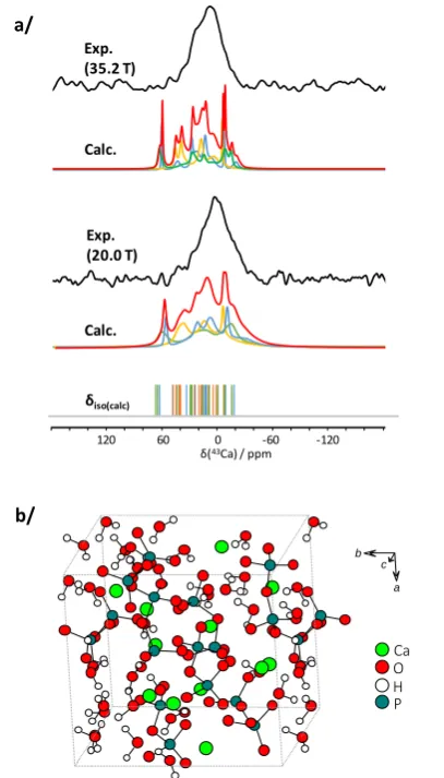
The natural abundance 43Ca MAS NMR spectrum of a-CPP was
recorded at 35.2 T (Figure 3a). Thanks to the very fast relaxation
of 43Ca in the sample, less than 4 hours were needed to record
the spectrum, which is all the more remarkable when
considering that the signal is 10 times broader in comparison
to those of the crystalline phases reported above.
In order to gain greater ‘chemical insight’ into the origin of these distributions, computational models of the amorphous compound were developed, using Monte Carlo (MC) based simulations with bond constraints followed by relaxation with Ab-Initio Molecular Dynamics (AIMD) simulations and geometry
optimization with DFT calculations (see supporting
information). It should be noted that in contrast to
non-hydrated calcium derived glasses,12 the commonly used
melt-quench approach for MD could not be used to model
amorphous Ca2P2O7.4H2O, as it would have led to condensed
phosphate chains with various lengths and would not have been able to tackle the presence of water molecules. Here, using combined MC, AIMD, and DFT calculations, three models of this amorphous phase with hydrated calcium pyrophosphate groups
were generated (each with 12 Ca sites, see Figure 3b).§ For each
model, calculated Pair Distribution Function (PDF) analyses were compared to the experimental data, showing that the agreement is reasonable (see supporting information, Figure S3).
Figure 3: a/ 43Ca MAS NMR spectra of amorphous-Ca
2P2O7.4H2O (a-CPP) at
20.0 and 35.2 T, together with the GIPAW-DFT 43Ca NMR calculations related
to the three AIMD derived models (sum in red, contributions of Models I, II and III in blue, orange and green respectively; calculated isotropic shifts are shown as vertical bars); b/ AIMD derived computational model of a-CPP (Model I) used for DFT calculations (see supporting information for further
details as each of three models provides 12 calcium sites).
The analysis of the Ca local environments in these models demonstrates that coordination numbers range between 4 and
9, that up to 5 water molecules can be bound to Ca2+, and that
the average Ca…O bond distances are between 2.3 and 2.6 Å (see Supporting Information, Table S4). These values are in agreement with the structural parameters of the crystalline calcium pyrophosphate phases (dihydrate and tetrahydrate). Coordination numbers range from 6 to 7 for calcium atoms in these structures and bond distances are between 2.257 and
2.668 Å.13,14,15 DFT calculations of the 43Ca NMR parameters
were then carried out, using the GIPAW-DFT approach (see Supporting Information for computational details). The range of variation of the calculated isotropic chemical shift (-16.1 ppm
< calcδiso(43Ca) < 64.3 ppm; average value of 21.6 ppm) and
quadrupolar coupling constants (1.2 MHz < |calcCQ(43Ca)| < 6.5
MHz ; average value of 3.4 MHz) is consistent with the experimental data (Figure 3a). More specifically, it is worth
noting that the calculated values calcδiso(43Ca) spread out across
the experimental lineshape obtained at 35.2 T, with more of the
a/
b/
a b
c
Ca O H P
δ(43Ca) / ppm
-120 -60 0 60 120
Exp. (35.2 T)
Calc.
Calc. Exp. (20.0 T)
[image:4.595.328.532.67.424.2]COMMUNICATION Journal Name
calculated values positioned where the signal has maximum intensity. Despite the lack of sufficient statistics at this stage, which are visible from the simulated sums of the models (Figure 3a, red curves), these AIMD models appear as the first realistic starting point for describing Ca local environments in the amorphous materials. In Figure S4, simulations including Czjzek distributions of quadrupolar parameters (Gaussian Isotropic
Model, GIM) as well as Gaussian distributions of 43Ca isotropic
chemical shifts16 are presented. At ultra-high magnetic field, it
is likely that simple distributions of isotropic chemical shifts are favoured.
All in all, the 1.5 GHz series connected hybrid instrument paves
new avenues for natural abundance 43Ca MAS NMR. Significant
gains in both resolution and sensitivity were demonstrated in the case of crystalline calcium pyrophosphate and oxalate phases. Most importantly, thanks to favorable relaxation
characteristics, the natural abundance 43Ca MAS NMR spectrum
of an amorphous hydrated calcium pyrophosphate was successfully obtained, which could be compared for the first time to computational models of this phase. This point is of crucial importance as amorphous precursors of calcium
carbonate,17 calcium oxalate18 and calcium (pyro)phosphates19
are meant to play a fundamental role in biomineralization processes, which have not been fully characterized so far
(notably regarding Ca environments). Moreover, such 43Ca NMR
experiments at ultra-high magnetic field may also provide complementary clues regarding the polyamorphism of
biomaterials.2,20
The French National Research Agency (ANR) is acknowledged for financial support ("CAPYROSIS" project – ANR-12-BS08-0022-01; "PYVERRES" project – ANR-16-CE19-0013). A portion of this work was performed at the National High Magnetic Field Laboratory, which is supported by the National Science Foundation Cooperative Agreement No. 1157490 & DMR-1644779, and the State of Florida. The UK 850 MHz solid‐state NMR Facility used in this research was funded by EPSRC and BBSRC (contract PR140003), as well as the University of Warwick including part funding through Birmingham Science City Advanced Materials Projects 1 and 2 supported by Advantage West Midlands (AWM) and the European Regional Development Fund (ERDF). NMR spectroscopic calculations were performed using HPC resources from GENCI-IDRIS (Grant 097535).
Conflicts of interest
There are no conflicts to declare.
Notes and references
§ Due to the high computational cost of the AIMD simulations (see supporting information for further details), only 3 models of the a-CPP phase have been generated so far.
1 C. M. Widdifield, Ann. Rep. NMR Spectrosc, 2017, 92, 227; D.
Laurencin and M.E. Smith, Prog. Nucl. Magn. Res. Spectrosc. 2013, 68, 1; D. Bryce, Dalton Trans 2010, 39, 8593.
2 P. Gras, A. Baker, C. Combes, C. Rey, S. Sarda, A. J. Wright, M.
E. Smith, J. V. Hanna, C. Gervais, D. Laurencin and C. Bonhomme, Acta Biomater. 2016, 31, 348.
3 H. Colas, L. Bonhomme-Coury, C. Coelho Diogo, F. Tielens, F.
Babonneau, C. Gervais, D. Bazin, D. Laurencin, M. E. Smith, J. V. Hanna, M. Daudon and C. Bonhomme, CrystEngComm, 2013, 15, 8840.
4 K. M. N. Burgess, F. A. Perras, I. L. Moudrakovski, Y. Xu, and
D. L. Bryce, Can. J. Chem. 2015, 93.
5 D. Laurencin, A. Wong, J. V. Hanna, R. Dupree and M. E.
Smith, J. Am. Chem. Soc. 2008, 130, 2412; A. Wong, D. Laurencin, R. Dupree and M. E. Smith, Sol. St. Nucl. Magn.
Res. 2009, 35, 32; D. Laurencin, C. Gervais, A. Wong, C.
Coelho, F. Mauri, D. Massiot, M. E. Smith and C. Bonhomme,
J. Am. Chem. Soc. 2009, 131, 13430.
6 D. Lee, C. Leroy, C. Crevant, L. Bonhomme-Coury, F.
Babonneau, D. Laurencin, C. Bonhomme and G. De Paëpe,
Nature Comm. 2017, 8, 14104.
7 Z. Gan, I. Hung, X. L. Wang, J. Paulino, G. Wu, I. M. Litvak, P.
L. Gor'kov, W. W. Brey, P. Lendi, J. L. Schiano, M. D. Bird, L. R. Dixon, J. Toth, G. S. Boebinger and T. A. Cross, J. Magn.
Reson. 2017, 284, 125.
8 C. Gervais, D. Laurencin, A. Wong, F. Pourpoint, J. Labram, B.
Woodward, A. P. Howes, K. J. Pike, R. Dupree, F. Mauri, C. Bonhomme and M. E. Smith, Chem. Phys. Lett. 2008, 464, 42.
9 C. J. Pickard and F. Mauri, Phys Rev B, 2001, 63, 245101.
10 A. Pedone, T. Charpentier and M. C. Menziania, Phys. Chem.
Chem. Phys. 2010, 12, 6054.
11 K. Shimoda, Y. Tobu, Y. Shimoikeda, T. Nemoto and K. Saito,
J. Magn. Reson. 2007, 186, 156; F. Angeli, M. Gaillard, P.
Jollivet and T. Charpentier, Chem. Phys. Lett. 2007, 440,324.
12 E. Gambuzzi, A. Pedone, M. C. Menziani, F. Angeli, P. Florian
and T. Charpentier, Solid St. Nucl. Magn. Reson. 2015, 68-69, 31.
13 N. S. Mandel, Acta Cryst. B 1975, 31, 1730.
14 T. Balić-Žunić, M. R. Christoffersen and J. Christoffersen, Acta
Cryst. B 2000, 56, 953.
15 P. Gras, C. Rey, G. André, C. Charvillat, S. Sarda and C.
Combes, Acta Cryst. B 2016, 72, 96.
16 D.R. Neuville, L. Cormier and D. Massiot, Geochim.
Cosmochim. Acta 2004, 68, 5071.
17 Y. Wang, S. Von Euw, F.M. Fernandes, S. Cassaignon, M.
Selmane, G. Laurent, G. Pehau-Arnaudet, C. Coelho, L. Bonhomme-Coury, M.-M. Giraud-Guille, F. Babonneau, T. Azaïs, N. Nassif, Nature Mater., 2013, 12, 1144; S. Weiner, Y. Levi-Kalisman, S. Raz and L. Addadi, Connect Tissue Res. 2003, 44, 24.
18 E. Ruiz-Agudo, A. Burgos-Cara, C. Ruiz-Agudo, A.
Ibanez-Velasco, H. Cölfen and C. Rodriguez-Navarro, Nature
Commun. 2017, 8, article 768.
19 K. Ley-Ngardigal, C. Combes, S. Teychene, C .Bonhomme, C.
Coelho-Diogo, P. Gras, C. Rey and B. Biscans, Cryst. Growth &
Design, 2017, 17, 37.
20 J.H.E. Cartwright, A.G. Checa, J.D. Gale, D. Gebauer and C.I.