During locust flight, the modulation of motor patterns underlying flight behaviour is mediated by a set of sense organs (Wendler, 1974). In particular, the wing hinge stretch receptors (Kutsch, 1974; Pearson et al., 1983), the antennae (Gewecke, 1975), the system of frontal wind-sensitive hairs (Gewecke, 1975; Horsmann et al., 1983) and the tegula organs (e.g. Kien and Altman, 1979; Büschges and Pearson, 1991) have been studied. Afferent information from such proprioceptive sensory organs participates in flight pattern generation: studies of tethered flying animals have demonstrated that information from the wing-associated sense organs modulates muscle activity patterns (Neumann et al., 1983; Möhl, 1985; Wolf and Pearson, 1988).
The tegulae are complex sense organs on the anterior base of each wing; they consist of a knob-shaped hair field (the cupula) and internal chordotonal sensilla (Kutsch et al., 1980). The tegulae are excited in parallel with the downstroke movement of the wings (Neumann et al., 1983). Tegula afferents project onto identified interneurones related to the central pattern generator (Pearson and Wolf, 1988). Phasic input from the the tegulae was found to be partly responsible for the initiation of elevator activity and, therefore, for the timing of the elevator muscles within the wingbeat cycle (Wolf and Pearson, 1988). The tegula organs of the fore- and hindwings are not equally important in generating flight patterns: partial deafferentation experiments
have shown that the forewing organs are of minor, if any, importance for the neural generation of basic elevator–depressor flight patterns (Büschges and Pearson, 1991). The flight system can recover from the loss of wing proprioceptive sense organs (Kutsch, 1974; Büschges et al., 1992a,b; Gee and Robertson, 1996), suggesting synaptic plasticity within the nervous system (Wolf and Büschges, 1997).
Although detailed information is available about the interaction between tegula afferent input and the centrally generated flight pattern, there are few reports concerning tegula function. In experiments involving tethered flying locusts, Wolf (1993) pointed out that tegula input may be involved in the control wing movement and in wing-related aerodynamic force production. Investigating the characteristics of free flight in locusts recovering from tegula ablation, Gee and Robertson (1998) reported an overall deficit in free flight ability following 3–4 weeks of recovery from hindwing tegula ablation and impairment of free flight ability in locusts recovering from forewing tegula removal. These observations are inconsistent with previous studies of tethered flight, which have demonstrated complete recovery of the flight motor pattern following ablation of the hindwing tegulae (Büschges and Pearson, 1991) and no significant effects of the removal of the forewing tegulae on the flight motor pattern (Büschges et al., 1992a,b).
Printed in Great Britain © The Company of Biologists Limited 1999 JEB1824
Tegulae are complex proprioceptors at the wing base of locusts. Deafferentation of the tegulae causes a lack of specific phasic information related to the wing downstroke and the timing of the upstroke. Employing telemetry during free flight of the locust Schistocerca gregaria, we investigated the consequences of tegula ablation on free flight parameters including motor patterns (wingbeat frequency and the relationship between the activation of flight muscle antagonists), free flight speed and aerodynamic output. We investigated the role of the tegula pairs of both wings on the motor pattern generated in free-flying locusts. We show that the tegula organs are not essential for generating the motor patterns necessary for
free flight. However, they are required for increasing the motor output to give additional effective lifting power during adaptive behaviour. We also investigated long-term changes in the free flight parameters after tegula ablation. The recovery of the adult flight system revealed in the present study suggests that there is adaptation to the loss of proprioceptive information; this argues for a full functional and behavioural recovery of the flight system of the locust under closed-loop conditions.
Key words: tegula, telemetry, locust, Schistocerca gregaria, free flight, motor pattern, aerodynamic output.
Summary
Introduction
TEGULA FUNCTION DURING FREE LOCUST FLIGHT IN RELATION TO MOTOR
PATTERN, FLIGHT SPEED AND AERODYNAMIC OUTPUT
HANNO FISCHER* ANDECKHARD EBERT
Fakultät für Biologie, 78457 Universität Konstanz, Germany
*Author for correspondence and present address: Zoologisches Institut der Universität zu Köln, Weyertal 119, 50923 Köln, Germany (e-mail: [email protected])
In seeking an explanation for these different findings, it is necessary to examine the behavioural consequences of the changed motor output that follows sensory deafferentation during locust free flight. Among the questions to be answered are the following. (a) Does the decrease in the wingbeat frequency after tegula removal (e.g. Büschges and Pearson, 1991) cause a lowering of motor output to levels inadequate for maintaining the general free-flight capability? (b) Is the phasic information mediated by the tegula involved in, or even essential for, the regulation of behavioural parameters such as the free flight speed? (c) Does the recovery of the central flight patterns following tegula ablation accompany a recovery of the behavioural parameters such as speed and lift generation during free flight?
In the present study, therefore, we focus on changes in the behavioural parameters of free-flying locusts following tegula ablation. Telemetric acquisition of electromyographs (e.g. Kutsch et al., 1993) by a modified low-weight device enables analysis of motor patterns during free flight of intact and sensory-ablated locusts. This approach is used for a simultaneous investigation of the motor output of the locust flight system and related behavioural parameters, such as speed and aerodynamic force production during free flight. We determined the motor and behavioural output during free flight and evaluated the significance of the tegula organs in generating the motor pattern underlying the capability for free flight. In addition, we investigated the recovery of free flight behaviour following removal of either the forewing or the hindwing tegulae and after ablation of all tegula organs.
Materials and methods
Animals and flight conditions
All tests were carried out using female locusts Schistocerca
gregaria Forskål. The animals were bred at 42 °C and 70 %
relative humidity in crowded populations of both sexes with a 12 h:12 h light:dark cycle. Maturation occurred 18 days after final ecdysis (determined by observations of copulating pairs). Individual flight sequences were investigated in a room measuring 10 m×5.5 m×2.2 m. The room temperature could be adjusted within the range 18–40 °C. Unless stated otherwise, the experiments were performed at 34 °C. The first flight activity of each individual was induced by horizontal acceleration (v0≈2.0 m s−1, where v0 is the initial flight velocity), which was immediately followed by spontaneous flight sequences by the animal. Flight duration varied between 5 and 30 s per flight sequence. In each type of experiment, each individual was restricted to three induced and five spontaneous flight sequences (summed flight time per individual averaged 1.5 min). Wherever possible, spontaneous flight sequences have been evaluated.
Telemetric device for recording electromyographs during free flight of locusts
For telemetric acquisition of electromyographs (EMGs), the
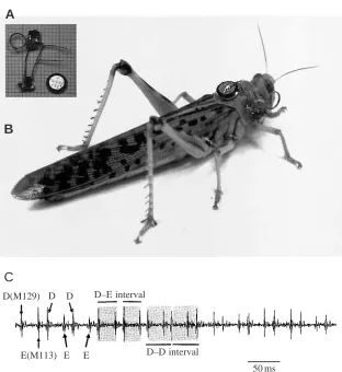
one-channel transmitter design of Kutsch et al. (1993) has been modified: the transmitter mass has been reduced to 0.3 g including the power supply using 0605 and 0805 SMD components (Bürklin, Germany) and a lithium battery (2.2 V, 3.3 mA; Renata, Switzerland). The transmitter design and its attachment to the pronotum are shown in Fig. 1A,B. The transmitter-specific VHF carriers were adjusted in the range from 128 to 145 MHz. An HB9CV aerial (32 dB amplification) was used for signal detection. A wide-band receiver (consisting of Harris modules, UK) was used to demodulate the signals.
Electromyograph wires (Manganin, Isabellenhuette Dillenburg, Germany; 50µm in diameter, insulated except at the tip) were inserted into one of the hindwing depressor (D) muscles, M129, and one of the hindwing elevator (E) muscles, M113 (nomenclature after Snodgrass, 1929), both electrodes being connected to the preamplifier input. A further electrode wire was inserted in the abdominal haemocoel as a reference. Typical telemetric EMGs from these antagonistic muscles during free flight are shown in Fig. 1C.
Tegula ablation and post-experimental control
To remove the hindwing tegulae, the animals were taped ventral side down onto a cork platform with the forewings unfolded. The exposed hindwing tegulae were ablated using forceps with sharpened spoon-shaped tips specially designed for tegula ablations (see also Wolf, 1993). Ablation of the forewing tegula organs required turning the animal sideways and enlarging the junctions between the pro- and mesothorax. Manipulations were carried out under a dissecting microscope to avoid damage to the wing base or other related structures. Animals from control groups were treated identically except that the tegula organs were not removed (sham-operation). Removal of the cupula in the fore- and hindwing cut N1C1a, but N1C1b remained intact in all the experimental animals (for nerve nomenclature, see Campbell, 1961; Kien and Altman, 1979).
In addition, the successful removal of the forewing tegulae was checked after the free-flight experiments by recording extracellularly from nerve N1C1 (Fig. 2). The animals were fixed to a holder by their ventral surface, and the episternal cuticle was opened by cutting a narrow window immediately below the first basalar sclerite. Recordings from N1C1 were made proximal to branches N1C1a and N1C1b using a silver hook electrode (30µm in diameter). Stimulation of the nerve was carried out by passive movement of the forewing. In the case of the hindwing tegulae, post mortem checks were carried out to confirm that N1C1a had been cut and that N1C1b was intact. We did not record from the metathoracic N1C1 since it was not possible to evoke adequate stimulation by moving the complete hindwing, which has a soft morphology.
Recording technique and analysis of free flight in locusts
frequency 50 Hz, shuttering time 1 ms, frame interval 20 ms). The EMGs were displayed on an oscilloscope (Tektronics D11) and stored on tape (Racal Store 4DS). The oscilloscope screen was filmed using a second video camera (Revue Video 8; frame frequency 50 Hz, shuttering time 1 ms), and the synchronous video outputs of both cameras were mixed on-line (Panasonic Digital Mixer WJ-AVE5) and recorded on a video master tape.
Free flight parameters
Before the experiments, the mass and body length of each individual were determined. Free flight parameters were acquired by frame-by-frame analysis of the continuous displacement of the body silhouettes stored on the video master tape (Fischer, 1998). The centre of gravity (at approximately 50 % of body length; Weis-Fogh, 1956b; Fischer, 1994) was chosen as a reference point on the body. The parameters obtained were, if necessary, transformed with reference to the parallax of the video recordings (see also Baker and Cooter, 1979). All parameters are considered as ‘instantaneous’ flight parameters since they were constant within the frame interval of 20 ms.
Parameters describing the motor patterns during free flight
Instantaneous D–D (i.e. cycle length), D–E and E–D intervals (see Fig. 1C) (all measured in ms) were determined
from simultaneous telemetric EMGs of M129 (D) and M113 (E). Instantaneous wingbeat frequencies (Hz) were deduced from the D–D intervals. The instantaneous elevator phase (φ) within the depressor cycle was calculated from φ=(D–E)/(D–D) and describes the relationship between the activation of the two antagonists during a wingbeat cycle.
Parameters monitoring behaviour during free flight
The following behavioural parameters were investigated during free translatory movement of an animal: (a) ascent angle
αto the horizontal (degrees) (i.e. vertical flight path); and (b) flight speed v (m s−1). Here, flight capability is defined as the
ability to maintain at least continuous level flight (i.e. α⭓0 ° within the evaluated flight sequences), indicating that the animal is able to generate sufficient lifting power to support its body mass.
Parameters monitoring the aerodynamic output of the flight system during free flight
Unlike the tethered flight approach, which allows direct ‘weighing’ of vertical and horizontal force components (e.g. Weis-Fogh, 1956b; Kutsch and Gewecke, 1979; Wolf, 1993), it is not possible to acquire measurements of aerodynamic forces during free flight. During free flight, the aerodynamic output of the flight system is dependent on the ascent angle α
A
B
C
D(M129) D D
E(M113) E E
D–E interval
D–D interval
50 ms Fig. 1. (A) Advanced telemetric device for
recording electromyograms (EMGs) of locusts during free flight (mass 0.3 g, battery included) using SMD components (scale: small squares 1 mm2). For basic circuitry and a detailed
[image:3.609.251.563.67.407.2]and can be monitored as the instantaneous effective lifting power PEL(W), calculated as:
PEL=m*gvsinα,
where m* is the body mass of the animal in kg, transmitter mass included, g is the gravitational constant (g=9.81 m s−2), v
is the instantaneous flight speed (m s−1) and α is the
instantaneous ascent angle to the horizontal (degrees). PELis
the instantaneous vertical power component additionally generated by the animal when changing from level flight [α=0 °; PEL(0 °)=0 W] to ascending flight at a given value of α.
Fixing the transmitter device to the pronotum increased the original body mass m of the animals (ranging from 2.8 to 3.3 g for the adult females used here) by 8–12 %. Adult females of
S. gregaria are able to carry approximately 30 % additional
body mass without losing the capability for horizontal flight (indicating the ability to support their body weight during flight, see Fig. 1A in Fischer et al., 1997). There was no significant difference between the free flight parameters (speed, body angle, ascent angle, PELmaxand fWB, where fWB
is wingbeat frequency) for freely flying adult female S.
gregaria either unloaded or loaded with a 0.3 g transmitter (for
details, see Fischer, 1998, p. 70). Although the body mass m*
(which, on average, increased by less than 10 % compared with
m) also contributes to PEL, values of PELare mainly dependent
on the observed flight speed (v) and the ascent angle (α).
Statistics
Independent samples of the animal populations were obtained by replacing the animals into the specific stem population after the measurements. In each specific set of measurements, the data from all animals were pooled. All sets of data were tested for normal distribution (χ2; P<0.01) and for
homogeneity (by analysis of variance, ANOVA; P<0.05). Comparisons of two homogeneous sets of data were performed using Student’s t-test; where inhomogeneity occurred, the mt-test of Dixon and Massey (1969) was used. For non-parametric comparisons, the Mann–Whitney U-test was applied. Comparisons of multiple data sets were made using the Kruskal–Wallis H-test. Regression analysis was performed using the least-squares difference method (LSD; e.g. Sachs, 1978). The significance of linear and non-linear regressions was tested using the coefficient of determination r2.
Mean phase values φ of the elevator activity within the depressor cycle were calculated using circular statistics (see Batschelet, 1981). Mean phase values are given as φ ± mean angular deviation, with r describing the mean vector. Circular
Intact
A
Up
Down
1 s
Wing position N1C1
N1C1b cut
B
1 s
Wing position N1C1
Tegula ablated (N1C1a cut)
C
1 s
[image:4.609.253.566.83.427.2]Wing position N1C1 Fig. 2. Extracellular recordings of the mesothoracic
two-sample comparison was performed using the Watson–Williams test. Results are presented as means ± S.D. (N).
Results
Free flight motor pattern after partial and complete tegula ablation
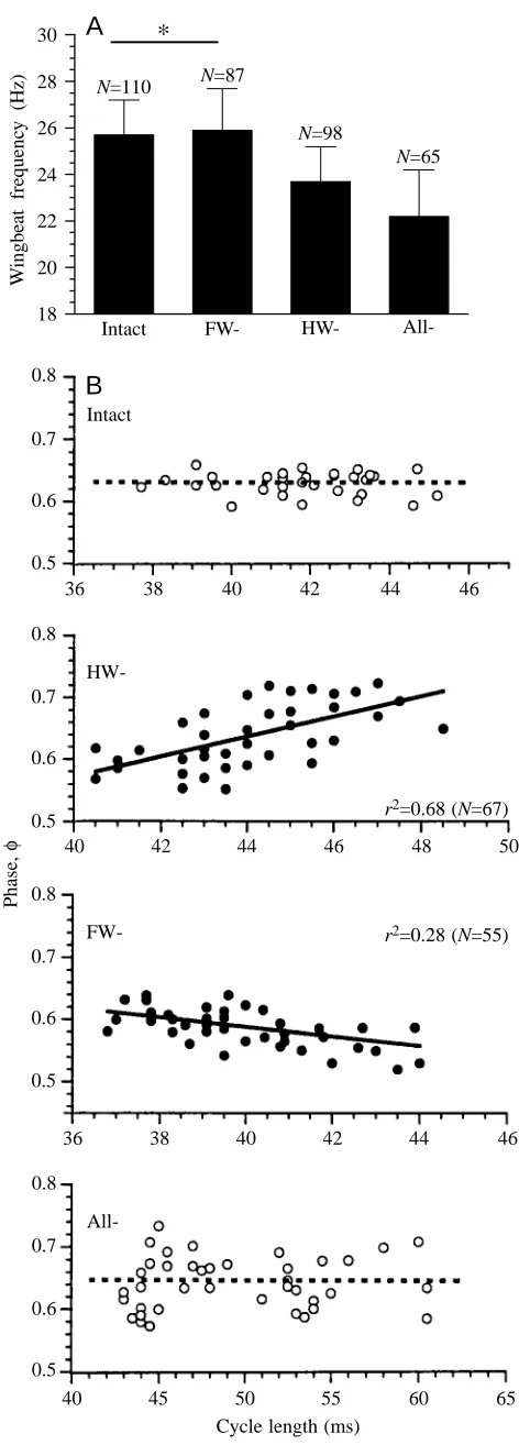
Effects on wingbeat frequency
The instantaneous wingbeat frequencies (fWB) at 34 °C of 12
animals were determined by analyzing the telemetric EMGs. The mean fWB after ablation of the specific tegula organs are
given in Fig. 3A. After removal of the hindwing tegulae, fWB
decreased significantly from 25.7±1.5 Hz (mean ±S.D.; N=110) to 23.7±1.5 Hz (N=98). The removal of all tegulae caused a further decrease in fWBto 22.2±2.0 Hz (N=65). After removal
of the forewing tegulae, fWBdid not differ significantly from
the control value (25.9±1.8 Hz, N=87). The capability for free flight was maintained for all these animals.
Effects on phase relationships of muscle antagonists
The phase (φ) relationships during free flight after partial and complete tegula ablation were determined by analysing the telemetric EMGs of the metathoracic antagonists M129 (D) and M113 (E) (Fig. 3B). Ablation of the hindwing tegulae caused a significant increase in the mean D–E interval from 24.2±2.5 ms (N=63) to 28.5±3.0 ms (N=45, P<0.05). The D–E interval increased significantly to 29.6±4.0 ms (P<0.05) following removal of all tegulae. No significant change in mean D–E interval was observed after ablation of the forewing tegulae. In controls, there was no significant dependence of the instantaneous elevator phase on the actual cycle length (i.e. D–D) (P>0.05). After ablation of the hindwing tegulae, φ became positively correlated with cycle length (P<0.05). This phase shift corresponds to an increase in the D–E interval and indicates a delay in elevator activation during the wingbeat cycle when cycle length is increased. After removal of the
18 20 22 24 26 28 30
Intact FW- HW-
All-N=110 N=87
N=98
N=65
*
W
ingbeat frequenc
y (Hz)
B
A
0.8
0.7
0.6
0.5
Phase,
φ
36 38 40 42
Intact
0.8
0.7
0.6
0.5
HW-r2=0.68 (N=67)
0.8
0.7
0.6
0.5
FW- r2=0.28 (N=55)
0.8
0.7
0.6
0.5
All-40 42 44 46
44 46
36 38 40 42
48 50
46 44
40 45 50 55
Cycle length (ms)
60 65
[image:5.609.324.560.89.746.2]remaining forewing tegulae, the elevator phase φ was found not to be significantly correlated with the actual cycle length (P>0.05). The flight muscle pattern after ablation of the forewing tegulae showed a slight but significant negative correlation between φ and cycle length (P<0.01), which corresponds to a reduction in the D–E interval and indicates an earlier activation of the elevator when cycle length is increased. No significant difference (P>0.05) in the mean phase relationship of the antagonists was found between controls (φ=0.629±0.018, r=0.994; N=75) and after ablation of all tegula organs (φ=0.636±0.0394, r=0.968; N=45).
Instantaneous effects of tegula ablation on free flight behaviour
Free flight speed
Instantaneous flight speeds of two populations (each with 12 individuals) were determined after specific tegula ablation (Fig. 4). After control flights (sensory input intact) of both populations, the hindwing tegulae of one population were ablated while the forewing tegulae of the other group were ablated. After flight speed had been measured again, the remaining tegulae were removed in both populations to give complete ablation.
The mean flight speed of the intact animals (4.1±0.5 m s−1,
N=80) did not change significantly (P>0.05) after removal of
the forewing tegulae (4.0±0.6 m s−1; N=77), but subsequent
removal of the hindwing tegulae resulted in a significant decrease in the mean flight speed to 3.2±0.4 m s−1 (N=101,
P<0.05). A slightly but significantly reduced flight speed
(4.2±0.6 m s−1, N=50; intact mean 4.5±0.6 m s−1, N=64) was
observed when only the hindwing tegulae were removed
(P<0.05). In this population, removal of the remaining forewing tegulae caused a significant decrease in the mean flight speed (3.4±0.4 m s−1, N=78). Both populations showed
comparable mean flight speeds after deafferentation of all tegulae (means not significantly different, P>0.05). Flight capability was maintained in all these individuals.
Aerodynamic output
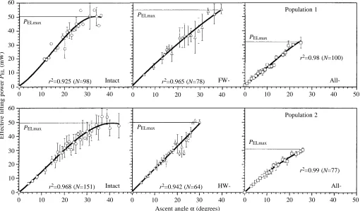
The non-linear characteristics of PELas a function of α(r2
significantly different from zero, P<0.01) for the populations investigated are shown in Fig. 5. Maximum effective lifting power PELmax at α=αmax was calculated from the equations
used to fit the data. The values obtained for PELmax after
ablation of the forewing tegulae (54.8 mW, population 1) or the hindwing organs (49.9 mW, population 2) did not differ significantly from the control values (50.4 mW for population 1; 49.8 mW for population 2). After ablation of all tegula organs, a similar decrease in PELmax was seen in both
populations (to 32.2 mW for population 1; to 30.9 mW for population 2). These findings indicate that even after the removal of all tegula organs (a) the aerodynamic output of the flight system is sufficient to maintain flight capability and to enable ascending flight; and (b) the observed range of ascent angles (i.e. the vertical flight paths) is limited to lower values than those for controls.
Long-term changes in free flight parameters after tegula ablation
Flight speed
Adult individuals (N=30) from one colony were investigated. Three days after final ecdysis, the hindwing tegula organs were removed from 10 animals, and all tegulae were ablated in a further 10 individuals. Ten animals were left intact to serve as controls. Sampling was first carried out 1 day after ablation; then, starting 9 days after ablation, consecutive measurements were perfomed at 3 day intervals until 18 days after ablation. The mean flight speeds of the intact and deafferented groups are shown in Fig. 6. During the period of observation, the speed of the sensory-intact animals increased significantly from 3.2±0.6 m s−1to 5.1±0.6 m s−1(mean ±S.D.;
P<0.05). For individuals without hindwing tegulae, flight
speeds over the first 15 days were significantly lower than those of the sensory-intact group (P<0.05); however, no significant difference was found on the eighteenth day of observation. The flight speed following hindwing tegula ablation increased significantly from 2.7±0.4 m s−1on day 1 to
4.9±0.7 m s−1 (P<0.05) on day 18. No significant increase in
flight speed was observed over 18 days (P>0.05) in individuals without any tegulae, and by the end of the observation period the flight speed was only 58 % of that of sensory-intact locusts. All individuals maintained their capability for free flight.
Aerodynamic output
Long-term changes in PELmaxhave been investigated (data
[image:6.609.52.278.462.633.2]not shown) and, depending on maturation, a general increase in this output was found in intact animals with values of PELmax Fig. 4. Instantaneous effects of tegula removal on the mean flight
speed during free locust flight (at 34 °C) investigated in two populations with a different test program (each with 12 adult females aged 18 days). An asterisk indicates that no significant decrease in flight speed was observed (P>0.05) following ablation of the forewing tegulae (FW-). Removal of all tegulae (All-) caused a significant decrease in flight speed in both populations. The flight capability of all the animals was maintained even after ablation of all tegulae. Values are means +S.D. I, intact animals.
3 3.5 4 4.5 5 5.5
Population 1 Population 2
Flight speed (m s
-1)
I
N=80 N=77
N=101
N=64
N=50
N=78
*
All-increasing from 57 mW on day 9 to 78 mW at 18 days. For individuals with all tegulae removed, no significant changes in the output were observed (PELmaxat 9 days, 41 mW; at 18 days,
46 mW, which is approximately 60 % of the intact value). After removal of the hindwing tegula organs, PELmaxwas 50 mW on
the ninth day of observation and this increased to 71 mW on day 18. Both values did not differ significantly from the values found in the control population.
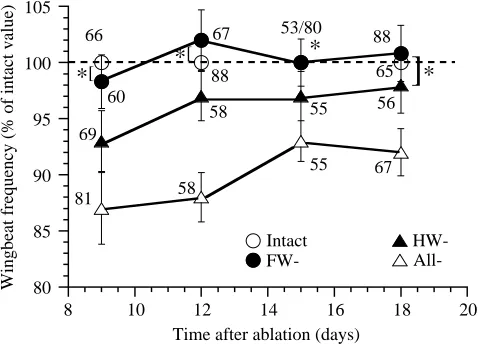
Wingbeat frequency
The experiments described here were carried out on a population of 80 females from one colony. This colony was initially divided into four groups, each containing 20 animals. Three days after final ecdysis, 20 individuals were deafferented by removal of either the fore- or the hindwing tegulae; in a further 20 individuals, all tegulae were ablated. Twenty animals served as controls. Five animals in each group were first tested 9 days after ablation, and five other individuals from each of these groups were tested every third day. The mean values of fWB were determined from telemetric EMGs of
M129. In all groups, fWB was observed to change with
maturation: fWB increased significantly from 23.9±1.6 Hz
(N=66, 9 days) to 25.9±1.8 Hz (N=88, 18 days, P<0.05) in controls, and from 20.8±1.9 Hz (N=81) to 24.5±1.4 Hz (N=67; values mean ± S.E.M., P<0.05) in animals with all tegulae
0 10 20 30
Ascent angle α(degrees) 60
50
10
0
Effective lifting power
PEL
(mW)
40
30
20
PELmax
40 r2=0.925 (N=98) Intact
0 10 20 30
PELmax
40 r2=0.965 (N=78)
FW-0 10 20 30
PELmax
40 50
r2=0.98 (N=100)
All-Population 1
0 10 20 30
60
50
10
0 40
30
20
PELmax
40 r2=0.968 (N=151) Intact
0 10 20 30
PELmax
40 r2=0.942 (N=64)
HW-0 10 20 30
PELmax
40 50
r2=0.99 (N=77)
[image:7.609.54.564.74.373.2]All-Population 2
Fig. 5. Effective lifting power PELin relation to the ascent angle αobserved in intact and tegula-ablated animals (two populations; see Fig. 4). When different values were obtained for a given α, the mean value ± S.D. is plotted. Maximum effective lifting power PELmax was either calculated from the characteristics of the relationship between αand PELor from the maximum observed PELat α=αmax. All r2values shown are significant (P<0.01).
Fig. 6. Changes in the free flight speed (mean ± S.D.) in intact animals (open circles), after ablation (A) of the hindwing organs (filled circles) or after removal of all tegulae (open triangles; each group contained 10 individuals). Deafferentation was carried out 3 days after final ecdysis, and first sampling was at 1 day after ablation (i.e. at 4 days of adult age). Each mean consists of N samples of all 10 individuals within each group, with the value of N given beside the data points. An asterisk indicates that means do not differ significantly (P>0.05). For details, see text.
2 2.5 3 3.5 4 4.5 5 5.5 6
0 A
5 10 15 20
69
73
90 65
63
38 55
98 67
66 63
65 66
66
All- HW-Intact
Time after ablation (days)
Flight speed (m s
-1)
*
*
[image:7.609.59.288.459.633.2]ablated. These changes in fWB(as a percentage of that of the
intact group) are plotted in Fig. 7. Over the period of observation, the mean fWB of individuals deafferented by
forewing tegula removal was not significantly different from that of the controls (P>0.05). For the group with the hindwing tegulae removed, the mean fWB was significantly lower than
that of controls over the first 15 days, but no significant difference was found on the eighteenth day of observation (P>0.05). The mean fWB of individuals after removal of all
tegulae was always significantly different (between 88 % and 93 %) from the value found in controls. Free flight capability was retained by these locusts throughout the period of observation.
‘Minimum’ wingbeat frequency required for free flight
To evaluate the role of the tegula organs in the generation of the motor pattern that underlies free flight (i.e. the motor output adequate to support the body weight), we first determined the ‘minimum’ fWB needed for free horizontal
flight (α=0 °).
During continuous level flight (α=0 °), fWBwas quantified
by telemetry of M129 activity at three different environmental temperatures (18, 27 and 34 °C). In a population of nine intact individuals, no ascents where α⭓0 ° were observed at a temperature lower than 18 °C. In most animals, no flight activity was documented at all below 18 °C. Continuous level flight (α=0 °) was first observed at 18 °C (but only in three of the nine animals). These animals exhibited a mean fWB of
17.3±1.9 Hz (N=45). For animals with a lower fWB, only
descents were observed at 18 °C. We conclude, therefore, that
the minimum fWB(i.e. the minimum motor output) required for
free flight in adult female S. gregaria is approximately 17 Hz. At 27 °C, six of the nine animals showed level flight, and the mean fWBwas determined to be 23.3±1.3 Hz (N=78). All
nine animals were capable of free flight at 34 °C, with a mean
fWB of 25.6±1.5 Hz (N=103). The mean fWB after removal of
all tegula organs was 22.9±1.8 Hz (N=77) at 34 °C. All the animals retained their capability for free flight after removal of all tegulae.
We conclude from these observations that, even after the loss of all afferent information from the tegulae, the capability for free flight (at 34 °C) is maintained because the wingbeat frequency is still approximately 5 Hz above the minimum value generally required to support the body weight.
Discussion
Significance of the tegulae for motor pattern generation during free flight
The tegula organs are wing proprioceptors that are considered to be necessary for the generation of a functional flight motor pattern (Wolf, 1993). It has been shown in tethered flying preparations that ablating the hindwing tegulae increases the time interval between activation of the wing depressor and elevator muscles, consequently decreasing the wingbeat frequency (Wolf and Pearson, 1988; Büschges and Pearson, 1991). Stimulation of the hindwing tegula afferents is able to reset the flight rhythm at any phase of the wingbeat cycle by depolarizing the elevator motoneurones (i.e. finishing the downstroke; Wolf, 1993). Since tegula activity is correlated with the downstroke (see Fig. 2), this indicates that the tegula organs have an important role in determining the timing of activity in the elevator muscles within the wingbeat cycle.
The present study on freely flying animals not only corroborates previous observations concerning the effects of tegula deafferentation in fixed or restrained preparations (Büschges and Pearson, 1991; Büschges et al., 1992a,b) but also provides further evidence of how afferent input from the tegulae may be involved in the timing of elevator activity within the wingbeat cycle (Fig. 3). In individuals with ablated hindwing tegulae, phase values for elevator activity depended on the actual wingbeat frequency/cycle length. Under free-flight conditions, and in contrast to the apparent lack of functional significance of the forewing tegulae in previous studies of tethered flight (Büschges et al., 1992a,b), a significant correlation was also found after ablation of the forewing tegulae, although the effect was smaller than when the hindwing tegulae were removed.
Phase did not depend on wingbeat frequency/cycle length in sensory-intact animals or in animals in which all tegulae had been ablated. This indicates that the tegulae of both wings may synchronously participate in elevator timing, probably having an antagonistic influence since phase shifts in elevator activity are observed when only one tegula pair is lacking but not when both pairs are intact or have been removed. In addition, hindwing muscles can be phasically influenced by forewing
80 85 90 95 100 105
8 10 12 14 16 18 20
Intact
FW- HW-
All-Wingbeat frequency (% of intact value)
Time after ablation (days) 66
60
67
88
53/80
88
65
*
[*
[
*
56
]
*
69
58 55
81 58
[image:8.609.48.287.73.245.2]55 67
tegula nerve stimulation (Neumann et al., 1983). This supports our finding that, in the absence of the hindwing tegulae, the forewing tegulae can influence the motor patterns phasically.
No significant phase shift in elevator activity was observed after removal of all tegulae, as has been reported for tethered preparations (Büschges and Pearson, 1991). This may be due either to sensory open loops caused by tethering (e.g. Gewecke, 1975) or to the generally lower wingbeat frequencies that occur during tethered flight (Baker et al., 1981) allowing discrimination of such effects in more detail.
Role of the tegulae in the generation of aerodynamic output
A major result of the present investigation was the finding that, even after the removal of all tegulae, the locust is able to maintain free flight. Apparently, in spite of the absence of any afferent information from the tegulae, the flight system is able to generate aerodynamic output sufficient to support the body weight. This might be explained by the observation that the wingbeat frequencies of animals capable of free flight (irrespective of whether they are intact or have undergone tegula ablation) were not below the value determined as the probable ‘minimum fWB’ enabling level
flight (i.e. sufficient aerodynamic output to support the body weight). However, other sense organs (e.g. frontal wind-sensitive hairs, stretch receptors) are still intact in animals with ablated tegulae, and it is probable that afferent information mediated by such organs is sufficient for the generation of an adequate output.
Only if all tegulae are removed is there a decrease in the maximum effective lifting power (Fig. 5). This is in agreement with a reduction in the range of observed ascent angle, α. Since PELis the aerodynamic output that has to be
generated by all four wings when changing from level flight to climbing flight, we conclude from this observation that tegula ablation does not affect the basic aerodynamic output of the flight system that is required for free flight capability (even after removal of all tegulae, climbing flight was observed). The ablation of all tegula organs causes a reduction in the wingbeat frequency, and this may result in a decrease in the effective lifting power that can be additionally generated at maximum wingbeat frequencies. In tethered preparations, tegula removal seems to change the angular setting of the wing, and this is probably responsible for the decrease in the observed net lift generated (Wolf, 1993; but see Gee and Robertson, 1998).
Significance of the tegula for the regulation of flight speed
In the present study, the observed free flight speed of sensory-intact S. gregaria ranged from 3 to 6 m s−1. This is
in agreement with field studies reporting comparable flight speeds in S. gregaria (Waloff, 1972) and Locusta migratoria (Baker et al., 1981). The removal of the forewing tegulae had no significant effect on the mean flight speed. Ablation of the hindwing tegulae caused a shift in the flight speed towards significantly lower values, but this decrease was smaller than the reduction in speed following the deafferentation of all
tegula organs. The results obtained using the experimental approach described here, however, do not allow us to conclude that the tegula organs participate in the instantaneous regulation of the actual flight speed since, for the individuals investigated here, a wide range of flight speeds was still observed after ablation of all tegula organs. Therefore, the presence of tegula afferent information seems to be required to generate the motor output that enables maximum flying speeds, but it is apparently not obligatory for the thrust production that underlies flight capability (Zarnack, 1999).
Recovery of the motor pattern and behavioural parameters after tegula ablation
During previous studies of tethered flight, the capacity of the locust flight system to recover from tegula deafferentation was investigated. Following hindwing tegula removal, the flight motor patterns returned progressively towards normal in both mature (Büschges and Pearson, 1991; Büschges et al., 1992a,b) and immature locusts (Gee and Robertson, 1996). This recovery of the motor patterns was also observed in the present study (Fig. 7). This reorganization of sensory regulation has to be considered as functional because it corresponds to a full recovery of the free flight parameters: following hindwing tegula ablation, free flight speed (Fig. 6) and aerodynamic output returned to normal values within 2 weeks of ablation. No recovery in flight speed (Fig. 6) and aerodynamic output was found following ablation of all the tegulae. This also reflects the inability of motor patterns to recover after the removal of all tegulae, as previously reported for tethered preparations (Büschges et al., 1992a,b).
Our results are inconsistent with some recent findings: in tethered flying locusts, it was observed that recovery was less extensive when monitored by stroboscopic measurements of wingbeat frequency than when monitored using electromyography (Gee and Robertson, 1996). Investigating the free-flight ability (but not simultaneously the motor pattern) of locusts recovering from partial deafferentation, Gee and Robertson (1998) observed an overall deficit, which was independent of recovery from forewing or hindwing tegula ablation. In our opinion, this inconsistency is mainly caused by the large difference in the environmental temperatures at which the two studies were performed (∆T=10 °C). Gee and
Robertson (1998) state that their experiments were carried out on ‘a warm day (approximately 25 °C)’ compared with our investigations at 34 °C. They report that a high proportion of control locusts were nonflyers. As shown in the present study, flight activity is strongly affected by the environmental temperature (see also Weis-Fogh, 1956a; Waloff, 1972). For the majority of S. gregaria investigated here, free flight activity occurred at temperatures above 27 °C. We do not believe that the inconsistent findings are due to the fact that Gee and Robertson (1998) investigated L. migratoria rather than S.
gregaria. Comparing the free flight behaviour of both of these
pattern or the free flight parameters in either species (for details, see Fischer, 1998, pp. 72–74). Furthermore, tegula removal affects the wingbeat frequency in a similar manner for tethered flying L. migratoria (e.g. Büschges and Pearson, 1991) and for freely flying S. gregaria (see Fig. 3A).
In conclusion, it is clear that phasic information mediated by tegula afferents modulates the motor pattern during free flight in a manner similar to that previously reported from fixed or restrained preparations (Büschges and Pearson, 1991; Büschges et al., 1992a,b). Furthermore, the present state of knowledge on pattern generation during free flight is insufficient to conclude that there is an altered ‘basic’ mechanism of motor pattern generation during tethered flight. In contrast to previous assumptions and observations, the capability for free flight is maintained following tegula removal. This indicates that, even in the absence of tegula-specific information, motor patterns are generated that must be considered as ‘functional’ in the sense of being sufficient to maintain the capability for free flight. During free flight, the ultimate role of the tegula organs is apparently less important than predicted from tethered studies (Büschges and Pearson, 1991; Wolf, 1993). However, phasic information from a complete set of tegula organs is required for the motor output that enables ‘peak values’ of speed and effective lifting power to be achieved: when one set of tegula organs is ablated, the locust flight system is able to restore the pattern, including those free flight parameters that are for a time influenced by ablation.
We would like to thank R. M. Robertson (Queens University, Kingston, Ontario) for critically reading early manuscript drafts, and H. Wolf (University of Ulm) for his continuous interest and for giving valuable advice during the experimental performance. We are especially grateful to W. Kutsch for providing most of the equipment and to M. van der Wall for help with the circular statistics. This project was supported by the DFG (Ku 240/17-1,2).
References
Baker, P. S. and Cooter, R. J. (1979). The natural flight of the migratory locust, Locusta migratoria. I. Wing movements. J. Comp. Physiol. A 131, 79–87.
Baker, P. S., Gewecke, M. and Cooter, R. J. (1981). The natural flight of the migratory locust, Locusta migratoria. III. Wingbeat frequency, flight speed and attitude. J. Comp. Physiol. A 141, 233–237.
Batschelet, E. (1981). Circular Statistics in Biology. London, New York: Academic Press.
Büschges, A. and Pearson, K. G. (1991). Adaptive modifications in the flight system of the locust after the removal of wing proprioceptors. J. Exp. Biol. 157, 313–333.
Büschges, A., Ramirez, J.-M., Driesang, R. and Pearson, K. G. (1992a). Connections of the forewing tegulae in the locust flight system and their modification following partial deafferentation. J. Neurobiol. 23, 44–60.
Büschges, A., Ramirez, J.-M. and Pearson, K. G. (1992b).
Reorganization of sensory regulation of locust flight after partial deafferentation. J. Neurobiol. 23, 31–42.
Campbell, J. (1961). The anatomy of the nervous system of the mesothorax of Locusta migratoria. Proc. Zool. Soc. Lond. 137, 403–432.
Dixon, W. J. and Massey, F. J. (1969). Introduction to Statistical Analysis, third edition. New York: MacGraw.
Fischer, H. (1994). Analyse des freien Fluges der Wanderheuschrecke Schistocerca gregaria mit optischen und radiotelemetrischen Methoden. Diploma thesis, University of Konstanz.
Fischer, H. (1998). Untersuchungen zur Verhaltensphysiologie frei fliegender Heuschrecken unter Einsatz von Telemetrie, vol. 347. Allensbach: UFO Atelier und Verlag GmbH.
Fischer, H., Ebert, E., Salva, J. and Kutsch, W. (1997). Adjustment of the free flight system of desert locust (S. gregaria) to changes in body mass. Proceedings of the 25th Goettingen Neurobiology Conference, vol. II, p. 270. Stuttgart, New York: Georg Thieme. Gee, C. E. and Robertson, R. M. (1996). Recovery of the flight
system following ablation of the tegulae in immature adult locusts. J. Exp. Biol. 199, 1395–1403.
Gee, C. E. and Robertson, R. M. (1998). Free-flight ability in locusts recovering from partial deafferentation. Naturwissenschaften 85, 167–170.
Gewecke, M. (1975). The influence of the air-current sense organs on the flight behaviour of Locusta migratoria. J. Comp. Physiol. A 103, 79–95.
Horsmann, U., Heinzel, H. G. and Wendler, G. (1983). The phasic influence of self-generated air current modulations on the locust flight motor. J. Comp. Physiol. A 150, 427–438.
Kien, J. and Altman, J. S. (1979). Connections of the locust tegulae with metathoracic flight motoneurones. J. Comp. Physiol. A 133, 299–310.
Kutsch, W. (1974). The influence of the wing sense organs on the flight motor patterns in maturing locusts, Locusta migratoria. J. Insect Physiol. 19, 763–772.
Kutsch, W. and Gewecke, M. (1979). Development of flight behaviour in maturing adults of Locusta migratoria. II. Aerodynamic parameters. J. Insect Physiol. 25, 299–304. Kutsch, W., Hanloser, H. and Reinecke, M. (1980). Light- and
electron microscopic analysis of a complex sensory organ: the tegula of Locusta migratoria. Cell Tissue Res. 210, 461–478. Kutsch, W., Schwarz, G., Fischer, H. and Kautz, H. (1993).
Wireless transmission of muscle potentials during free flight of a locust. J. Exp. Biol. 185, 367–377.
Möhl, B. (1985). The role of propioception in locust flight control. II. Information signalled by forewing stretch receptors during flight. J. Comp. Physiol. A 156, 103–116.
Neumann, L., Möhl, B. and Nachtigall, W. (1983). Quick phase-specific influence of the tegula on the locust flight motor. Naturwissenschaften 69, 393.
Pearson, K. G., Reye, D. M. and Robertson, R. M. (1983). Phase-dependent influences of wing stretch receptors on flight rhythm in the locust. J. Neurophysiol. 49, 1168–1181.
Pearson, K. G. and Wolf, H. (1988). Connections of the hindwing tegulae with flight interneurones in the locust, Locusta migratoria. J. Exp. Biol. 135, 381–409.
Sachs, L. (1978). Angewandte Statistik, fifth edition. Berlin: Axel Springer.
Waloff, Z. (1972). Observation on the airspeed of freely flying locusts. Anim. Behav. 20, 367–372.
Weis-Fogh, T. (1956a). Tetanic force and shortening in locust flight muscle. J. Exp. Biol. 33, 668–684.
Weis-Fogh, T. (1956b). Biology and physics of locust flight. II. Flight performance of the desert locust (Schistocerca gregaria). Phil. Trans. R. Soc. Lond. B 239, 459–510.
Wendler, G. (1974). The influence of proprioceptive feedback on locust flight co-ordination. J. Comp. Physiol. A 88, 173–200. Wolf, H. (1993). The locust tegula: significance for flight rhythm
generation, wing movement control and aerodynamic force production. J. Exp. Biol. 182, 229–253.
Wolf, H. and Büschges, A. (1997). Dynamic synaptic arrangement in sensory–motor pathways of the adult locust flight system. Naturwissenschaften 84, 177–197.
Wolf, H. and Pearson, K. G. (1988). Proprioceptive input patterns elevator activity in the locust flight system. J. Neurophysiol. 59, 1831–1853.