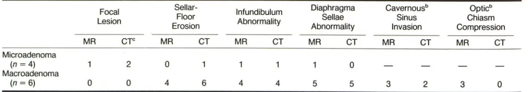
MR Imaging of Pituitary Adenoma: CT, Clinical, and Surgical Correlation
Full text
Figure


Related documents
Conclusion: The knowledge and practice levels of lifestyle modifications amongst type 2 diabetes mellitus patients attending.. tertiary care hospital in Lucknow were
We analyzed the miRNA expression of the patients with COPD and health controls for differentially expressed miRNAs in smokers versus nonsmokers.. We have not found any miRNAs
The Huh7 and HCCLM3 cells proliferation was measured using cell counting Kit-8 (CCK-8) assay following transfection with miR-519 mimics or mimics NC, respectively.. Flow
The designed study was to evaluate prescribing practices among private practitioners in urban. areas of
By analyzing the gene expression omnibus (GEO) database, we discovered that NME1 was more highly expressed in HCC tumor tissues than non-HCC liver tissues ( P < 0.001), and
The hydrolysis of primary sludge and secondary sludge were examined using biochemical methane potential (BMP) tests, with the monitoring of volatile solids concentrations and
The objective of this work is to investigate the effect of partial replacement of river sand with quarry dust on the compressive strength, flexural strength, split tensile
anp: anterior neural plate; nt: neural tube; PLE: presumptive lens ectoderm ef: eye field;.. rpe: retinal pigmented epithelium;
