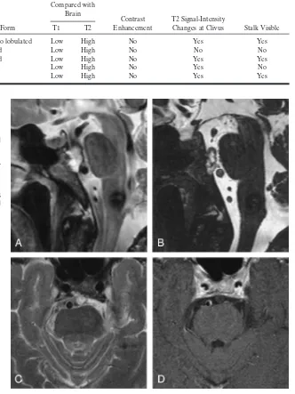
Retroclival Ecchordosis Physaliphora: MR Imaging and Review of the Literature
Full text
Figure

Related documents
Key words race ; black social workers; post-qualifying education; child care practice;
The cell e.s.d.'s are taken into account individually in the estimation of e.s.d.'s in distances, angles and torsion angles; correlations between e.s.d.'s in cell parameters are
Data collection: CAD- 4 Software (Enraf±Nonius, 1989); cell re®nement: CAD- 4 Software ; data reduction: HELENA (Spek, 2002); program(s) used to solve structure: SHELXS 97
Hence the structure is not de®ned by hydrogen bonds (to whose network other factors have to adapt) but rather by quadrupole±quadrupole and van der Waals interactions between
Figure 5:Comparative dissolution profile of formulation M1 in 2%w/v Myrj dissolution media and Biorelevent dissolution
related to the distorted structure of the complex and the different rigidity of the 2-(2-pyridyl)-4-methoxycarbonyl- quinoline ligand and the bipyridine.. Similar distortions have
As part of a study directed towards relating NMR chemical shifts to the degree of conjugation of substituents in phenan- threne, the structure of 4-nitrophenanthrene, (I) (Fig..
While there is no sub- stantial change of retinal structure after expo- sure to hypergravity (+10 Gz/3 min), the expres- sion levels of the Müller cell marker GFAP and the
![2 [(ρ Methoxyphenylcarbonyl)(1,2,4 triazol 1 yl)methyl]sulfanyl 4,6 dimethylpyrimidine](data:image/gif;base64,R0lGODlhAQABAIAAAP///wAAACH5BAEAAAAALAAAAAABAAEAAAICRAEAOw==)