Printed in Great Britain
CENTRAL PATTERNING AND REFLEX CONTROL
OF ANTENNULAR FLICKING IN THE HERMIT CRAB
PAGURUS ALASKENSIS (BENEDICT)
BY PETER J. SNOW*
Zoology Department, University of Alberta, Edmonton, Alberta, Canada, and The Friday Harbor Laboratories, Friday Harbor, Washington 98250, U.S.A.
(Received 8 November 1974)
SUMMARY
1. The effects of altering sensory input on the motoneuronal activity underlying antennular flicking have been tested.
2. Removal of the short segments of the outer flagellum results in a reduction of the number of spikes/burst in the fast flexor motoneurones A3iFand A32F.
3. During a flick the delay between the burst in motoneurone A31F and the burst in motoneurone A32F is insensitive to alteration of sensory input.
4. Sensory feedback from the flexion phase of a flick is necessary for the activation of either extensor motoneurone. Evidence is presented to suggest that this feedback is primarily from joint-movement receptors at the MS-DS and DS-OF joints.
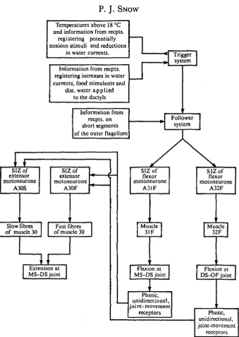
5. The results are incorporated into a model in which the patterns of flexor activity result from some specified properties of three components: a trigger system, a follower system, and the spike initiating zone of the flexor motoneurones. The trigger system determines when a flick will occur. The follower system determines the number of flexor spikes during a flick. Properties of the spike initiating zone determine the spike frequency and the timing between bursts in the flexor motoneurones. Extensor activity in the model is reflexively elicited by feedback from phasic, unidirectional receptors sensitive to joint flexion.
6. The functional significance of reflex control of extensor activity is discussed in relation to the form and proposed function of antennular flicking. It is suggested that this form of control is adapted to the function of antennular flicking because flexion at the MS-DS joint is not always necessary for the fulfilment of the function of a flick.
INTRODUCTION
It has been suggested that antennular flicking in the hermit crab Pagurus
alaskensis, might provide a means of sampling dissolved chemicals in the crab's
immediate surroundings (Snow, 1973 a). Flicking could thus be compared with sniffing in higher vertebrates. The unusual form and possible function of flicking
• Present address: Department of Physiology, Royal (Dick) School of Veterinary Studies, University of Edinburgh, Edinburgh EH9 iQH, Scotland.
18 P. J. SNOW
suggested that an understanding of its nervous control may provide some interesting contrasts with the neuronal mechanisms underlying other stereotyped, invertebrate activities. The objective of the present work was thus to determine the effects of altering sensory input on the motoneuronal activity underlying antennular flicking.
MATERIALS AND METHODS
Collection and maintenance of Pagurus alaskensis (Benedict) is described elsewhere (Snow, 19736). Data presented here were derived from experiments on 52 large crabs. Motoneuronal activity was monitored by recording electromyograms from the antennules of partially restrained animals or by extracellular recordings from the antennular nerves in the proximal segment (Snow, 1975). Extracellular recordings from the antennular nerves also permitted monitoring of sensory activity arising from antennular sensilla.
Two preparations were used for recording directly from the antennular nerves. Partially dissected preparations, described elsewhere (Snow, 1975), were used most frequently. Data from these were supplemented by recordings from isolated brain-antennular preparations. These involved cannulation of the dorsal artery and perfusion of the brain with oxygenated Cancer pagurus saline (Pantin, 1948). The brain and antennules were then dissected away from the rest of the body and the proximal antennular segment (containing the statocyst) was dissected away to expose the antennular nerves. Isolated brain-antennule preparations favourable for recording showed good antennular withdrawal reflexes and some antennular flicking.
During the present work it became necessary to rapidly flex the medial segment-distal segment joint through 5-150. This was achieved by securing the medial segment to a block of Sylgard 184 Encapsulating Resin (Dow Corning) (Snow, 1975) and by moving the distal segment with a glass probe attached to the slug of a Dormeyer P8-1L solenoid. This solenoid could be driven by a pulse from a Grass S4 stimulator.
RESULTS
(1) Flexor patterns following the alteration of sensory input
00 (b)
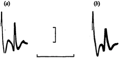
Fig. 1. Recordings from nerve z showing the first spikes in the fast flexor motoneurones A31F (large spike) and A32F during patterns of activity which, in an intact antennule, always result in a flick, (a) Superimposed flexor patterns of five successive flicks during which all antennular nerves were intact (6) Five superimposed flexor patterns recorded after all the antennular nerves have been cut distal to the recording electrode. The slight change in waveform between (a) and (b) resulted from a slight disturbance of the nerve-suction electrode seal during the cutting of the nerves. All recordings were taken from a partially dissected preparation. Scale: 50/iV, 6 msec.
During flicking in intact animals the first spike in motoneurone A31F precedes the first spike in motoneurone A32F by an almost constant interval, which in different animals lies in the range of 1-4-2-6 msec (Snow, 1975). No operation.upon, or manipulation of, the antennules, eyestalks, or antennae alters this interval and it remains unchanged following the excision of all ipsilateral antennular nerves in partially dissected preparations (cf. Fig. i(a) with Fig. 1(6)).
In intact animals, or animals from which the antennular flagella had been removed, increasing the rate of flow of sea water through the chamber, pipetting distilled water or, in some animals, pipetting filtered fish juice (the filtrate of 5-10 g of fish muscle macerated in 25 ml of sea water) into the inflow stream or over the dactylo-podites of the claws or legs, increased the mean frequency of flicking. Similarly, isolated brain-antennules preparations flicked more frequently in response to increased water currents or a few drops of distilled water introduced into the experimental chamber. Flicking could also be tonically inhibited by noxious stimu-lation of the antennules (Snow, 1973 a). In no preparation did these or any other stimuli have any effect on the number of flexor spikes during a single flick, although the most common number of spikes was sometimes seen to change during a long (over 10 h) recording session from intact animals. These observations are consistent with the observation that the number of flexor spikes is not related to the preceding inter-flick interval. It should be noted that the percentage of long flexor bursts is quite variable between animals (Snow, 1975).
In conclusion, it seems that the mean number of flexor spikes/flick is sensitive either to subtle and slow fluctuations in the environment or to endogenous changes in the physiological state of the crab or to both these factors. This conclusion, how-ever, does not infer that the number of flexor spikes is completely independent of any sensory input but just that any such dependency is under some form of long-term regulation.
Table I. The effects of altered smory input on the number of spikes in flexor motoneurones Ag IF and A32F during antennular Jlicks % of No. of ASIF spikeslno. of A32F spikes I/Z, 211 A , and 212 Animal Antennule condition
Free MS-DS
Im. MS-DS and DS-OF Im. DS-OF freed MS-DS freed
Free MS-DS
and
DS-OF
Im.
OF
excised
Free OF
excised
Free OF
excised
Free MS-DS
and DS-OF Im. DS-OF freed MS-DS freed OF excised
Total I00 I00 I00 50 27 I 80 I 80 170 70 70 I 28 I
18 60 60 60 60 60
patterns
I2 30 58
^
|(t
|U
M31F W ,
M32F X»L. «..,.»,
I
M31F M32F
Fig. 2. Simultaneous electromyogram recordings in flexor muscles 31F and 32F during single flicks: before (a) and after (6) excision of all but the basal segment of the outer flagellum. Note the reduction in the number of EJPs in each muscle which reflects a reduction in the number of spikes in flexor motoneurones A31F and A32F. Scale: 200 fiV, 30 msec
spikes in flexor motoneurones A31F and A32F (Table 1). In contrast, excision of all but the basal segment of the OF usually resulted in a considerable reduction in the percentage of long bursts in motoneurones A31F and A32F (cf. Fig. z(a) with Fig. 2(b) and Table 1). This effect was most clearly seen in crabs in which the number of spikes in motoneurones A31F and A32F was highly variable. Following this operation most flicks resulted from only 1 or 2 spikes in motoneurones A31F and A32F (Fig. 2, Table 1). This was true even during a series of flicks which had a short mean inter-flick interval. By far the most frequent pattern was two spikes in motoneurone A31F and 1 spike in motoneurone A32F, while 1 spike in motoneurone A31F was never followed by more than one spike in motoneurone A32F. Records from muscles 31F and 32F more than 2 weeks after this operation showed no recovery of the ability to produce an appreciable percentage of longer bursts in motoneurones A31F and A32F. Only one animal, which was initially showing a high percentage of 2:1 A3iF:A32F patterns, failed to show a clear reduction in the percentage of longer flexor bursts following this operation (animal no. 4, Table 1). In no case did this operation affect the number of spikes in motoneurones A31F and A32F of the contralateral antennule.
It therefore appears that information from receptors on the short segments of the OF (Snow, 1974) is necessary for the generation of most bursts of more than two spikes in flexor motoneurones A31F and A32F during flicking in intact animals.
(2) Extensor activity following the alteration of sensory input
The extension phase of a flick normally results from a burst of 1-4 spikes in the fast extensor motoneurone A30F and 1-6 spikes in the slow extensor motoneurone A30S. There is a 20-40 msec delay between the first spike in the flexor motoneurone A31F and the first spike in either extensor motoneurone. This delay is quite variable within and between preparations. These observations suggest that the mechanism underlying the timing of extensor activity is quite labile.
22 P. J. SNOW
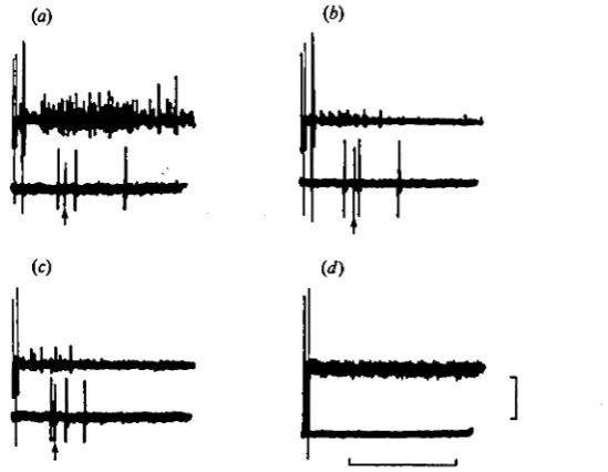
Fig. 3. Recordings of flexor (nerve 2, top trace) and extensor (nerve 2a, bottonr-'trace) motoneuronal activity during flexor activity which in the intact antennule al.vays results in a flick, (a) Recordings during a flick with all nerves intact. Arrows differentiate the spikes in the fast extensor motoneurone A30F from those in the slow extensor motoneurone A30S. (6) Recordings taken after the nerve containing the flexor motoneurones (nerve 2) has been cut distal to the recording electrode. The slight change in the waveform of the spikes in motoneurone A32F between (a) and (6) resulted from a slight disturbance of the nerve-suction electrode seal during the cutting of nerve 2. All recordings were taken from a single partially dissected preparation. Scale: 100 /tV, 30 msec.
(a) (ft)
( f )
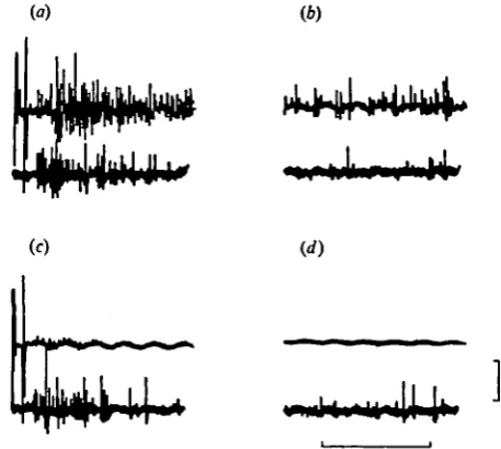
Fig. 4. Recordings from nerve 2 (top traces) and nerve 3 (bottom traces) during antennular flicking (a), (c) and in the resting antennule (b), (d); before (a), (6) and after (c), (rf) removal of the receptors of the antennular flagella (see text). This operation results in the marked reduction of the sensory activity normally seen in nerve 2 during flicking (compare top traces of (a) and (<:)) and the complete abolition of the sensory activity in nerve 2 normally seen in the resting antennule (compare top traces of (b) and (d)). Note that this operation has little effect on the sensory activity in nerve 3. All recordings were taken from a single partially dissected preparation. Scale: 60 msec, top traces 50/tV, bottom traces 25 fiV.
[image:6.451.111.340.262.467.2](a) (*)
Fig. 5. Recordings of flexor (nerve 2, top traces) and extensor (nerve 2 a, bottom traces) motoneuronal activity during flexor activity which in the intact antennule always results in a flick. Arrows differentiate the spikes in the fast extensor motoneurone A30F from those of the slow extensor motoneurone A30S. (a) Recordings during a flick in an intact antennule. (b) Recordings after the removal of the receptors of the antennular flagella (see text). Note the reduction of sensory activity in nerve 2. (c) Recordings during a flick, after the removal of the receptors of the antennular flagella and after nerves 1 and 3 have been cut. Note the maintenance of activity in the fast and slow extensor motoneurones. (d) Recordings after the nerve containing the flexor motoneurones (nerve a) has been cut distal to the recording electrode. Note the complete abolition of extensor activity. All recordings were taken from a single partially dissected preparation. Scale: 50 /*V, 60 msec.
recorded from the proximal stump of nerve 2. These bursts were similar to those recorded during antennular flicking prior to cutting nerve 2. Despite this, in five out of five preparations there was complete abolition of any extensor activity correlated with the bursts in the flexor motoneurones (Figs. 3 a, b, 5a, d). From this experiment it was concluded that extensor activity was either dependent on sensory feedback from the flexor activity or on tonic sensory activity in nerve 2 or both these factors.
In the resting antennule there is considerable sensory activity in nerve 2 (Fig. 46). Mechanical stimulation of hairs on the inner and outer flagella with a glass probe always results in increased sensory activity in nerve 2. Excision of the IF and all but the basal segment of the OF removes most if not all of the receptors on the antennular flagella (see Snow, 1974) and completely abolishes any sensory activity detectable in nerve 2 in the resting antennule (Fig. 4<f). This operation, however, does not result in the reduction of activity in the extensor motoneurones A30S and A30F during antennular flicking (Fig. 5^). It therefore seems likely that the extensor activity during flicking is dependent only on sensory feedback from the flexion phase of a flick.
24 P. J. S N O W
The DS bears only 2-4 setae on its dorsal surface. During flicking in partially dissected preparations there are thus three possible sources of sensory feedback from the flexion phase of a flick: (1) mechanosensitive hairs and internal pro-prioceptors on the antennular flagella; (2) receptors sensitive to muscle tension; (3) receptors sensitive to joint movements.
Even very light mechanical stimulation of either antennular flagella results in increased activity in nerve 2. In addition, intense mechanical stimulation of the IF is sufficient to excite both the fast and slow extensor motoneurones (Snow, 1975). During a flick a multi-unit burst of sensory activity can be recorded in nerves 2 and 3 (Fig. 4a). These bursts often begin only 10-15 msec after the first spike in flexor motoneurone A31F and usually have a duration of over 100 msec. Excision of the IF and all but the basal segment of the OF abolishes most but not all of this sensory activity in nerve 2 without appreciably altering the sensory activity in nerve 3 (Figs. 4a, c, 5 a, b) As mentioned above, this operation does not reduce the activity in either extensor motoneurone during a flick (Fig. 5 a, b). It thus seems that although feedback from receptors of the antennular flagella is elicited by the flexion phase of a flick, it is not necessary for the maintenance of normal activity in the extensor motoneurones A30F and A30S. Despite this, it is presently impossible to consider that feedback from flagellar receptors during a flick exerts absolutely no excitatory influence on the extensor motoneurones.
As pointed out above, removal of the flagellar receptors does not abolish all sensory feedback during a flick (Fig. 4a, c). The feedback which remains after this operation can be considered as arising from two possible sources: (1) receptors sensitive to muscle tension; (2) receptors sensitive to joint movements. Differentiation between muscle-tension receptors and joint-movement receptors in crustaceans requires the application of several rigorous tests (see Macmillian & Dando, 1972). For the present purposes, sensory units which are activated by muscle tension in the absence of visually observable angle change at the antennular joints (joint movement) will be tentatively defined as receptors responding to muscle tension.
Joint immobilization was achieved by stapling the MS, DS and the basal segment of the OF to a Sylgard block (Snow, 1975) in such a way that the MS-DS and DS-OF joints were in the partially extended position they adopt in the resting antennule. Complete immobilization of the MS-DS and DS-OF joints following the removal of the flagellar receptors abolished any feedback in nerve 2 but only slightly reduced the amount of feedback in nerve 3. These observations suggest two things. Firstly, that there exist antennular receptors with axons in nerve 3 which respond to tension development in the fast flexor muscles in the absence of changes in joint angle. Secondly, that there exist antennular receptors with axons in nerves 2 and 3 which respond to joint-flexion movements in the presence of tension in the fast flexor muscles. In relation to this second point, passive and rapid flexion at the MS-DS or the DS-OF joints elicits activity in nerves 2 and 3 in excised antennules. This suggests that antennular receptors exist which respond to joint-flexion movements in the complete absence of muscle tension.
U— lju,—
to
M31F(A31F)
M30(A30F)
M31F(A31F) A ^ , A | j L Hs
M30M31F(A31F) M30
(A30F)
^
Fig. 6. Simultaneous electromyogram recordings in flexor muscle 31F and extensor muscle 30 during flicks of a free antennule (a) and following immobilization of the MS—DS joint (6) and the MS-DS and DS-OF joints (c). The motoneurones considered responsible for the various patterns are shown in parentheses. Note the prognessive abolition of activity in the fast extensor motoneurone A30F through sets (a), (6) and (c). Scale: 30 msec, top traces 200 fiV, bottom traces 100 fiV.
movement is sufficient to excite the extensor motoneurones; (2) that feedback in receptors that respond only in the presence of joint movement is necessary for the excitation of the extensor motoneurones.
The effect of joint immobilization on the fast extensor motoneurone A30F was most easily tested by recording electromyograms in partially restrained animals (Snow, 1975). In unrestrained antennules there was no activity in motoneurone A30F during 0-27% of flicks. Following immobilization of the MS-DS joint, 10-92 % of flicks were not correlated with activity in motoneurone A30F. In this condition, flicks which were accompanied by more than a single spike in moto-neurone A30F were rare, even in preparations which in the unrestrained state usually showed 2 or more fast extensor spikes during a flick (compare Fig. 6(a) with (b)). When both the MS-DS and the DS-OF joints were immobilized there was no fast extensor activity in 60-100 % of flicks (Fig. 6c), all but one animal showing a 90-100 % abolition of activity in the fast extensor motoneurone. With the exception of a single animal, total joint immobilization resulted in the total abolition of flicks which were accompanied by more than a single extensor spike. In the absence of joint movement, a flick was considered to be represented by the appearance of bursts in the flexor motoneurones A31F and A32F which were identical to those seen during flicking in an unrestrained antennule (see Fig. 6c).
Releasing the DS-OF joint and then the MS-DS joint progressively restored activity in the fast extensor motoneurone A30F. The antennule could then be immobilized and released again with qualitatively identical results.
26
[image:10.451.72.380.45.144.2]to
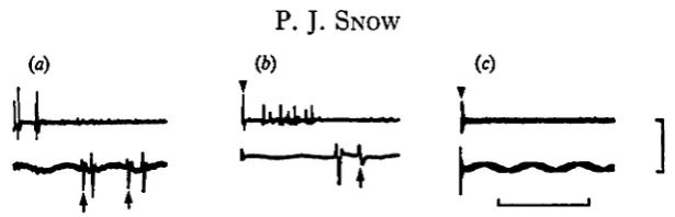
Fig. 7. Recordings from nerve 2 (top traces) and nerve 2 a (bottom traces), (a) Recordings during a flick in an intact antennule. (6) Recordings during passive and rapid flexion of the MS-DS joint following removal of the inner flagellum and the DS-OF joint. Note the burst of sensory activity in nerve 2. (c) Superimposed recordings during two successive, passive and rapid flexions of the MS-DS joint after cutting nerve 2 distal to the recording electrode. Arrowheads mark the artifact which results from activation of the solenoid. Arrows differentiate the spikes of the fast extensor motoneurone A30F from those of the slow extensor motoneurone»A3oS. Disturbance of the recording electrodes during severance of the distal segment accounts for the absence of sensory activity following the flexor spikes in (a) and for the change in waveform of the fast extensor motoneurone spikes seen in (a) and (6). All recordings were taken from a single partially dissected preparation. Scale: 30 msec, top traces 200 fiV, bottom traces 100 /tV.
good discrimination between the fast and the slow extensor motoneurone was possible. Attempts to test the effects of joint immobilization on the activity in the slow extensor motoneurone A30S during flicking were, therefore, not entirely successful. It was observed, however, that joint immobilization resulted in a reduction in the number, and sometimes a complete abolition, of spikes in the extensor motor nerve (nerve 2 a).
The fact that abolition of joint movement suppresses and often abolishes extensor activity suggests that receptors responding only in the presence of joint movement generally provide the necessary feedback which activates the extensor motoneurones during a flick. Feedback from receptors responding to muscle tension in the absence of joint movement might have some excitatory influence on the extensor motoneurones but this does not seem sufficient for the maintenace of normal extensor activity.
The above observations cannot be used to imply that feedback from the joint movements during the flexion phase of a flick is sufficient to excite the extensor motoneurones. There are, however, two lines of evidence that strongly suggest the sufficiency of this source of feedback. Firstly, normal activity in the fast and slow extensor motoneurones persists after the removal of the flagellar receptors and the severance of nerves 1 and 3 (Fig. 5 a, c). It has been argued above that nerve 2 contains only the axons of receptors which respond to joint movement, the flagellar receptors and the antennular motoneurones (see also Snow, 19736). The removal of the flagellar receptors and the severance of nerves 1 and 3 would therefore reduce feedback from the flexion phase of a flick to that carried by receptors which respond only in the presence of joint movement (Fig. 5 c).
during a flick in the intact antennule (Fig. 7 a, b). Furthermore, extensor activity was preceded by a burst of sensory activity in nerve 2 (Fig. jb). After cutting nerve 2 but leaving nerves 1 and 3 intact, no activity could be recorded from the proximal stump of nerve 2 or from the extensor motor nerve (nerve 2 a) on rapid and passive flexion of the MS-DS joint (Fig. 7c).
In conclusion, it seems that feedback from joint-movement receptors which have axons in nerve 2 is sufficient and necessary for the generation of normal extensor activity during a flick, whereas feedback from other sources is not sufficient or necessary, although it may have some subthreshold excitatory influence on the extensor motoneurones. Similarly, it is entirely possible that the extensor motoneurones receive some subthreshold excitation from a central mechanism underlying antennular flicking (cf. Mellon, 1969).
DISCUSSION
(1) Flexor activity
The dependence of the flexor patterns on central mechanisms v. sensory input will be considered in relation to three major questions: (1) How is activity in the flexor motoneurones initiated? (2) How is the number of spikes/burst determined? (3) How is the timing between bursts in motoneurone A31F and bursts in moto-neurone A32F determined?
Flicking occurs at irregular intervals but, as yet, single stimuli have been un-successful in consistently eliciting single flicks. Furthermore, no stimulus or combi-nation of stimuli appear to influence the number of spikes/burst or the timing between bursts in the flexor motoneurones. Thus the flexion movements at the MS-DS and the DS-OF joints during a flick fulfil the major requirement of a triggered movement that 'the intensity of the initiating stimulus must reach "threshold" but the movement itself will not vary as a function of suprathreshold variations in stimulus intensity' (Bizzi & Evarts, 1971).
As there is no clear coordination of flicking between left and right antennules (Snow, 1973 a) it also seems likely that the flexor activity in each antennule is the result of activation of a separate trigger system (Fig. 8). Each trigger system may be a single interneurone, a group of interneurones or even part of one or both of the flexor motoneurones. Stimuli which increase the mean flicking frequency are con-sidered as having an excitatory effect on the trigger systems while stimuli which decrease the mean frequency are considered as having an inhibitory effect (Fig. 8) (Snow, 19730)- Observations on isolated brain-antennule preparations further sug-gest that inputs from antennular receptors are' sufficient to excite or inhibit the trigger system.
28 P. J. SNOW
Temperatures above 18 °C and information from recpts.
registering potentially noxious stimuli and reductions
in water currents.
Information from recpts. registering increases in water currents, food stimulants and
dist. water applied to the dactyls
Information from recpts. on short segments of the outer flagellum
SIZof extensor motoneurone
A30S
SIZof extensor motoneurone
A30F
Slow fibres of muscle 30
Fast fibres of muscle 30
H
Extension at MS-DS joint
SIZof
flexor motoneurone
A31F
SIZof flexor motoneurone
A32F
Muscle 31F
[Muscle 32F
Flexion at MS-DS joint
Flexion at DS-OF joint
Phasic, unidirectional, joint-movement
receptors Phasic,
unidirectional, joint-movement
[image:12.451.65.405.48.525.2]receptors
a group of interneurones or part of one or both flexor motoneurones. I propose that only activation of the trigger system can drive the follower system to threshold but that information from receptors on the short segments of the outer flagellum is necessary for suprathreshold excitation of the follower system (Fig. 8). At threshold levels of excitation the follower system drives only 1 or 2 spikes in the flexor motoneurones. Suprathreshold excitation of the follower system results in longer bursts in the flexor motoneurones. The low proportion of bursts of more than 2 spikes in some intact animals suggests that certain physiological states result in a total suppression of a large number of flexor spikes despite the presence of the short segments of the outer flagellum.
Activity in motoneurones A31F and A32F has only been observed during flicking (Snow, 1975). Furthermore, activity in one of these motoneurones has not been observed in the absence of activity in the other motoneurone. It thus seems appropriate to suggest that excitation of the follower system always results in a threshold depolarization of the spike initiating zone of each flexor motoneurone (Fig. 8). At the level of the proximal antennular segment this results in the first spike in moto-neurone A31F preceding the first spike in motomoto-neurone A32F by i-4-2<6msec (Snow, 1975). Within any animal this delay is quite constant. Furthermore, it is unrelated to the number of spikes/burst in the flexor motoneurones and is not changed by alteration of sensory input. It is possible that the delay between the burst in each flexor motoneurone results from the spike initiating zone of moto-neurone A32F having a higher threshold to activation of the follower system than the spike initiating zone of motoneurone A31F. Activation of the follower system might then cause synchronous depolarization of the spike initiating zones of both motoneurones but earlier spiking of motoneurone A31F. Differences in threshold between the flexor motoneurones could also account for the observations that the shortest inter-spike intervals in motoneurone A31F are usually less than those in motoneurone A32F, and that in motoneurone A32F there are never more and often less spikes than in motoneurone A31F (Snow, 1975).
Longer inter-spike intervals often occur in the middle of the bursts in the flexor motoneurones (Snow, 1975). Such long intervals in motoneurone A31F were always accompanied by a long interval of approximately similar duration in motoneurone A32F (Snow, 1975). It therefore seems probable that these long intervals do not result from differences in the properties (e.g. threshold or refractoriness) between the spike initiating zones of the flexor motoneurones. One possibility is that the long intervals represent brief periods of subthreshold activity in the follower system. This would also account for the observation that following a long inter-spike interval, the first spike in motoneurone A31F precedes the first spike in motoneurone A32F by a delay approximately equal to the delay between the first spikes in the flexor motoneurones on the initiation of a flick (Snow, 1975).
30 P. J, SNOW
(2) Extensor activity
Evidence described above suggests that the activity in both the fast and the slow extensor motoneurone is usually dependent on feedback from the movements at the MS-DS and DS-OF joints during the flexion phase of a flick. It seems likely that activation of phasic, unidirectional movement receptors, sensitive to joint flexion, is necessary for the activation of both extensor motoneurones (Fig. 8). Firstly, phasic, unidirectional movement receptors have been reported in the chordotonal organs of the MS-DS and DS-OF joints of the lobster antennule (Wyse & Maynard, 1963, 1965). Secondly, these receptors would theoretically be inactivated by immobilizing the antennular joints. In contrast, this manipulation would not abolish feedback from any receptors responding to muscle tension (see Macmillian & Dando, 1972) and does not abolish bursts in the flexor motoneurones normally seen only during flicking. Thirdly, rapid and passive flexion at the MS-DS joint is sufficient to excite both extensor motoneurones, provided a specific nerve (nerve 2) containing the axons of sensory neurones which respond to joint movements, is intact. Fourthly, in crustacean chordotonal organs, phasic, unidirectional movement receptors are con-sidered as having the largest axons and thus the highest conduction velocities (Burke, 1954; Wyse & Maynard, 1965; Hartman & Austin, 1972). This would be advantageous for the reflexive excitation of the extensor motoneurones, which often occurs in as little as 20 msec after activation of the flexor motoneurones (Snow,
1975)-In only one other system is there good evidence for the total dependence of an entire phase of the motor output on phasic information from proprioceptors. This system controls the rhythmical contractions of the fast adductor muscles of the scallop, which are the basis of its escape-swimming response (Mellon, 1969).
(3) The functional significance of reflex control
:hus be useful in ensuring that extension at the MS-DS joint only occurs during flicks which resulted in appreciable flexion at this joint.
A second possibility for the functional significance of reflex control during flicking stems from the observation that the extensor motoneurones are often not active during flicks which occur while the MS-DS joint is being held in a flexed posture (Snow, 1975). This absence of extensor activity might be expected, as in this posture there is little flexion at the MS-DS joint during flicking (Snow, 1973 a). The reduction of flexion probably arises from the orientation of fast muscle 31F being such that the amount of flexion it can produce is reduced when the MS-DS joint is partially flexed (Snow, 1973 b). In addition, joint resistance to flexion probably increases with the degree of initial flexion. In crabs that have withdrawn into their shells, flicking is often maintained despite a high degree of tonic flexion at the MS-DS joint. The result of this antennular posture would be to reduce the flexion movement at the MS-DS joint during a flick and thus probably to reduce or abolish activity in the extensor motoneurones. While the crab is withdrawn into the shell, extension at the MS-DS joint would result in the outer flagellum hitting the inside of the shell. If this did occur, motoneurone A31S and possibly motoneurone A31F-S would be excited, resulting in the re-establishment of tonic flexion at the MS-DS joint (Snow, 1975).
It seems likely that the reflex control of extension during a flick can be tolerated because circulation of water around the aesthetasc hairs may be achieved in spite of considerable modification of the component movements of a flick. Ttye contri-bution of reflex control to flicking can thus be regarded as complementary to the popular concept that cycle-to-cycle load fluctuations are usually correlated with a greater utilization of phasic feedback information in forming the motor output (Pearson, 1972; Kater & Rowell, 1973).
I would like to thank the Director and Staff of the Friday Harbor Laboratories for the provision of facilities, and Drs K. G. Pearson, P. A. Getting and S. Thompson for their critical reading of the manuscript of this and the preceding paper (Snow, 1975). I would also like to thank Drs T. H. Bullock, R. B. Stein and S. L. Tamm and Messrs R. Condrey and J. Buckland-Nicks for their suggestions. Work reported in these papers was supported by NRC grant A-1445 to Dr D. M. Ross and a University of Alberta Dissertation Fellowship to Peter Snow.
REFERENCES
BlZZl, E. & EVARTS, E. V. (1971). III. Translational mechanisms between input and output.
Neuro-sciences Res. Prog. Bull. 9, 31-59.
BURKE, W. (1934)- An organ for proprioception and vibration sense in Carcinus maenat. J. exp. Biol. 31, 127-38.
HARTMAN, H. B. & AUSTIN, W. D. (1972). Proprioceptor organs in the antennae of decapod Crustacea. I. Physiology of a chordotonal organ spanning two joints in the spiny lobster, Panulirus interruptus (Randall). J. amp. Pkytiol. 81, 187-202.
KATER, S. B. & ROWBIX, C. H. F. (1973). Integration of sensory and centrally programmed com-ponents in generation of cyclical feeding activity of Heluoma trivolvis. J. Neuropkysiology 36,
32 P. J. SNOW
MELLON, D E F . (1969). The reflex control of rhythmic motor output during swimming in the scallop.
Z. vergl. Pkysiol. 6a, 318-36.
PANTIN, C. F. A. (1948). Notes on Microscopical Techniques for Zoologists. Cambridge University Press.
PEARSON, K. G. (1972). Central programming and reflex control of walking in the cockroach. J. exp.
Biol. 56, 173-93.
SNOW, P. J. (1973 a). The antennular activities of the hermit crab, Pagurus alaikensis (Benedict).
J. exp. Biol. 58, 745-65.
SNOW, P. J. (19736). The motor innervation and musculature of the antennule of the hermit crab,
Pagurus alaskensis (Benedict). J. exp. Biol. 58, 767-84.
SNOW, P. J. (1974). Surface structures of the antennular flagella of the hermit crab, Pagurus alaskensis (Benedict) - A light and scanning electron microscopy study. J. Morph. 144, 195-216.
SNOW, P. J. (1975). Patterns of activity in the antennular motoneurones of the hermit crab, Pagurus
alaskensis (Benedict). J. exp. Biol. 63, 1-15.
WYSE, G. A. & MAYNARD, D . M. (1963). Joint proprioceptore in the antennule of the spiny lobster
(Panulirus argus). Am. Zool. 3, 513.
WYSE, G. A. & MAYNARD, D . M. (1965). Joint receptors in the antennule of Panulirus argus, Latreille.