pH GRADIENTS IN LEPIDOPTERAN MIDGUT
BY JULIAN A. T. DOW
Department of Cell Biology, University of Glasgow, Glasgow G12 8QQ, UK
Summary
Lepidopteran larvae demonstrate several remarkable specialisations of the alimentary canal: the most active epithelial transport known; a unique cell type, called a goblet cell; and the highest pH values known to be generated by a biological system. The electrogenic K+ pump in midgut is now known to be energised by a H+-pumping V-ATPase, and net alkali metal transport is achieved by linking it to a «H+/alkali metal exchanger, which recycles H+ into the cytoplasm.
Generation of high luminal pH is modelled as a passive (Nernstian) distribution of protons in the electrical field generated by the V-type ATPase. Electrode impalements show that the potential difference across the goblet cavity membrane is extremely high. Measurements of pH gradients generated in vitro confirm that the midgut itself generates such a gradient, that this process relies on metabolic energy, and that the differential ability of midgut subregions to perform acid-base transport maps to their differing morphologies and to the pH profiles observed along the gut in vivo.
During larval/larval moults, K+ transport is suppressed. The transepithelial potential difference (PD) across the gut collapses and recovers in phase with the loss and recovery of the gut pH gradient, and with tissue V-ATPase activity, confirming that these processes are intimately linked.
Acridine Orange partitions into acidic compartments and might be expected to be concentrated in goblet cavities, as these are the compartments toward which the V-ATPase pumps protons. However, under normal conditions, Acridine Orange is excluded from the cavities. Red metachromasia of the cavities (implying low pH) is only observed when the ion transport status of the tissue is compromised. It thus seems likely that, under physiological conditions, K+/H+ exchange is tight enough to produce a neutral or alkaline, rather than acidic, cavity.
Molecular analysis of the 16000Mr subunit from Manduca midgut reveals it to be
closely similar to other known 16000MT sequences, particularly that from Drosophila
brain. It is thus likely to be a true H+ channel, rather than one modified for K+ transport. The cavity can be modelled in two ways: (i) to isolate the site of proton equilibration electrically from the main gut lumen, and thus allow larger pH gradients to develop, or (ii) to buffer the V-ATPase from the alkaline pH in the gut lumen, which would otherwise destroy the gradient driving the exchange of H+ for alkali metal cations. The first model would predict a high cavity pH, whereas the second would predict a near neutral pH and would imply a non-cavity route for transport of base equivalents. Work with both pH-sensitive dyes and pH-pH-sensitive electrodes so far tends to support the second model.
Caterpillars
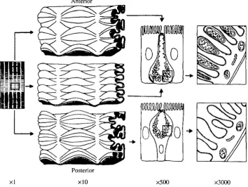
Physiology can often be studied most profitably in systems which display a property of interest in the extreme. Holometabolous insects, for example, divide their life cycle into two distinct phases; a larval growth phase and an adult sexual phase. It is not surprising, therefore, that the larvae of lepidopteran insects display spectacular adaptations of the alimentary canal for feeding and growth. In this study, the model insect is the caterpillar of the tobacco hornworm, Manduca sexta, which grows from egg to 10 g larva in about 2 weeks. Its internal anatomy is thus dominated by the gut (Fig. 1).
K+ pump
The physiological specialisation which initially attracted attention was the presence of an electrogenic K+ pump (Harvey and Nedergaard, 1964). This has since been shown to be ubiquitous among insects, being found in midgut, Malpighian tubules, salivary glands and cuticular sensillae (Harvey et al. l9S3a,b). Of these, the caterpillar midgut is probably the most accessible to physiological study, as it can be mounted in an Ussing chamber. This reveals an electrical signature unparalleled in transporting epithelia; the tissue can develop open-circuit potential differences in excess of 150mV and short-circuit currents in excess of 1 rnAcm"2. As far as is known, this property is general among larval Lepidoptera (Dow, 1986).
Anterior
[image:2.451.49.404.350.616.2]xl x500 X3000
The pump is found in the apical membrane of a unique goblet cell (Dow et al. 1984; Wieczorek et al. 1986), which is distinct from the mucus-secreting vertebrate intestinal cell of the same name (Fig. 1). The lepidopteran goblet cell is characterised by an apically directed cavity, vacuolar in origin (Hakim et al. 1988), with a spectacularly complex valve joining - or isolating - it from the lumen. The valve appears to be formed of fused, microvillate apical projections from the goblet cell, with conspicuous membrane decorations visible by freeze fracture (Flower and Filshie, 1976). It is not clear whether the valve opens and shuts cyclically or whether the open valves sometimes seen under the electron microscope reflect poor fixation, but there appears to be a tighter, more tortuous, seal during moulting, when K+ pumping is inhibited (Cioffi, 1984).
The minimum cost of the ion transport can be calculated at 10% of the larva's total ATP production (Dow and Peacock, 1989). So why does a lepidopteran larva spend such an enormous fraction of its energy on such a prodigiously expensive activity? Historically, this expenditure was argued to represent a specific adaptation to rapid growth on a K+-rich plant diet (Harvey and Zehran, 1972; Maddrell, 1971). However, there are several problems with this argument (Dow, 1986), not least of which is the presence of a valve at the exit from the cavity, which adds a significant overhead [estimated at 5 kJ mol"1 K+ (Dow and Peacock, 1989)] to K+ transport. Additionally, the major component of the electrochemical gradient driving K+ from gut lumen into the blood (and thus the gradient against which the pump must work) is the electrical gradient produced by the pump itself (Dow and Harvey, 1988; Dow and Peacock, 1989). Accordingly, if the pump did not exist, it would not need to!
HighpH
Another remarkable property of certain insect guts is a very high luminal pH. Waterhouse (1949) showed that a range of lepidopteran larvae possessed high luminal pH values within their guts. However, pH determinations in such small compartments were based on concentrated dye in the water or diet and were necessarily approximations. Such determinations were also sensitive to any coloration present in the diet or in the insect's cuticle, and pH gradients were found to be notoriously sensitive to experimental manipulations, such as dissection (Dadd, 1975, 1976). Although confined to insects, such high pH values are not unique to lepidopteran larvae; similar high pH values have been recorded in the midguts of certain dipteran larvae (Dadd, 1975), in termites (Bignell and Anderson, 1980) and in the beetle, Oryctes nasicornis (Bayon, 1980).
likely that the insect benefits by virtue of a limited protection against tannins, a higher rate of enzymatic hydrolysis (the enzymes have correspondingly alkaline pH optima) and some protection against microbial pathogens. The major disadvantage of a high gut pH is its obligate role in activating parasporal 5-endotoxins of Bacillus thuringiensis, thus rendering the insect liable to natural or human attack by this natural insecticidal agent (Bullaera/. 1980).
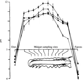
With improving technology, estimates for luminal pH became steadily higher. Using pH semi-micro electrodes, it recently became feasible to measure the pH profile at multiple sites along the length of the alimentary canal. This task was undertaken for four species of insects (Dow, 1984), revealing a characteristic profile in each (Fig. 2). The peak values recorded (in excess of pH 12) are the highest known in biology. So, whereas vertebrates have evolved a highly acidic gut, lepidopteran larvae have developed a fluid resembling oven cleaner.
Obviously, both the function and the mechanism of generation of this gradient demanded explanation. At one level, the concept of strong ion difference (SID) (Stewart, 1981) seems to render the explanation trivial. Simply by producing an excess of a strong ion (such as K+) in a compartment, the solution is necessarily alkaline; and so a K+
-12-,
X
Q.
7 -Diet
4
[image:4.451.92.372.322.596.2]transporting epithelium in which counterion flux is not facilitated (like the midgut) is a classical exemplar of the SID theory. However, SID is a macroscopic accounting system, rather than an explanation; electrogenic transport of K+ into a compartment would simply polarise it to the electromotive force (EMF) of the pump and prevent anything further from happening, unless transmembrane fluxes of other species were allowed to occur. Thus, although the pH gradient may be driven by electrogenic K+ flux, there must also be flux of one or more weakly ionised species (H+, OH~, HCC"3~, CC"32~) at the membrane level.
What is the nature of the companion process to K+ transport? Luminal CO2 has been determined as 5mmoll~' in the blood and 50mmoll~' in the midgut lumen in Hyalophora cecropia (Turbeck and Foder, 1970). At pH 11-12, this would exist mainly asCO32~; accordingly, the gut contents are effectively a 5 0 m m o l l " ' solution of K2CO3. This, of itself, does not imply active transport of CO32~ or HCO3", since metabolic CO2 could permeate freely through the gut tissue and would be trapped in a high-pH (i.e. high OH~) solution as carbonate. It is also worth noting that processes which generate the weakly alkaline fluids observed in some vertebrate body fluids are insufficient to explain the high values in lepidopteran midgut, as they are based on bicarbonate transport; a lOOmmoll"1 bicarbonate solution has a pH of approximately 9. Accordingly, a novel mechanism must be invoked.
The shape of the pH profile (Fig. 2) was also highly suggestive, as it mapped to multiple morphological and biochemical discontinuities along the gut.
(a) General folding (Fig. 1). The midgut can be divided by inspection into three regions, based on the general level of folding. In the anterior region, the gut is heavily folded; in the middle midgut, it is lightly folded; and in the posterior midgut, the folds are themselves folded (Cioffi, 1979).
(b) Goblet morphology (Fig. 1). In the anterior and middle regions, the goblet cells show reduced cytoplasm, with squashed, disc-like nuclei. In the posterior midgut, nuclei are spherical, and the goblet cells have a normal cytoplasmic volume (Cioffi, 1979).
(c) Mitochondrial placement (Fig. 1). In the anterior and middle midgut, every apical infolding or microvillus (but not sensu strictu) of the plasma membrane of the goblet cavity contains a mitochondrion, a specialisation found in certain insects at sites of particular metabolic activity (Cioffi, 1979). In the posterior midgut, the mitochondria lie just below the infoldings.
(d) Carbonic anhydrase (CA). The midgut is a rich source of CA (Turbeck and Foder, 1970), and K+ transport is sensitive to Pco. and to the CA inhibitor, cardrase (Haskell et al. 1965). More detailed histochemical tests showed that CA was confined to the anterior and middle midgut goblet cells, but to columnar cell apical microvilli in the posterior midgut (Ridgway and Moffett, 1986). Given that this enzyme is a marker for tissues involved in acid-base balance, it would be tempting to hypothesise that the goblet cells in anterior and middle midgut are involved in alkalising the gut lumen, whereas the columnar cells in posterior midgut bring the pH back down, as required for homeostasis at the organismal level.
and posterior midgut, whereas the soluble form of the enzyme is concentrated in the goblet cells of the posterior region (Azuma and Eguchi, 1989; Azuma et a I. 1991).
(f) Bismuth accumulation. Insect gut lumen is also characterised by wide swings in redox potential. Dietary additions of bismuth subnitrate resulted in accumulation of insoluble bismuth salts in the goblet cavities of only the posterior midgut, implying that goblet cells might be secreting sulphides (Appel and Martin, 1990).
(g) Electrical properties. Although the properties of the K+ pump seem similar along the length of the alimentary canal, there are subtle differences. The agreement of net 4 2K+ flux with short-circuit current is better in posterior midgut than in anterior or middle midgut (Cioffi and Harvey, 1981). The short-circuit flux ratio in posterior midgut is 43:1, three times higher than in anterior and middle midgut (Cioffi and Harvey, 1981).
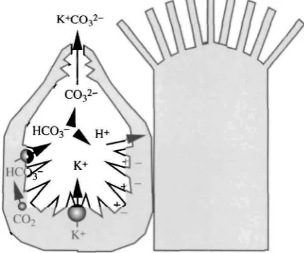
Taken together, these data suggest overwhelmingly that, although ion transport occurs with a broadly similar electrical signature along the length of the midgut, other processes show distinct regional localisations and that, in particular, the distinction between the anterior/middle midgut regions and the posterior region is marked. Additionally, the distribution suggests models for the basis of ion transport which might lead overall to a high luminal pH (Fig. 3). Electrogenic K+ transport (as originally hypothesised) would polarise the goblet cavity membrane to such an extent that Nernstian (passive) proton distribution would produce the luminal pH values observed (Dow, 1984). It was hypothesised that the cavity membrane was highly permeable to protons and was electrically isolated from the main midgut lumen by the complex apical valve. The permeability properties of the valve could thus be explained as a dynamic compromise between a very loose valve (high flux, but lower PD and thus lower ApH) and a perfectly tight valve (zero flux, but PD approaching pump EMF, and thus maximal ApH).
It is also necessary to explain the presence of carbonic anhydrase in goblet cells. A simple explanation would be that it catalyses conversion of metabolic CO2 into
[image:6.451.127.343.428.607.2]
bicarbonate and protons. The bicarbonate might then be translocated across the cavity membrane either by an ATPase or via a Cl~/HC03~ exchanger. A bicarbonate ATPase has been identified in lepidopteran midgut (Deaton, 1984), but its precise location and role remain uncertain. Once in the lumen, at any time a small fraction of bicarbonate would be present as carbonate and as protons; if these protons were near the membrane, the electric field would be expected to drive them into the goblet cytoplasm. This would thus pull the HCO3~/(H++CO32~) equilibrium towards CO32~.
Model for insect pump
It has recently become clear that the alkali metal ion (M+) pump is in fact a combination of two distinct transport processes: a vacuolar H+-ATPase, together with a nH+M+ antiporter (Klein et al. 1991; Schweikl et al. 1989; Wieczorek et al. 1989). This seems to be general to all insect epithelia where this pump has been described. For example, fluid secretion in insect Malpighian tubules is sensitive to dicyclohexylcarbodiimide (DCCD) (S. H. P. Maddrell, personal communication) and to amiloride, an inhibitor of the sodium/proton antiport (Bertram etal. 1991).
This model offers a pleasing parsimony, as no new processes need be invented to explain this unusual ion transport process. The linkage between the V-ATPase and the cotransport is tight: 100% of the short-circuit current across the midgut is carried by K+ (Cioffi and Harvey, 1981); thus, the macroscopic signature presented is one of an alkali metal ATPase. Nernst potentials, like short-circuit currents, are macroscopic phenomena and are not intimately affected by membrane-level exchange processes. Accordingly, the original model for high pH generation can accommodate the newly discovered nature of the ATPase, and K+ pump is adopted here as an epithelial shorthand for the distinct molecular processes that underlie the transport at the membrane level.
Electrode evidence
Luckily, it was possible to see the gut in great detail, either with Nomarski or phase-contrast optics (see Fig. 9), and to impale goblet or columnar cells, or goblet cavities, at will. The resting potentials of the two cell types were entirely normal, but there was a massive potential gradient at the proposed site of the pump (Fig. 5A). This potential was entirely consistent with the model that high pH could be generated by Nernstian distribution of protons across the cavity. A similar, if smaller, potential gradient was also observed under short-circuit conditions (Moffett and Koch, 19886). The size of this potential gradient is unusual, but not shocking to those acquainted with V-ATPases; potentials approaching 300 mV have been measured in plant systems. Although there is some discrepancy in precise estimates for the goblet cavity membrane, it is clear that the PD in vivo is very large (Dow and Peacock, 1989; Moffett and Koch, 1988/?). This can be demonstrated by simple arithmetic; the in vivo TEP is agreed to lie between 100 and 150 mV, the resting potential of the goblet cells is 50-70 mV negative relative to the basal side, and the cavity is 50 mV positive with respect to the apical side. Accordingly, addition of these figures produces estimates in the range 200-270 mV.
Fig. 4. Apparatus for guided impalements of intracellular sites of Manduca sextet midgut with simultaneous measurement of transepithelial PD under double perfusion in vitro. V denotes a high-impedance voltmeter for measurement of transepithelial PD. (Redrawn from Dow and Peacock, 1989.)
A Electrical potential differences Apical lumen +150
B Electrochemical potential differences Apical lumen
Blood side
Additionally, it was possible to calculate from electrochemical gradients for K+ that the presence of an apical valve separating the goblet cavity from the gut lumen carried a metabolic overhead of 5 kJmol~' K+(Fig. 5B) (Dow and Peacock, 1989). It is interesting to note that the cavity valve was impermeable to Lucifer Yellow (see Fig. 9B), implying a fairly tight seal; in this context, the residence time for K+ within a cavity can be calculated to be only a few seconds (J. A. T. Dow, unpublished results), and so a rapid flux of smaller solutes must be possible through the valve.
In vitro assay
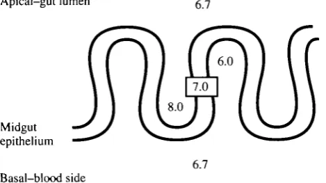
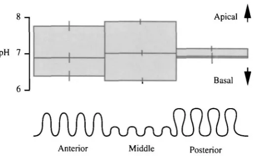
So it seemed as if the cavities can generate the PD needed to produce a high luminal pH. But was the midgut itself generating the gradient? The evidence was persuasive, but indirect. It was also necessary to demonstrate that the midgut itself could generate and sustain a pH gradient in vitro. Additionally, it was predicted that the three midgut subregions would differ in their abilities to sustain a pH gradient in vitro, as would be implied by the gradients determined earlier in vivo. Using a similar double-perfused gut preparation, but with double-barrelled pH microelectrodes, it was possible to measure pH profiles within the infoldings of the midgut under defined conditions (Dow and O'Donnell, 1990). This showed that the extracellular environment within apical infoldings was alkaline and within basal infoldings correspondingly acidic, when the gut was double-perfused with oxygenated saline at pH 6.7 (Fig. 6). The cytoplasmic compartment had a pH of 7.0 under such conditions. Furthermore, the anterior and middle midguts both showed substantial standing gradients, whereas the posterior midgut failed to demonstrate a significant gradient (Fig. 7), exactly as would have been predicted by the pH profiles previously determined in vivo (Fig. 2).
Similar results were obtained using a pH-stat technique (Chamberlin, 1990); in this case, a small alkalisation by posterior midgut was demonstrable, but was dwarfed by the rate of alkalisation by the anterior and middle subregions.
To preclude the possibility that the gradients detected by pH microelectrodes were artefacts produced by poor washout of gut contents, the gut was briefly perfused with hypoxic saline, then returned to normoxic saline, a manipulation which rapidly and
Apical-gut lumen g 7
Midgut epithelium
[image:9.451.109.344.488.619.2]6.7 Basal-blood side
pH 7
-6 J
Apical
Basal
t
[image:10.451.103.361.65.223.2]Anterior Middle Posterior
Fig. 7. Comparison of pH gradient across the three regions of Manduca sexta midgut in vitro. For each column, the upper line represents luminal pH, the centre line cytoplasmic pH and the lower line basal pH. Error bars show S.E.M. (Ate4): the gradients across anterior and middle midgut are significantly greater than zero (P<0.05). (Data from Dow and O'Donnell, 1990.)
reversibly inhibits K+ transport (Dow et al. 1985). Under these conditions, the pH gradient generated by the middle subregion fell significantly, then recovered to normal levels (Fig. 8). The gradient across the posterior midgut, by contrast, remained insignificant throughout. Furthermore, the time course of loss and recovery of the pH gradient across the midgut throughout the manipulation corresponded closely with that predicted from the measured transepithelial PD by the Nernst equation (Dow and O'Donnell, 1990).
These results demonstrated that the midgut itself generated a pH gradient by a process which required metabolic energy and that the shape of the pH profile observed in vivo could be explained by the differential abilities of the midgut regions to move acid-base equivalents.
Indicator dyes
How is the pump controlled? As far as is known, this pump runs flat out during each instar, with no obvious control in response to feeding. Given the large metabolic cost inherent in running the ATPase, this might seem surprising.
non-A Middle midgut Apical
pH 7
-6 J B Posterior midgut
pH 7
6
-Basal
Apical
Before anoxia
After 3 min of anoxia
Basal
After 3 min of recovery
[image:11.451.92.353.63.244.2]T
Fig. 8. Effect of brief anoxia on pH gradients across Manduca sexta midgut in vitro. Explanation as for Fig. 7. Anoxia produces a significant reduction in the pH gradient in middle midgut, and recovery produces a significant increase to levels not significantly different from control. Gradients in the posterior midgut are insignificant throughout. (Data from Dow and O'Donnell, 1990.)
invasive measurement, by adding pH dyes to the diet and monitoring gut luminal pH by inspection. It was found that the TEP developed by an isolated gut could be predicted non-invasively, and with complete reliability, by inspection of the intact insect's colour by transmitted light, thus opening new avenues for endocrinological research (Fig. 10), as the technique allows the precise determination of the time at which the insect's haemolymph can be presumed to contain a humoral factor which switches the ion transport status of the tissue, without experimental intervention or trauma.
At what level does the control of ion transport lie? Is it transcriptional, a post-translational modification, a stoichiometric inhibition/activation by an extra subunit, or simply the insertion/removal of pump-containing vesicles from the goblet cavity membrane, as has been demonstrated in kidney intercalated cells (Bastani et al. 1991; Brown et al. 1991)? There is some evidence for the latter model in insects: hormonal stimulation of Malpighian tubules leads to extensive remodelling of the apical plasma membrane (Bradley, 1989).
Cioffi (1984) showed, by electron microscopy, that goblet membrane portasomes were not lost during moulting. Accordingly, we found that isolated membrane preparations from midguts of feeding and moulting fourth-instar Manduca larvae show similar overall levels of V-ATPase protein. However, bafilomycin-sensitive ATPase activity disappears during the period when the pump becomes inactive (J.-P. Sumner, F. Earley and J. A. T. Dow, in preparation). So it seems as if- in contrast to the case in vertebrates (Bastani et al. 1991; Brown et al. 1991)-control of the V-ATPase in this tissue is by modification of pumps which are at all times present in the plasma membrane. Very recently, further possible mechanisms have been suggested for V-ATPase regulation (this volume); pump activity has been shown to be sensitive to the state of a labile disulphide bridge on the Mx
inhibitor peptides on the cytoplasmic face (Gluck, 1992), or to binding of an inhibitor to the extracytoplasmic face of the Vo complex. Accordingly, we are at present investigating
the possibility of stoichiometric shifts in the composition of the V-ATPase holoenzyme during moulting.
Acridine Orange
Acridine Orange is a useful probe for the function of V-ATPase, as it is trapped with red metachromasia in acidic compartments, such as lysosomes (Moriyama et al. 1982). A simple understanding of the Wieczorek model might suggest that the goblet cavities should be acid. In isolated cavity vesicles, this is indeed the case when ATP is added (Schweikl et al. 1989; Wieczorek et al. 1989), and this was the original basis for identifying the V-ATPase in the tissue. However, in intact, pumping midgut, Acridine Orange is excluded from the cavities of all three midgut regions (Fig. 11 A). This is supported by ion-selective microelectrode data from posterior midgut (Chao et al. 1991), which show that cavities are neutral or slightly alkaline.
Interestingly, red cavities are sometimes observed in vitro, but only after prolonged anoxia or at the rims of the short-circuit aperture (where the tissue has impaired access to saline) (Fig. 1 IB), or in prepupal (non-transporting) guts. It is thus clear that an acidic goblet cavity is pathological and is not a normal component of gut transport.
The V-ATPase in the goblet cavity should also be unmasked by K+-free saline, as the K+/H+ antiport would run down, yielding a low cavity pH: this is indeed the case, as revealed by red metachromasia of the cavities (Fig. 11C). This finding is important, as it is a direct demonstration of the Wieczorek model for K+ pumping in an intact tissue, rather than a membrane system. However, the data are also slightly puzzling: under K+ -free conditions, the PD across the midgut rapidly reverses to around — 30 mV (lumen negative). In principle, removal of the counterion for the antiport should either have no effect on the PD or, in the case that the antiporter is electrogenic (K+/nH+), it should allow the PD to increase slightly. The likeliest explanation is that low K+ levels shut down the highly electrogenic V-ATPase by an ill-defined process, unmasking other, less potent, active transporters, such as those for chloride, calcium or magnesium.
Fig. 9. Guided impalements of Manduca sexta midgut with verification of impalement site by Lucifer Yellow CH ionophoresis. (A) Phase contrast view of middle midgut, showing goblet cells. Out-of-focus horizontal striations are longitudinal muscle bands; vertical striations are radial muscle bands. The goblet cells at the top of the photograph are thrown out of focus by the folding of the epithelium; above the longitudinal muscles, the epithelium is flat. (B) Same view as A, but under epifluorescence with fluorescein optics (Leitz), showing two Lucifer Yellow fills of goblet cavities. Lucifer fills of cytoplasmic compartments (either goblet or columnar) resulted in rapid lateral diffusion to neighbouring cells via gap junctions; by contrast, Lucifer Yellow remained in cavities for as long as the experiment lasted. Final magnification for both plates X230.
Cell culture
In an effort to characterise these tissues more clearly, we have evolved a system for cell isolation and short-term culture. We can obtain isolated cells which retain their morphology for relatively long periods in culture (Fig. 1 ID). Viability of those cells that survive the trauma of dissociation is also high over a period of days, as assessed with fluorescein diacetate staining. A wide range of proteolytic enzymes was tried, with similar overall results; collagenase-dispase is slightly the best, but even trypsin is adequate. During isolation, the columnar cells are very fragile, as their microvilli can be broken off by even gentle pipetting; accordingly, their viability tends to be slightly lower than that of goblet cells (J. M. Peacock and J. A. T. Dow, in preparation).
Interestingly, goblet cavities of such isolated cells also reveal red metachromasia upon staining with Acridine Orange, implying (by comparison with the data for intact tissue above) that these cells are effete (Fig. 1 ID). In principle, this differential labelling could lead to a useful technique for the separation of the cells by fluorescence-activated cell sorting (FACS), although we have yet to attempt this. Acridine Orange is harmful in the long term, and so such purified cells may be more useful in biochemical studies than for long-term culture.
Cloning in Manduca sexta
How similar is a V-ATPase on a plasma membrane to one found in an intracellular compartment? Is there anything special about the insect V-ATPase? For example, a tight regulatory role for K+ must be invoked in midgut, as the TEP generated by the V-ATPase is drastically reduced in low-K+ bathing solution. Perhaps the interaction is so tight that alkali metal ions can actually substitute for protons in the ATPase.
Clearly, it was of interest to clone the subunits for the ATPase in order to demonstrate that the ATPase was present in midgut and to see how closely it resembled known sequences. Furthermore, given the close conservation of sequences among other species, it might prove possible to reconstitute cloned insect subunits in a heterologous expression system, and thus to demonstrate that the subunit was derived from a bonafide V-ATPase, rather than a related, but non-identical, ATPase. In this case, the most likely subunits to show differences would be those that formed the catalytic site (70000+58000 Mr), and
the one that formed the transmembrane channel (16000Mr). We have cloned the
160O0A/r subunit in Manduca (Dow etal. 1992) and the 57 000Mr subunit in Drosophila
(J. A. T. Dow and K. Kaiser, in preparation). Both are highly similar to published sequences. The peptide sequence of the 16 000 Mr subunit is shown in Fig. 12. There are
four putative transmembrane domains, and the N-terminal sequence agrees exactly with
1 50
Manduca MAENPIYGPF
Dmvhatp MSSEV SSDNPIYGPF Bovatpplc MSE AKNGPEYASF Hspchsuca MSE SKSGPEYASF Miranvp MAD IKNNPEYSSF Torpedo MS TPGAPEYSAF Schizo MS TDLCPVYAPF Oat MSS VFSGDETAPF Lumbric MSYDLA TAERAAYAPF Ysctfp3 MSTQLAS NIYAPLYAPF Ysvmall MSTQLAS NIYAPLYAPF Yscppal MNKESKDDDM SLGKFSFSHF L Y Y L V L I W I VYGLYKLFTG HGSDINFGKF
51 1
Manduca FG VMGAASAIIF SALGAAYGTA KSGTGIAAMS VKRPELIMKS
Dmvhatp FG VMGAASAIIF SALGAAYGTA KSGTGIAAMS VMRPELIMKS Bovatpplc FA VMGASAAMVF SALGAAYGTA KSGTGIAAMS VMRPEMIMKS Hspchsuca FA VMGASAAMVF SALGAAYGTA KSGTGIAAMS VMRPEQIMKS Mmmvp FG VMGASSAMVF SAMGAAYGTA KSGTGIAAMS VMRPELIMKS Torpedo FG VIGASAAMVF SALGAAYGTA KSGTGIAAMS VMRPELIMKS Schizo FG VMGCTAAIVF ASFGAAYGTA KAGVGISAMG VLRPDLIVKN Oat FG FLGAAAALVF SCMGAAYGTA KSGVGVASMG VMRPELVMKS Lumbric FG YMGAASAQIF TVLGAAYGTA KSAVGISSMG VMRPELIMKS Ysctfp3 FG FAGCA AAIGTA KSGIGIAGIG TFKPELIMKS Ysvmall FG FAGCAAAMVL SCLGAAIGTA KSGIGIAGIG TFKPELIMKS Yscppal LLRTSPYMWA NLGIALCVGL S W G A A W G I F ITGSSMIGAG VRAPRITTKN
2
Manduca I I P W M A G I I A I Y G L W A V L IAGSLDSPSN NY TLY RGFIHLGAGL
Dmvhatp I I P W M A G I I A I Y G L W A V L IAGALEEPS . KY SLY RGFIHLGAGL Bovatpplc I I P W M A G I I A I Y G L W A V L IANSLND. . . GI SLY RSFLQLGAGL Hspchsuca I I P W M A G I I A I Y G L W A V L IANSLND... DI SLY KSFLQLGAGL
Mmmvp I I P W M A G I I A I Y G L W A V L IANSLTD... GI TLY RSFLQLGAGL Torpedo I I P W M A G I I A I Y G L W A V L IANSLTE . . . DI SLF KSFLQLGAGL
Schizo T I P W M A G I I A I Y G L W S V L ISGNLKQI.. .L SLY SGFIQLGAGL Oat I V P W M A G V L GIYGLIIAVI ISTGINPKAK PY FLF DGYAHLSSGL Lumbrxc VIPVIMAGII G I Y G L W A M V LRGKVTSASA GY TLD KGFAHLAAGL Ysctfp3 L I P W M S G I L A I Y G L W A V L IAGNL.SPTE DY TLF NGFMHLSCAA Ysvmall L I P W M S G I L A I Y G L W A V L IAGNL.SPTE DY TLF NGFMHLSCGL Yscppal L I S I I F C E W AIYGLIIAIV FSSKLTVATA ENMYSKSNLY TGYSLFWAGI
3 * 4
Manduca AVGFSGLAAG FAIGIVGDAG VRGTAQQPRL FVGMILILIF AEVLGLYGLI
Dmvhatp AVGFSGLAAG FAIGIVGDAG VRGTAQQPRL FVGMILILIF AEVLGLYGLI Bovatpplc SVGLSGLAAG FAIGIVGDAG VRGTAQQPRL FVQMILILIF AEVLGLYGLI Hspchsuca SVGLSGLAAG FAIGIVQDAG VRGTAQQPRL FVQMILILIF AEVLGLYGLI Mmmvp SVGLSGLAAG FAIGIVGDAG VRGTAQQPRL FVQMILILIF AEVLGLYGLI Torpedo SVGLSGLAAG FAIGIVGDAG VRGTAQQPRL FVGMILILIF AEVLGLYGLI Schizo SVGLAGLAAG FAIGIVGDAQ VRGTAQQPRL FVAMILILIF AEVLQLYGLI Oat ACGLAGLAAG MAIGIVGDAG VRANAQQPKL FVGMILILIF AEALALYGLI Lumbric TCGLCGLGAG YAIGIVGDAG VRGTAQQPRL FVGMILILIF SE...LYGMI Ysctfp3 LCGICLFE *
Ysvmall CVGFACLSSG YAIGMVGDVG VRKYMHQPRL FVGIVLILIF SEVLGLYQMI Yscppal TVGASNLICG IAVGITGATA AISDAADSAL FVKILVIEIF GSILGLLGLI
* 215 Manduca VAIYLYTKQ Dmvhatp VAIYLYTK Bovatpplc VALILSTK Hspchsuca VALILSTK Mmmvp VALILSTK Torpedo VALILSTK
Schizo VALLLNTRAT DNVTC Oat VGIILSSRAG QSRAD Lumbric VALILGTSZT Ysctfp3
Ysvmall VALILNTRGS E.... Yscppal VGLLMAGKAS EFQ..
Fig. 12. Comparison of deduced peptide sequences for the 16000Mr subunit of the
V-ATPase. Sequences shown: Manduca, Manduca sexta midgut (Dow et al. 1992); Dmvhatp, Drosophila melanogaster larvae (Meagher et al. 1990); Bovatpplc, bovine chromaffin granules (Mandel et al. 1988); Hspchsuca, human kidney (Gillespie et al. 1991); Mmmvp, mouse cerebellum (Hanada et al. 1991); Torpedo, Torpedo marmorata electric organ (Birman et al. 1990); Schizo, Schizosaccharomyces pombe genomic DNA (Toyama et al. 1991); oat, Avena sativa roots (Lai et al. 1991); Lumbric, Ascaris lumbricoides suum (Hannon et al. 1990); Ysctfp3, yeast trifluorperazine-resistance gene (Shih et al. 1990); Ysvmall, yeast VMA11 gene (Umemoto et al. 1991); Yscppa 1, yeast PPAI gene (Apperson et al. 1990). The Manduca sequence has been published on the EMBL database under the accession number X65051. \-4, transmembrane domains.
direct protein microsequence obtained from a 16000Mrproteolipid of Manduca midgut
(M. E. Finbow, personal communication). Conservation of the four transmembrane domains, including the glutamate that is the target for DCCD binding, is almost absolute, as is conservation of the cytoplasmic loops between domains I and II, and III and IV. However, the regions believed to face out of the cell (N-terminal loop between domains II and LQ, and C terminus) are rather poorly conserved (Dow et al. 1992). Given the close similarity between the Manduca 70000Afr subunit and analogous subunits in other
species (Graf et al. 1992), and similar conservation of the 57OOOMr subunit in another
lepidopteran (Gill and Ross, 1991), it is clear that the important parts of this protein are almost indistinguishable from those found in other species, whether it is found on a plasma membrane or in an endomembrane system.
We have constructed a transgene and transformed VMA3 ~ mutants of Saccharomyces, which completely lack the 16000Mr subunit and are unable to grow at pH7, and are
investigating the completeness of complementation of the mutation by the Manduca gene. In this way, we will be able to assess whether the Manduca gene product is completely normal in all its properties or whether it shows some specialisation for its unique epithelial role.
Revised models for K+ transport
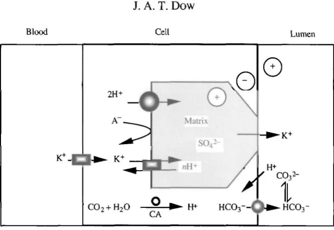
In summary, it is clear that (a) K+ transport through the goblet cavity is energised by a plasma-membrane V-ATPase, which is very closely homologous with endomembrane ATPases; (b) the midgut is very highly polarised electrically; (c) the midgut is functionally subdivisible into (at least) three zones; (d) the anterior and middle regions generate a very high pH by a process dependent on metabolism; (e) K+ transport, high pH and V-ATPase activity are inseparable under physiological conditions; and (f) goblet cavities are not normally acidic.
Blood Lumen
Fig. 13. A model for generation of high gut pH which imbues goblet cells with special properties, but without requiring a high cavity pH. In this case, negative charges on the sulphated glycosaminoglycan matrix within the cavity provide a temporary charge balance for cations within the cavity, preserving near neutral pH. As K+ leaves the cavity, charge balance is maintained by HCO3~, generated by goblet cell carbonic anhydrase. Protons are then stripped from the bicarbonate by the intense electric field near the membrane. The pathway for anion flux might be via columnar cells, but it is tempting to assign the goblet apical valve as the route (see text). Transmembrane transport proteins are shaded with gradients. CA, carbonic anhydrase; A~, anion (unspecified, but chloride in particular).
electrochemical gradient driving it into the gut lumen: Fig. 5), negative charges must follow immediately to maintain charge balance. Perhaps the site for bicarbonate secretion and proton exchange is the columnar microvilli, or better still the goblet cavity valve? The structure of the latter is enormously complex (Flower and Filshie, 1976), and the tortuosity of the path through the valve would allow ample opportunity for transmembrane exchange to occur. Goblet cell carbonic anhydrase would supply H+ to the V-ATPase and HCO3" to a C r / H C O3~ exchanger or HCO3~-ATPase at the goblet
valve. In this case, the function of the cavity would still be to isolate the goblet membrane electrically, as originally hypothesised (Dow, 1984) and later demonstrated (Dow and Peacock, 1989), but now it would be allowing pumped H+ to exchange efficiently with K+, without exposing the site of exchange to the very high pH in the midgut, as this would destroy the local H+ electrochemical gradient, essential for K+ transport, in the immediate vicinity of the membrane. In this case, the cavity is construed as an adaptation to allow electrogenic K+ transport to occur despite a high luminal pH.
drives H+ through the K+/nH+ exchanger in the cavity membrane, and it is precisely this electrical gradient which must be overcome in order to move K+ into the cavity. If the goblet lumen encountered even a subtle hint of the highly alkaline conditions which pertained in the midgut lumen, then the overall exchange in the antiport would become energetically unfavourable.
Is the antiporter electrogenic, either in lepidopteran midgut, or in other insect epithelia? Data from ion-selective electrode experiments on posterior midgut have been argued to demonstrate that a 1:1 K+/H+ exchanger could not operate in the goblet cavity membrane, and that at least 2 protons must exchange for each K+: that is, the exchanger must be electrophoretic (Chao et al. 1991; Wieczorek et al. 1991). However, this may represent an overinterpretation of the data. The enthalpy change (AC) for such an exchanger can be written:
AC =
where A/LH and A/IK are the electrochemical potential differences for protons and potassium ions, respectively, and n is the number of protons simultaneously transported in each cycle.
Considering an electroneutral exchanger (n=l), and dividing by zFto produce values in millivolts, the equation can be expressed:
AC / ak,cyt = 60 ApHcav-cyt - log •
ZF \ ak.cav
where z is the valency (1 for both ions), F is Faraday's constant and ak.cyt and ak.cav are the activities of potassium in cytoplasm and cavity, respectively.
Substituting for published values of pH and OK under short-circuit conditions (shown in Table I), the enthalpy change for a 1:1 exchanger seems slightly unfavourable at around +5 mV, but this considers only the means of the four vital determinations and not their variances. A simultaneous excursion of 1 standard deviation from the mean values embraces a range of energetic possibilites for the antiport, from strongly disfavoured at +85 mV (i.e. running in reverse) to strongly favoured at -75 mV. Under the more physiologically relevant open-circuit conditions, this range is even wider. Although it is beyond dispute that, for n=2 (an electrogenic 2H+:1K+ exchanger), the enthalpy change would be monotonically favourable under this mathematical treatment, it may be premature to discount an electroneutral exchanger on the basis of electrode data.
Table 1. Estimates for goblet cell and cavity pH and &K in posterior midgut, under short-circuit conditions
pH aK(mmoll"')
Cavity 7.23±0.63 94±18 Cytoplasm 7.14±0.50 95±29
What other experimental evidence is there for an electrogenic antiporter? Wieczorek et al. (1991) showed that, in the absence of ATP, vesicle potential could be altered by varying external K+ concentration, and that this effect was abolished in the presence of a non-amiloride Na+/H+ antiport inhibitor. However, these effects were small relative to a paradoxical and unexplained increase in oxonol fluorescence on addition of vesicles, making the data harder to evaluate. By contrast, the PD across the goblet cavity in vivo is extremely high, probably around 250 mV (see discussion in this review). Given that the EMF for the V-ATPase is likely to be around 300 mV, we are left with two explanations: (i) there is an electrogenic exchange process which efficiently dissipates the proton chemical gradient but which is very ineffective in dissipating the PD generated by the ATPase; or (ii) there is an electroneutral exchange process which is extremely effective in exchanging K+ for H+, without dissipating the cavity membrane PD. In this latter case, we might not expect to see a 'smoking gun' in the form of a large standing gradient of either ion. Given that the transepithelial PD is a major determinant of both luminal pH (see discussion above) and of the gradient driving amino acid uptake through the columnar cells, and that these are the best explanations for the existence of the transport in lepidopteran midgut, there would seem to be clear selective advantage in an electroneutral exchanger.
The goblet cavity antiporter shows a similarly high sensitivity to amiloride (Km=
O^mmoll"1) as the electroneutral Na+/H+ exchanger of vertebrate systems. In evolutionary terms, it might be expected that this sensitivity would be more likely to hold after the relatively subtle shift in conformation from a lNa+/l H+ exchanger to a 1M+/1H+ exchanger, than after the extensive remodelling implicit in a shift to a 2M+/1H+ exchanger.
So, according to this model for the role of the goblet cavity, high pH requires a low transmembrane conductivity to strong anions, such as Cl~; a facilitated pathway for movement of H+, OH~, HCO3~ or CO32~; and the isolation of the V-ATPase/antiporter complex from the high pH in the apical lumen, a task which might accurately explain goblet cavity shape. From this analysis, it can also be predicted that there will be analogous adaptations in other insect species with high gut pH, but lacking goblet cells. For example, the site of H+ pumping and M+/H+ exchange may be isolated from the main gut lumen by deep, probably mucus-filled, crypts. It will be interesting to extend these studies to other species with high gut luminal pH.
Additionally, organismal homeostasis requires that one consider a recycling of acid-base equivalents involving blood, anterior/middle midgut, gut lumen and the posterior midgut/tubules/hindgut. However, despite these reservations, it already seems clear that transepithelial PD is a major determining factor of luminal pH (Dow, 1984; Dow and O'Donnell, 1990; Dow and Peacock, 1989), and it is hoped that, by proposing alternative models as working hypotheses for further experiment, a complete solution will be reached more quickly.
This work was funded by the Science Research Council, the Royal Society, the Medical Research Council, and by general Funds of the University of Glasgow. The author is most grateful to Drs F. Lyall, S. H. P. Maddrell and M. J. O'Donnell for their critical reading of the manuscript, and to Professor W. R. Harvey for his helpful discussions on transport models.
References
APPEL, H. M. AND MARTIN, M. M. (1990). Gut redox conditions in herbivorous lepidopteran larvae. J. chem. Ecol. 16, 3277-3290.
APPERSON, M., JENSEN, R. E., SUDA, K., WITTE, C. AND YAFFE, M. P. (1990). A yeast protein,
homologous to the proteolipid of the chromaffin granule proton-ATPase, is important for cell growth. Biochem. biophys. Res. Commun. 168, 574-579.
AZUMA, M. AND EGUCHI, M. (1989). Discrete localization of distinct alkaline phosphatase isozymes in the cell surface of silkworm midgut epithelium. J. exp. Zool. 251, 108-112.
AZUMA, M., TAKEDA, S., YAMAMOTO, H., ENDO, Y. AND EGUCHI, M. (1991). Goblet cell alkaline phosphatase in silkworm midgut epithelium: its entity and role as an ATPase. J. exp. Zool. 258, 294-302.
BASTANI, B., PURCELL, H., HEMKEN, P., TRIGG, D. AND GLUCK, S. (1991). Expression and distribution of
renal vacuolar proton-translocating adenosine triphosphatse in response to chronic alkali loads in the rat. J. din. Invest. 88, 126-136.
BAYON, C. (1980). Volatile fatty acids and methane production in relation to anaerobic carbohydrate fermentation in Oryctes nasicornis larvae (Coleoptera: Scarabaeidae). J. Insect Physiol. 26, 819—828. BERENBAUM, M. (1980). Adaptive significance of midgut pH in larval lepidoptera. Am. Nat. 115,
138-146.
BERTRAM, G., SHLEITHOFF, L., ZIMMERMANN, P. AND WESSING, A. (1991). Bafilomycin-Ai is a potent
inhibitor of urine formation by Malpighian tubules of Drosophila hydei - is a vacuolar-type ATPase involved in ion and fluid secretion? J. Insect Physiol. 37, 201-209.
BIGNELL, D. E. AND ANDERSON, J. M. (1980). Determination of pH and oxygen status in the guts of lower and higher termites. J. Insect Physiol. 26, 183-188.
BIRMAN, S., MEUNIER, F.-M., LESBATS, B., LE CAER, J.-P., ROSSIER, J. AND ISRAEL, M. (1990). A 15 kD proteolipid found in mediatophore preparations from Torpedo electric organ presents high sequence homology with the bovine chromaffin granule protonophore. FEBS Lett. 261, 303-306.
BRADLEY, T. J. (1989). Membrane dynamics in insect Malpighian tubules. Am. J. Physiol. 257, R967-R972.
BROWN, D., SABOLIC, I. AND GLUCK, S. (1991). Colchicine-induced redistribution of proton pumps in kidney epithelial cells. Kidney Int. 40, S79-S83.
BULLA, L. A., BECHTEL, D. B., KRAMER, K. J., SHETNA, Y. I., ARONSON, A. I. AND FITZ-JAMES, P. C. (1980). Ultrastructure, physiology and biochemistry of Bacillus thuringiensis. CRC crit. rev. Microbiol. 8, 147-204.
CHAMBERLIN, M. (1990). Ion transport across the midgut of the tobacco hornworm (Manduca sexta). J. exp. Biol. 150,403-414.
CIOFFI, M. (1979). The morphology and fine structure of the larval midgut of a moth (Manduca sexta) in relation to active ion transport. Tissue & Cell 11,467^-79.
CIOFFI, M. (1984). Comparative ultrastructure of arthropod transporting epithelia. Am Zool. 24, 139-156.
CIOFFI, M. AND HARVEY, W. R. (1981). Comparison of K+ transport in three structurally distinct regions of the insect midgut. J. exp. Biol. 91, 103-116.
DADD, R. H. (1975). Alkalinity within the midgut of mosquito larvae with alkaline-active digestive enzymes./ InsectPhysiol. 21, 1847-1853.
DADD, R. H. (1976). Loss of midgut alkalinity in chilled or narcotized mosquito larvae. Ann. ent. Soc. Am. 69, 248-254.
DEATON, L. E. (1984). Tissue K+-stimulated ATPase and HCCvT-stimulated ATPase in the tobacco hornworm, Manduca sexta. Insect Biochem. 14, 109-114.
Dow, J. A. T. (1984). Extremely high pH in biological systems: a model for carbonate transport. Am. J. Physiol. 246, R633-R635.
Dow, J. A. T. (1986). Insect midgut function. Adv. Insect Physiol 19, 187-328.
Dow, J. A. T., BOYES, B., HARVEY, W. R. AND WOLFERSBERGER, M. G. (1985). An improved chamber for the short-circuiting of epithelia. J. exp. Biol. 116, 685-689.
Dow, J. A. T., GOODWIN, S. F. AND KAISER, K. (1992). Analysis of the gene encoding a 16-kDa proteolipid subunit of the vacuolar H+-ATPase from Manduca sexta midgut and tubules. Gene (in press).
Dow, J. A. T., GUPTA, B. L., HALL, T. A. AND HARVEY, W. R. (1984). X-ray microanalysis of elements in frozen-hydrated sections of an electrogenic K+ transport system: the posterior midgut of tobacco hornworm {Manduca sexta) in vivo and in vitro. J. Membr. Biol. 77, 223-241.
Dow, J. A. T. AND HARVEY, W. R. (1988). The role of midgut electrogenic K+ pump potential difference in regulating lumen K+ and pH in larval lepidoptera. J. exp. Biol. 140, 455-463.
Dow, J. A. T. AND O'DONNELL, M. J. (1990). Reversible alkalinization by Manduca sexta midgut. J. exp. Biol. 150, 247-256.
Dow, J. A. T. AND PEACOCK, J. M. (1989). Microelectrode evidence for the electrical isolation of goblet cavities of the middle midgut of Manduca sexta. J. exp. Biol. 143, 101-114.
FLOWER, N. E. AND FILSHIE, B. K. (1976). Goblet cell membrane differentiations in the midgut of a lepidopteran larva. J. Cell Sci. 20, 357-375.
FORGAC, M. (1992). Structure, function and regulation of the coated vesicle H+-ATPase. J. exp. Biol. 172, 155-169.
GILL, S. S. AND ROSS, L. S. (1991). Molecular-cloning and characterization of the B-subunit of a vacuolar H+-ATPase from the midgut and Malpighian tubules of Helicoverpa virescens. Archs Biochem. Biophys. 291, 92-99.
GILLESPIE, G. A., SOMLO, S., GERMINO, G. G., WEINSTAT-SASLOW, D. AND REEDERS, S. T. (1991). CpG island in the region of an autosomal dominant polycystic kidney disease locus defines the 5' end of a gene encoding a putative proton channel. Proc. natn. Acad. Sci. U.S.A. 88, 4289—4293.
GLUCK, S. L. (1992). Biochemistry of renal V-ATPases. J. exp. Biol. 172, 29-37.
GOLDSTEIN, J. L. AND SWAIN, T. (1965). The inhibition of enzymes by tannins. Phytochem. 4, 185-192. GRAF, R., NOVAK, F. J. S., HARVEY, W. R. AND WIECZOREK, H. (1992). Cloning and sequencing of cDNA
encoding the putative insect plasma membrane V-ATPase subunit A. FEBS Lett. 300,119-122. GUPTA, B. L. (1989). The relationship of mucoid substances and ion and water transport, with new data
on intestinal goblet cells and a model for gastric secretion. Symp. Soc. exp. Biol. 43, 81-110. HAKIM, R. S., BALDWIN, K. M. AND BAYER, P. E. (1988). Cell differentiation in the embryonic midgut of
the tobacco hornworm, Manduca sexta. Tissue & Cell 20, 51-62.
HANADA, H., HASEBE, M., MORIYAMA, Y., MAEDA, M. AND FUTAI, M. (1991). Molecular cloning of
cDNA encoding the 16 kDa subunit of vacuolar H+-ATPase from mouse cerebellum. Biochem. biophys. Res. Comm. 176, 1062-1067.
HANNON, G. J., MARONEY, P. A., DENICER, J. A. AND NILSEN, T. W. (1990). Trans splicing of nematode pre-messenger RNA in vitro. Cell 61, 1247-1255.
HARVEY, W. R., CIOFFI, M., DOW, J. A. T. AND WOLFERSBERGER, M. G. (1983a). Potassium ion transport ATPase in insect epithelia. J. exp. Biol. 106, 91-117.
HARVEY, W. R. AND NEDERGAARD, S. (1964). Sodium-independent active transport of potassium in the isolated midgut of the Cecropia silkworm. Proc. Natn. Acad. Sci. U.S.A. 51, 757-765.
HARVEY, W. R. AND ZEHRAN, K. (1972). Active transport of potassium and other alkali metals by the isolated midgut of the silkworm. Curr. Topics Membr. Transp. 3, 367-410.
HASKELL, J. A., CLEMONS, R. D. AND HARVEY, W. R. (1965). Active transport by the Cecropia midgut. I. Inhibitors, stimulants and potassium-transport. J. cell. comp. Physiol. 65, 45—56.
KLEIN, U., LOFFELMANN, G. AND WIECZOREK, H. (1991). The midgut as a model system for insect K+
-transporting epithelia: immunocytochemical localization of a vacuolar-type H+ pump. J. exp. Biol.
161,61-75.
LAI, S., WATSON, J. C , HANSEN, J. N. AND SZE, H. (1991). Molecular cloning and sequencing of cDNAs encoding the proteolipid subunit of the vacuolar H+-ATPase from a higher plant. J. biol. Chem. 266,
16078-16084.
MADDRELL, S. H. P. (1971). The mechanisms of insect excretory systems. Adv. Insect Phvsiol. 8, 199-331.
MANDEL, M , MORIYAMA, Y., HULMES, J. D., PAN, Y. E., NELSON, H. AND NELSON, N. (1988). cDNA sequence encoding the 16kD proteolipid of chromaffin granules implies gene duplication in the evolution of H+ATPases. Proc. natn. Acad. Sci. U.S.A. 85, 5521-5524.
MARTIN, J. S. AND MARTIN, M. M. (1984). Surfactants: their role in preventing the precipitation of proteins by tannins in insect guts. Oecologia 61, 342-345.
MEAGHER, L., MCLEAN, P. AND FINBOW, M. E. (1990). Sequence of a cDNA from Drosophila coding for the 16 kD proteolipid component of the vacuolar H+-ATPase. Nucleic Acids Res. 18, 6712.
MOFFETT, D. F. AND KOCH, A. R. (1988a). Electrophysiology of K+ transport by midgut epithelium of lepidopteran insect larvae. I. The transbasal electrochemical gradient. J. exp. Biol. 135, 25-38. MOFFETT, D. F. AND KOCH, A. R. (1988b). Electrophysiology of K+ transport by midgut epithelium of
lepidoteran insect larvae. II. The transapical electrochemical gradient. J. exp. Biol. 135, 39-49. MORIYAMA, Y., TAKANO, T. AND OHKUMA, S. (1982). Acridine Orange as a fluorescent probe for
lysosomal proton pump. J. Biochem., Tokyo 92, 1333-1336.
RIDGWAY, R. L. AND MOFFETT, D. F. (1986). Regional differences in the histochemical localization of carbonic anhydrase in the midgut of tobacco hornworm (Manduca sexta). J. exp. Zool. 237,407-412.
SCHWEIKL, H., KLEIN, U., SCHINDLEBECK, M. AND WIECZOREK, H. (1989). A vacuolar-type ATPase,
partially purified from potassium transporting plasma membranes of tobacco hornworm midgut. J. biol. Chem. 264, 11136-11142.
SHIH, C.-K., KWONG, J., MONTALVO, E. AND NEFF, N. (1990). Expression of a proteolipid gene from a high copy-number plasmid confers trifluoperizine resistance to Saccharomyces cerevisiae. Molec. cell. Biol. 10, 3397-3404.
STEWART, P. A. (1981). How to Understand Acid-Base. New York: Edward Arnold.
TOYAMA, R., GOLDSTEIN, D. J., SCHLEGEL, R. AND DHAR, R. (1991). A genomic sequence of the
Schizosaccharomyces pombe 16 kDa vacuolar H+-ATPase. Yeast 7, 989-991.
TURBECK, B. O. AND FODER, B. (1970). Studies on a carbonic anhydrase from the midgut epithelium of larvae of lepidoptera. Biochim. biophys. Acta 212, 134-138.
UMEMOTO, N., OHYA, Y. AND ANRAKU, Y. (1991). VMA1I, a novel gene that encodes a putative proteolipid, is indispensable for expression of yeast vacuolar membrane H+-ATPase activity. J. biol. Chem. 266, 24526-24532.
WATERHOUSE, D. F. (1949). The hydrogen ion concentration in the alimentary canal of larval and adult lepidoptera. Aust. J. Sci. Res. B 132,428-437.
WIECZOREK, H., PUTZENLECHNER, M., ZEISKE, W. AND KLEIN, U. (1991). A vacuolar-type proton pump energizes K+/H+ antiport in an animal plasma membrane. J. biol. Chem. 266, 15340-15347.
WIECZOREK, H., WEERTH, S., SCHINDLEBECK, M. AND KLEIN, U. (1989). A vacuolar-type proton pump in a vesicle fraction enriched with potassium transporting plasma membranes from tobacco hornworm midgut. J. biol. Chem. 264, 11143-11148.
WIECZOREK, H., WOLFERSBERGER, M. G., CIOFFI, M. AND HARVEY, W. R. (1986). Unique ATPase