PHYSIOLOGICAL MODULATION OF GAP JUNCTION
PERMEABILITY
BY JACQUES NEYTON AND ALAIN TRAUTMANN
Laboratoire de Neurobiologie, Ecole Normale Superieure, 46, rue d'Ulm, 75005 Paris, France
SUMMARY
In many tissues cells communicate directly through arrays of intercellular channels which are organized to form gap junctions. These channels are permeant to inorganic ions as well as to small hydrophilic molecules up to Mr 2000. The electrical
and chemical coupling provided by such junctions is under the control of intra-cellular and, in many cases, extraintra-cellular substances. The latter (hormones or neurotransmitters) function via the activation of intracellular second messengers. These can rapidly affect the state of opening of the junctions, or induce long-term modulation of the coupling. What are the second messengers and how do they control the functional state of the junctions? These questions' remain largely unanswered, although several internal molecules are thought to be involved in these modulations (e.g. Ca2+, H+ or cyclic AMP). The double patch-clamp technique which enables control of both the intracellular milieu and high resolution measure-ment of transjunctional currents, has recently been applied to study these problems. In particular, it is now possible to examine at the single channel level how junctional conductance is modulated in terms, for example, of the number of open channels or channel elementary properties.
INTRODUCTION
In most animal tissues, direct intercellular communication occurs through gap junctions (see Bennett & Goodenough, 1978; Larsen, 1983). This form of communi-cation can be temporarily increased, reduced or even interrupted. This review will consider where, how and why such modulations take place. Gap junctions were first defined morphologically as specialized intercellular junctions with a characteristic extracellular space (the gap) of 2-3 nm between the membranes of the two adjoining cells. In freeze fracture preparations, these junctions appear as typical aggregates of particles on the p face, and as complementary pits on the e face. There is a good correlation between the presence of morphologically identified gap junctions and the existence of intercellular communication (demonstrated by electrical coupling or by the passage of tracer molecules) between coupled cells, and it is now generally agreed that the gap junction is the structure which mediates this communication and that the intercellular channel is most probably contained in the gap junction particles (see below). Only rarely can electrical coupling not be correlated with the observation of a
classical gap junction structure (Azarnia & Loewenstein, 1977; Williams & De Haan, 1981). Nevertheless, in these exceptional cases, sparse particles, similar to those found as clusters in gap junctions, have been observed to cross the extracellular space between the cells, and may constitute junctional channels (Larsen, Azarnia & Loewenstein, 1977). Thus, to avoid systematic circumlocutions, we will use the term 'gap junction' without limitation to specific morphological data, and even in cases where morphological identification of gap junctions has not been performed.
The first part of this paper will describe the heterogeneity of gap junction structure and function. Succeeding sections will deal with the long-term regulation (e.g. action of hormones) and the short-term modifications (e.g. those induced by neuro-transmitters).
Both morphological and functional methods have been used to study the modu-lations of gap junctions. The morphological data have been reviewed and discussed elsewhere (e.g. Larsen, 1983; Peracchia & Bernardini, 1984; but see also the recent results of Page, Karrison & Upshaw-Earley, 1983; Hanna, Pappas & Bennett, 1984; Green & Severs, 1984; Miller & Goodenough, 1985). Functional studies deal with the transfer of molecules through gap junctions. The transfer of tracer molecules from one cell to another can be followed by optical techniques, for fluorescent compounds, or by autoradiography, for radiolabelled markers. These methods give interesting, but mainly qualitative information on the permeability state of a gap junction. They must therefore be used with caution when studying the mechanisms of gap junction modulation. As shown by Zimmerman & Rose (1985), the size of an open gap junction channel cannot be derived with certainty from measurements of cell-to-cell transfer of fluorescent dyes of various sizes, because the measures of this transfer also depend on the detection threshold and on the possibility of loss and sequestration of the tracers. Another approach is to study electrical coupling (i.e. the transfer of small ions between coupled cells). The simplest method used is to measure the coupling coefficient, the ratio V2/V1, between a voltage VI applied in one cell and the resulting change V2 measured in a coupled cell. This technique does not allow a quantitative characterization of the properties of the gap junction channel because the coupling coefficient depends on the resistances of both the junctional and the nonjunctional membranes. Indeed, as pointed out by Socolar (1977), with this technique a marked change in the junctional conductance may leave the coupling coefficient almost unchanged and, conversely, a change in the nonjunctional mem-brane resistance can modify the coupling coefficient, even in the absence of gap junction modification. Examples of this latter case have been reported (Spira & Bennett, 1972; Carew & Kandel, 1976; Spira, Spray & Bennett, 1980).
This limitation can now be overcome in some cases, with the whole cell tight-seal recording method (Marty & Neher, 1983), which can be applied simultaneously to both coupled cells (Neyton & Trautmann, 1985). With this method, as in the case of a double voltage-clamp system (using four electrodes), the transjunctional current can be measured by stepping the voltage in one cell and measuring the induced current, at constant voltage, in the other. There are, however, several differences between the two techniques. For the double patch-clamp, only two electrodes are used instead of four. As a result, the voltages are selected, but not measured, and there may be important series resistance artifacts which have to be corrected for. It is possible to use this method with pairs of small cells (10—20 ^zm diameter) and to obtain such a low background noise from the whole cell membrane that, in many cases, single channel currents through the gap junctions are measurable, when only a few channels are simultaneously open (Fig. 1). The double patch-clamp method was used on a preparation of isolated pairs of lacrimal gland cells to measure the ionic selectivity of the junctional channel, its elementary conductance and to study its
2s
+50 -50
400
0 +50
mV
PA
mV - 5 0 •
0i 0
IT °J
[image:3.451.40.414.290.506.2]u -200 \- 1 0 pA
Fig. 2. Kinetics of a junctional channel in a pair of rat lacrimocytes. With a steady transjunctional voltage (V2 = —40mV, Vj = +50mV), all the junctional channels were closed before one opened. It then oscillated for 5 6 s between the open and closed states (A). At two instants marked by the arrows, the current trace I2 was expanded, showing that transitions may be either much slower (exponential time constant = 14 ms in B) or faster (C) than the time detection threshold (0-5 ms in this experiment).
voltage sensitivity (Neyton & Trautmann, 1985). It was found that transitions between the open and closed states are often unusually slow, extending over tens of milliseconds. Besides these slow transitions, fast transitions (in the submillisecond range) can occur (Fig. 2).
THE INTERCELLULAR CHANNEL
can be found elsewhere (Loewenstein, 1981; Peracchia, 1985; Revel, Nicholson & Yancey, 1985; Spray & Bennett, 1985).
Structure of the junctional channel
It is generally admitted that the gap junction particles observed in freeze fracture are the morphological counterpart of the intercellular channel. X-ray diffraction (Caspar, Goodenough, Makowski & Phillips, 1977; Makowski, Caspar, Phillips & Goodenough, 1977) and Fourier analysis of electron micrographs (Unwin & Zampighi, 1980) have shown that a junctional channel consists of two hexameric proteins, one contributed by each membrane. These paired hexamers form along their central axis an aqueous pore which connects the cytoplasms of the two cells.
This general scheme raises different questions. What are the subunits of this dodecameric protein complex? Are they identical in different gap junctions? What is the three-dimensional structure of the complex? Partial answers have been given to these questions. The isolation and characterization of the subunit of the gap junction channel complex is still a subject of controversy. Most authors, nevertheless, agree that in several tissues the subunit is a protein of Mr = 26 000-28 000 (e.g. review by
Revel et al. 1985; but for other values see also Finbow, Shuttleworth, Hamilton & Pitts, 1983; Manjunath, Goings & Page, 1984; Warner, Guthrie & Gilula, 1984). Peptide maps (Gros, Nicholson & Revel, 1983) and partial sequencing (Nicholson
et al. 1983; Revel, Nicholson & Yancey, 1984) indicate that the primary sequence of
the subunits appears to vary widely accordingly to the tissues, even in the same species. Such a broad variation was not observed between different species for the
same tissue (Nicholson et al. 1981; Zigler & Horwitz, 1981). Nevertheless, the
subunits isolated from different tissues often share common antigenic properties (for review see Hertzberg, 1985). Thus, parts of these proteins have probably been conserved, while other parts have somehow become tissue specific. Consequently, heterogeneity is expected in the three-dimensional structure of the junctional channels of different origins. Such heterogeneity has, in fact, been observed in isolated gap junctions from heart and liver (see Page & Manjunath, 1985). The cytoplasmic surfaces of cardiac gap junctions (isolated in the presence of proteolysis inhibitors) are covered with a layer of fuzzy material which was never seen in liver junctions. The presence of this material is associated with the isolation of a major protein (presumably the subunit) of MT = 44 000-47 000. This protein is cleaved to A/r = 29 000 when proteolysis is not inhibited. Thus, a large part of the cardiac
junction subunit appears to protrude on the cytoplasmic face of the junction. An equivalent of this protruding part is absent in liver junctions. In this tissue, however, the cytoplasmic part of the subunits has a complex organization. In particular, the hexamers together form a small structure which can plug the mouth of the channel (Makowski et al. 1984). This result supports the idea that gating mechanisms involve changes in the structure of the cytoplasmic part of the channel (Makowski et al.
1984) and may not be identical in all gap junctions.
their permeability directly: transjunctional or transmembrane voltage, intracellular calcium concentration and internal pH. Yet the sensitivities to these parameters vary among gap junctions of different tissues.
Voltage dependence
Some junctions are highly sensitive to voltage applied across the junction. This is particularly true for certain electrical 'rectifying' synapses (Furshpan & Potter, 1959; Auerbach & Bennett, 1969; Nicholls & Purves, 1972), where a strong rectification can occur (i.e. currents pass through the junction more easily in one direction than in the other). At these junctions, dye transfer also depends on the polarization of the junction (Margiotta & Walcott, 1983; Giaume & Korn, 1984). These channels thus possess a dipole-like, voltage-sensitive structure which may move in the electric field. Other junctions present a symmetrical, high sensitivity to voltage applied either across the junction (Spray, Harris & Bennett, 1979; White, Spray, Campos de Carvalho & Bennett, 1982; Spitzer, 1982) or between the interior and the exterior of each of the coupled cells (Obaid et al. 1983; Spray et al. 1984). Such junctions probably possess two voltage-sensitive structures, instead of one as in the rectifying electrical synapse. More commonly, other junctions are apparently devoid of such structures since they are totally voltage insensitive (see review by Spray, White, Verselis & Bennett, 1985).
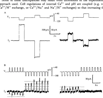
An intermediate situation is found in lacrimal glands. Fully open junctions are always voltage insensitive. This is also the case for some partly closed junctions. In Fig. 3A, the junction exhibited a similar voltage insensitivity at the beginning of the recording (G = 40 nS) as it did 9 min later, when it was almost closed (G = 600 pS). However, a voltage sensitivity was observed in some junctions that were almost completely closed. In these cases, the number of open channels depended upon the transjunctional voltage (but not on the membrane potential). In the experiment illustrated in Fig. 3B, a transjunctional voltage of one polarity caused more channels to open, whereas voltage jumps of the other polarity induced channel closure. When one or two channels at a time were open, their conductance could be measured, and apparently it was unaffected by voltage (see dotted lines). Thus, in gap junctions between lacrimocytes, when a voltage dependence is present, the potential appears to affect the number of open channels and not their elementary conductance. A similar situation seems to exist in the Chironomus salivary gland (Zimmerman & Rose, 1985). At least three kinetic processes can be revealed by the voltage dependence in the lacrimocyte preparation. They can occur in the submillisecond range (apparently instantaneous rectification), and in the range of tens of milliseconds and of tens of seconds (Fig. 4, see the legend for more details). The variability in the voltage sensitivity of lacrimal gland gap junctions suggests that this is not an intrinsic property of the lacrimocyte junctional channels.
pH and calcium dependence
Lowenstein, 1981; Spray & Bennett, 1985). However, the range of Caz+ and H+ intracellular concentration at which these modulations do occur, and the indepen-dency of their mechanisms, are still a matter of debate. For example, the minimum Ca,+ concentration inducing a decrease in gap junction permeability has been claimed to be either in the micromolar range (Dahl & Isenberg, 1980) or in the millimolar range (Spray, Stern, Harris & Bennett, 1982). Junction gating, by a decrease in pH;, seems to occur in a more restricted range with a pKa for the gating
varying between 6-4 for mammalian liver junctions (Spray & Hertzberg, 1985) and 7-3 for coupled fish blastomeres (White et al. 1982). An exception is nevertheless found in the lens, where coupling between the fibres appears to be poorly sensitive to CO2 exposure (Scheutze & Goodenough, 1982).
Some of these discrepancies may result from differences in the experimental approach used. Cell regulations of internal Ca2+ and pH are coupled (e.g. via Ca +/ H+ exchange, or Ca +/ N a+ and N a+/ H+ exchanges) so that increasing the
+40
-120
500pA I 50pA 2s
mV +40
[image:7.451.44.404.222.565.2]-160
nnni
B
-150
30 pA
400 ms
j^rr
. . r u 4 - V t W * ^
40s Fig. 4. Voltage dependence of junctional channels in pairs of lacrimocytes. (A) An example of a junction where a transjunctional voltage of one polarity increases the number of open channels, whereas the other polarity causes a channel closure, by two different kinetic processes. The first one, apparently completed in a few milliseconds, reduced the number of open channels from five (at the end of the negative voltage jump) to two (at the beginning of the positive voltage jump). During that later jump, another channel closed, with a time constant of 118 ms. Processes on the same time scale have been described in Fig. 2. (B) The level of coupling was too high in this other junction to allow a resolution of the transjunctional current at the single channel level. A transjunctional voltage of + 100mV slowly reduced the junctional current, with a more-or-less exponential time course (time constant of the exponential fit = 51 s). The meaning of the current changes observed on one channel and on a population of channels are, of course, completely different.
a protein which shows conformational changes upon binding with calcium or protons (see e.g. Pundak & Roche, 1984), could be this intermediate. This protein indeed binds to lens and liver gap junction proteins (Hertzberg & Gilula, 1981; Welsh et al. 1982). Moreover, calmodulin inhibitors inhibit the CO2-induced uncoupling of amphibian embryonic cells (Peracchia, Bernardini & Peracchia, 1983; Peracchia, 1984) and can also produce uncoupling in the epidermis of an insect (Lees-Miller & Caveney, 1982). Lastly, calmodulin is required to confer a calcium-sensitive gating to channels obtained by reincorporation into liposomes of lens isolated gap junction proteins (Girsch & Peracchia, 1985). Calmodulin may thus be directly involved, at least in the lens, in the gating mechanism of junctional channels by calcium. However, an indirect involvement of calmodulin (e.g. through the control of a protein kinase), or even the possibility of a direct action of H+ or Ca2+ ions on the channel structure itself cannot be excluded.
LONG-TERM MODULATION OF GAP JUNCTIONS
Long-term modulation of direct cell-to-cell communication has been described in several tissues, during development or in response to extracellular messengers. These modulations could result from changes in the properties of the elementary channels or in their number (by acting in the latter case on their turnover mech-anisms). Gap junctions form in the so-called formation plaques (Johnson, Hammer, Sheridan & Revel, 1974) where channel assembly and functional maturation is thought to occur (see Larsen, 1985). Gap junctions may disappear, and their removal from the surface of contact between the coupled cells takes place through an endocytosis by one of the two cells (see Larsen, 1985). We will review some examples of long-term modulations, illustrating and discussing these different mechanisms.
Uterine muscle
In the uterine muscle of pregnant mammals, a change in the concentration of circulating hormones appears to occur just prior to parturition and may be respon-sible for it (see Thorburn & Challis, 1979). It consists of an increase in oestrogen and prostaglandins F2, El and E2, and a decrease in progesterone and prostaglandin 12. These hormonal changes are paralleled by the appearance of large and numerous gap junctions between myometrial cells (Garfield, Sims & Daniel, 1977), an increase in electrical coupling (Sims, Daniel & Garfield, 1982) and metabolic cooperation (Cole, Garfield & Kirkaldy, 1985). This correlation strongly suggested that gap junction formation was hormonally controlled. This hypothesis is supported by the in vivo and in vitro application of exogenous analogues of the hormones (see the review of Cole & Garfield, 1985).
oestrogen might activate the neosynthesis of macromolecules involved in the control of other stages of the gap junction formation process (e.g. affecting cell surface properties or channel subunit assembly). Such an alternative has indeed been found in a very different preparation: the C1-1D cell line (a malignant subline of mouse L-cells), in which the formation of gap junctions is induced by an increase in intracellular cyclic AMP (Azarnia, Dahl & Loewenstein, 1981). Even if, in this case, the junction formation requires protein synthesis, the amount of 27-kD junctional channel subunit, measured with specific antibodies, does not change when the junction formation is induced (see Hertzberg, 1985).
Ovarian follicle
In the mammalian ovarian follicle, the oocyte is enclosed by a population of granulosa cells whose multiplication and differentiation is controlled by gonado-tropins and steroid hormones. At different stages of follicular growth, gap junctions are found between granulosa cells and between granulosa cells and the oocyte (see e.g. Burghardt & Matheson, 1982). Interestingly, the level of intercellular commun-ication in the follicle appears to be hormonally controlled through a modulation of the processes of gap junction formation and removal. During the early growth of ovarian follicles in hypophysectomized animals, the appearance of gap junctions between granulosa cells does not require any hormonal stimulation, but the number and size of the junctions are markedly enhanced by follicle-stimulating hormone (FSH) (Burghardt & Matheson, 1982). In the later stages of follicular growth, FSH also stimulates the internalization of gap junctions in granulosa cells.
In contrast, a few hours after the application to hypophysectomized animals of ovulatory doses of hCG (human chorionic gonadotropin, a hormone which binds to granulosa cell receptors for LH, the luteinizing hormone), the coupling between granulosa cells and oocyte decreases, and a dissociation of granulosa cells is observed in preovulatory follicles (Gilula, Epstein & Beers, 1978; Moor, Smith & Dawson, 1980; Eppig, 1982). These hCG effects are paralleled by a decrease in the number of gap junctions between oocyte and granulosa cells (Gilula et al. 1978) and by a stimulation of gap junction removal in granulosa cells (see Larsen, 1985). The hCG effects were mimicked in some in vitro preparations by applications of LH (Dekel, Lawrence, Gilula & Beers, 1981) and in other preparations by FSH (Moor et al. 1980). It should be emphasized that ovulation in vivo is preceded by a peak in the plasma oestrogen concentration, followed rapidly by a surge in LH and FSH concentrations.
and, in some preparations, pharmacological treatments that increase internal cyclic AMP mimic the hormones' actions on granulosa cell gap junctions (Dekel et al. 1981). An increase in intracellular cyclic AMP stimulates gap junction formation in other preparations including various mammalian cell lines (Flagg-Newton, Dahl & Loewenstein, 1981; Azarnia et al. 1981; Radu, Dahl & Loewenstein, 1982) and cultured rat sympathetic neurones (Kessler, Spray, Saez & Bennett, 1984). In these preparations, the cyclic-AMP-induced gap junction formation requires RNA and protein synthesis (Azarnia et al. 1981; Kessler et al. 1984) and may result from protein phosphorylation by a cyclic-AMP-dependent protein kinase (Wiener & Loewenstein, 1983). The target(s) of this kinase and the consequences of this protein phosphorylation are unknown.
Insect larval tissues
In the salivary glands of Drosophila hydrei larvae (Hax, van Venroogi & Vossenberg, 1974) and in the larval epidermis of the beetle Tenebrio tnolitor (Caveney & Blennerhasset, 1980), the moulting hormone 20-hydroxyecdysone induces, within a few hours after application, an increase in electrical coupling. This results from modifications of both the junctional and nonjunctional membranes. In contrast with the two examples given above (uterine muscle and ovarian follicle), the increase in coupling conductance observed in these cases is due to a modification of the properties of pre-existing junctional channels (presumably a change in their gating kinetics, see Caveney & Safranyos, 1985) rather than to an addition of new ones to the junctions (Caveney, Berdan & McLean, 1980). The hormonal action does not involve protein synthesis, but, surprisingly, can be prevented by actinomycin D, an inhibitor of RNA synthesis (Caveney et al. 1980). An increase in internal cyclic AMP has been claimed both to reverse the effect of 20-hydroxyecdysone, in the beetle epidermis (Caveney, 1978), and to have the contrary action, by mimicking the action of the hormone in the Drosophila salivary gland (Haxet al. 1974). In the latter case, however, a rise in internal cyclic AMP induces a hyperpolarization of the cells which, per se, elicits an increase in the voltage-sensitive junctional conductance (see above).
SHORT-TERM MODULATION OF GAP JUNCTION CONDUCTANCE
either pancreas or lacrimal glands (Iwatsuki & Petersen, 1978a; Neyton & Trautmann, 1986) and dopamine (DA) can uncouple horizontal cells of turtle or fish retina (Gerschenfeld, Neyton, Piccolino & Witkovsky, 1982; Teranishi, Negishi & Kato, 1983; Piccolino, Neyton & Gerschenfeld, 1984).
Modulation of the permeability of horizontal cell gap junctions by dopamine
The axon terminals of the HI horizontal cells (HIATs) in the turtle retina, like the HI cell bodies in the fish retina, are electrically coupled by extensive gap junctions (see e.g. Witkovsky, Owen & Woodworth, 1983) and constitute large functional networks. The hyperpolarizing responses of these cells to light spot stimuli still increases when the diameter of the spot enlarges far beyond their anatomical arborization under control conditions. Thus these cells possess a very large receptive field. Preliminary experiments by Negishi & Drujan (1979) showed that DA causes a narrowing of the receptive field profile of fish horizontal cells. A similar effect of DA was also found in the turtle retina (Gerschenfeld et al. 1982; Piccolino et al. 1984). The hyperpolarization provoked in an H1AT by a light spot of small diameter (i.e. which covers only the central part of its receptive field) is increased in the presence of DA at micromolar concentrations (maximal effect at 2— lO/imolP1). When the light stimulates the periphery of the receptive field of an HI AT (annulus of light) the light response of the peripheral axon terminals is less well transmitted to the central one in the presence of DA. The onset of the DA action (2-5 min) is related to the DA diffusion in the tissue and, possibly, to the production of second messengers (see below). Such a reduction of the size of the HI AT receptive field indicates that DA reduces the coupling in the HI AT network. This decrease of coupling could result either from an increase in the nonjunctional membrane conductance of the axon terminals, or from a decrease in their junctional conductance. The latter case has been demonstrated by two in-dependent methods. First, the spread of intracellu-larly injected current in the H1AT network was examined. The potential change measured in an H1AT, in response to the injection of current in another H1AT (located at less than 0-1 mm from the first one), was increased by DA. This result may appear paradoxical. Yet reduction of gap junction conductance induces a shrinkage of the functional network. This will reduce the spread of current in the furthest HIATs, but produce in the injected axon terminal and its closest neighbours larger voltage changes for a given current injection. In contrast, an increase of the nonjunctional conductance would also shrink the network, but the amplitude of the electrotonic potentials evoked by the current injection would decrease in all the cells of the network.
receptive field to light stimuli, spread of current, spread of a dye) thus give convergent results, suggesting that DA closes the gap junctions between HIATs in the turtle retina. The DA receptors involved in that modulation are certainly localized on the horizontal cell itself and not in another interneurone, because the effects of DA persist when synaptic transmission is blocked by 2mmoll~1 cobalt. Moreover, it was shown that an endogenous release of DA may physiologically modulate the coupling in the turtle HI AT network. Indeed, the effects produced by DA can be mimicked by the application of drugs known to stimulate the release of DA by nerve terminals: both amphetamine and DOPA reduce the H1AT receptive field.
Cyclic AMP appears to be involved as a second messenger in the action of DA on the gap junctions of HIATs. Its inhibitory action on gap junction conductance is quite different from that previously described on long-term modulation. The evidence for this is as follows.
(1) The effects of DA on HI AT gap junctions are blocked by DA antag-onists specific for the Dl receptor type; in the retina, as in the brain, the acti-vation of dopamine Dl receptors stimulates the adenylate cyclase (Kebabian, Petzold & Greengard, 1972; Brown & Makman, 1972; Watling & Dowling, 1981).
(2) Forskolin (a compound known to directly activate the cyclase) causes a closure of gap junctions between HIATs as measured by the three methods described above (see Fig. 5C).
(3) Similar effects are obtained by inhibiting the degradation of cyclic AMP: if the action of the phosphodiesterase is inhibited by isobutylmethylxanthine (IBMX) or theophylline, the HIATs gap junctions appear to close (see Fig. 5D).
Finally, the intercellular communication in the network of the axon terminals of turtle HI horizontal cells may be physiologically modulated by dopaminergic terminals, which, by releasing DA, could evoke an increase in the concentration of cyclic AMP in the H1 ATs. A similar conclusion has also been reached in experiments with an isolated fish retina preparation (Teranishi et al. 1983), or on pairs of cultured, isolated horizontal cells (Lasater & Dowling, 1985). However, it is still not known if cyclic AMP acts directly on the gap junction channels of horizontal cells, or if another intermediate, like a cyclic-AMP-dependent protein kinase, is needed in its action.
Fig. 5. Diffusion of Lucifer Yellow in the network of axon terminals of turtle HI horizontal cells. (A) The dye injection was performed in a retina bathed in normal Ringer solution. The Lucifer Yellow diffused from the injected HI AT into a complex network of interconnected HIATs. The dye also backfilled some HI cell bodies through the fine axon fibres which connect the cell bodies to their axon terminal. (B) In this retina, after a bath application of lOjwnoll" dopamine, the diffusion of the dye was restricted to the injected HI AT and its cell body. (C) A similar restriction is observed with a bath application of lOf/moll"1 forskolin. (D) Restriction of Lucifer Yellow diffusion induced by SO/imoll"1 isobutylmethylxanthine. (Results from Piccolino, Neyton & Gerschenfeld, 1984). Scale bar, 100/un.
via an increase in intracellular cyclic AMP and a subsequent phosphorylation of the
junctional channels.
Modulation of a gap junction permeability by acetylcholine
description of gap junction closure by ACh in these glands was based on the coupling coefficient method (Iwatsuki & Petersen, 1978a) with its unavoidable uncertainties. The apparent simultaneity of junction closure and increase in internal calcium provoked by ACh suggests a causal link between the two phenomena (Iwatsuki & Petersen, 19786). We have re-examined this problem using the double patch-clamp technique on isolated pairs of lacrimal gland cells (see above). We first studied the effect of internal calcium on the junction by measuring, under various intracellular calcium concentrations, the stability of the coupling as a function of time after double dialysis. The possible interactions between Ca2+ and pH, could be avoided in this system, because the concentrations of both ions were strongly buffered. The accuracy of the measurements was limited to some extent by a systematic and spontaneous rundown of the coupling following double-cell dialysis, even when Ca2+ and H+ internal concentrations were kept low. At first sight this is a disturbing phenomenon, but it reveals more interesting processes. There is a high level of noise in the transjunctional current during this rundown, indicating that a number of channels are fluctuating between two or more levels of conductance. The functional elimination of channels during the rundown is thus not an instantaneous process: the junctional channels appear to be open most of the time at the beginning of an experiment, then oscillate for some time between the open and closed states, and finally shut.
The effect of increased Caf+ on the coupling was estimated, statistically, by measuring the rate of junctional conductance rundown. As expected, increasing Caf+ could speed up this rundown, but quite high concentrations were needed to get an effect: l/Umoll"1 was ineffective; 10/xmoll"1 Ca2+ caused a closure of the junction within a few seconds. These values, which are similar to those reported in other preparations (Rose & Loewenstein, 1976; Rose & Rick, 1978; seem too large to be likely in a living cell.
closure by ACh. This suspicion became a certainty with a second series of exper-iments in which the internal calcium concentration was strongly buffered in both cells at a low value (pCa8, see Fig. 6B). Under these conditions, ACh failed to increase Ca, (as judged by the absence of calcium-dependent currents), but uncoupling (preceded by a transient increase in coupling) still occurred. The uncoupling was obviously not calcium- or proton-dependent (since the internal pH of these cells was strongly buffered at pH 7-2).
Do the effects of ACh result from changes in the number of open junctional channels or alteration in their elementary properties? This is a difficult question to answer at the single channel level, because several levels of conductance occur which correspond to the closed state, one, or possibly more, fully open state in the range
l
nA 05
nS 5
-0 L
70—180 pS, and several intermediate states (partially opened or partially obstructed). Despite this complexity, it is clear that closure of the junction by ACh does not result simply from an increased probability of the channel being in an intermediate, low-conductance state. It seems that during ACh action some channels close completely (but reversibly) while others remain fully open. Thus, the mechanism which had been previously suggested for calcium-induced closure of gap junctions (i.e. decrease of the diameter of the junctional channels: Rose, Simpson & Loewenstein, 1977) does not apparently occur in the case of the ACh-induced modulation of the gap junctions between lacrimocytes. It should also be emphasized that the experimental evidence for the hypothesis of partial channel closure, which is based on measure-ment of the diffusion of various fluorescent molecules, does not appear to be reliable (Zimmerman & Rose, 1985). The mechanism of the transient increase in gap junction conductance at the beginning of the ACh action is still unclear. It might result from a slight increase in the elementary conductance of the open junction channels.
In summary, an application of ACh on acinar cells from lacrimal glands causes an increase in Caf+. This increase controls the secretion of both enzymes, by exocytosis, and of ions, through channels and pumps (Marty et al. 1984). On the other hand, ACh induces a delayed and long-lasting closure of gap junction channels which can be reversible. We ignore at the moment the nature of the second messenger involved in this latter phenomenon, but it clearly does not result from a rise in Ca, + . Trisphosphoinositol is apparently involved in the internal calcium release induced by ACh in these cells (Evans & Marty, 1986). This raises the question of the role of phosphoinositide breakdown in the ACh-induced uncoupling. What the function of this junction closure could be is a puzzling question. We suggest that it could be used during prolonged gland stimulation, which probably requires a lot of energy and produces important concentration changes inside the cells. By disconnecting the lacrimocytes, ACh might allow a discontinuous secretion by each individual cell with resting periods. But to answer this question it is necessary to know the extent and the kinetics of junction closure in vivo (i.e. under the action of nerve-released ACh, in nondialysed cells).
CONCLUDING REMARKS
a syncitial behaviour. Indeed, functional differences between two coupled cells may be preserved by a reversible closure of the junctional channels. It is likely that a limited number of intracellular mechanisms are directly involved in the control of junctional channel gating, and that different physiological modulators are able to activate or inhibit such common mechanisms.
We wish to thank Dr M. Piccolino, who kindly gave us Fig. 5, and Dr H. M. Gerschenfeld for his comments on the manuscript. Supported by CNRS (ATP Pharmacologie des re'cepteurs des neurome'diateurs), by the Ministere de la Recherche et de la Technologie (Aides 83.C.0914 et 85.C.1138) and by the University Pierre et Marie Curie.
REFERENCES
AUERBACH, A. A. & BENNETT, M. V. L. (1969). A rectifying electrotonic synapse in the central nervous system of a vertebrate. J. gen. Physiol. 53, 211-237.
AZARNIA, R., DAHL, G. & LOEWENSTEIN, W. R. (1981). Cell junction and cyclic AMP: I I I . Promotion of junctional permeability and junctional membrane particles in a junction-deficient cell type. J. Membrane Biol. 63, 133-146.
AZARNIA, R. & LOEWENSTEIN, W. R. (1977). Intercellular communication and tissue growth. VIII. A genetic analysis of junctional communication and cancerous growth. J. Membrane Biol. 34, 1-37.
BENNETT, M. V. L. & GOODENOUGH, D. M. (1978). Gap junctions, electrotonic coupling and intercellular communication. Neurosci. Res. Prog. Bull. 16, 373-486.
BENNETT, M. V. L . , SPIRA, M. E. & SPRAY, D . C. (1978). Permeability of gap junctions between embryonic cells of Fundulus: a reevaluation. Devi Biol. 65, 114—125.
BROWN, J. H. & MAKMAN, M. H. (1972). Stimulation by dopamine of adenylate cyclase in retinal homogenate and of adenosine 3',S'-cyclic monophosphate formation in intact retina. Proc. natn.
Acad. Sci. U.SA. 69, 539-543.
BuRGHARDT, R. C. & MATHESON, R. L. (1982). Gap junction amplification in rat ovarian granulosa cells. I. A direct response to follicle-stimulating hormone. Devi Biol. 94, 206—215. CAREW, T . J. & KANDEL, E. R. (1976). Functional effects of decreased conductance EPSP:
synaptic augmentation and increased electrotonic coupling. Science 192, 150-153.
CASPAR, D . L. D . , GOODENOUGH, D . A., MAKOWSKI, L. & PHILLIPS, W. C. (1977). Gap junction structures. I. Correlated electron microscopy and X-ray diffraction. J. Cell Biol. 74, 605-628. CAVENEY, S. (1978). Intercellular communication in insect development is hormonally controlled.
Science 119, 192-195.
CAVENEY, S., BERDAN, R. C. & MCLEAN, S. (1980). Cell-to-cell ionic communication stimulated by 20-hydroxyecdysone occurs in the absence of protein synthesis and gap junction growth.
J. Insect Physiol. 26, 557-567.
CAVENEY, S. & BLENNERHASSET, M. G. (1980). Elevation of ionic conductance between insect epidermal cells by /J-ecdysone in vitro. J. Insect Physiol. 26, 13-25.
CAVENEY, S. & SAFRANYOS, R. (1985). Control of molecular movement within a developmental compartment. In Gap Junctions (ed. M. V. L. Bennett & D. C. Spray), pp. 265-273. Cold Spring Harbor Laboratory.
COLE, W. C. & GARFIELD, R. E. (1985). Alterations in coupling in uterine smooth muscle. In Gap
Junctions (ed. M. V. L. Bennett & D. C. Spray), pp. 215-230. Cold Spring Harbor Laboratory. COLE, \V. C , GARFIELD, R. E. & KIRKALDY, J. S. (1985). Gap junctions and direct intercellular
communication between rat uterine smooth muscle cells. Am. J. Physiol. 249, C20—C31. DAHL, G. & ISENBERG, G. (1980). Decoupling of heart muscle cells: correlation with increased
DEKEL, N., LAWRENCE, T . S., GILULA, N. B. & BEERS, W. H. (1981). Modulation of cell-to-cell
communication in the cumulus-oocyte complex and the regulation of oocyte maturation by L.H.
Devi Biol. to, 356-362.
D E MELLO, W. C. (1984). Effect of intracellular injection of cAMP on the electrical coupling of mammalian cardiac cells. Biochem. biophys. Res. Comrnun. 119, 1001 — 1007.
DUFAU, M. L. & CATT, K. (1978). Gonadotropin receptors and regulation of steroidogenesis in the testes and ovary. Vit. Horm. 36, 461-592.
EPPIG, J. J. (1982). The relationship between cumulus cell-oocyte coupling, oocyte meiotic maturation and cumulus expansion. Devi Biol. 89, 268-272.
EVANS, M. G. & MARTY, A. (1986). Potentiation of muscarinic and alpha-adrenergic responses by analogue of guanosine triphosphate. Proc. natn. Acad. Set. U.SA. (in press).
FINBOW, M. E., SHUTTLEWORTH, J., HAMILTON, A. E. & PITTS, J. D. (1983). Analysis of vertebrate gap junction protein. EMBOJ. 2, 1479-1486.
FLAGG-NEWTON, J. L., DAHL, G. & LOEWENSTEIN, W. R. (1981). Cell junction and cyclic AMP: I. Upregulation of junctional membrane permeability and junctional membrane particles by administration of cyclic nucleotide or phosphodiesterase inhibitor. J. Membrane Biol. 63, 105-121.
FURSHPAN, E. J. & POTTER, D. D. (1959). Transmission at the giant motor synapses of the crayfish. J. Physioi, hand. 145, 289-325.
GARFIELD, R. E., MERRETT, D. & GROVER, A. K. (1980). Gap junction formation and regulation in myometrium. Am. J. Physioi. 239, C217-C228.
GARFIELD, R. E., SIMS, S. & DANIEL, E. E. (1977). Gap junctions: their presence and necessity in myometrium during parturition. Science 198, 958-960.
GERSCHENFELD, H. M., NEYTON, J., PICCOLINO, M. & WITKOVSKY, P. (1982). L-horizontal cells
of the turtle: network organization and coupling modulation. Biomed. Res. (Suppl.) 3, 21-32. GlAUME, C. & KORN, H. (1984). Voltage-dependent dye-coupling at a rectifying electrotonic
synapse of the crayfish. J. Physioi., Lond. 356, 151-167.
GILULA, N. B., EPSTEIN, M. L. & BEERS, W. H. (1978). Cell-to-cell communication and ovulation: a study of the cumulus-oocyte complex. .7. Cell Biol. 78, 58-75.
GIRSCH, S. J. & PERACCHIA, C. (1985). Lens cell-to-cell channel proteins: I. Self-assembly into liposomes and permeability regulation by calmodulin. J. Membrane Biol. 83, 217-225. GREEN, C. R. & SEVERS, N . J. (1984). Gap junction connexon configuration in rapidly frozen
myocardium and isolated intercalated disks. J . Cell Biol. 99, 453-463.
GROS, D. B., NICHOLSON, B. J. & REVEL, J. P. (1983). Comparative analysis of the gap junction protein from rat heart and liver: is there a tissue specificity of gap junctions? Cell 35, 539-549. HANNA, R. B., PAPPAS, G. D. & BENNETT, M. V. L. (1984). The fine structure of identified
electrotonic synapses following increased coupling resistance. Cell Tissue Res. 235, 243-249. HAX, W. M. A., VAN VENROOGI, G. E. M. P. & VOSSENBERG, J. B. (1974). Cell communication: a
cychc-AMP mediated phenomenon. J. Membrane Biol. 19, 253-266.
HERTZBERG, E. L. (1985). Antibody probes in the study of gap junctional communication. A. Rev.
Physioi. 47, 305-318.
HERTZBERG, E. L. & GILULA, N. B. (1981). Liver gap junctions and lens fiber junctions: comparative analysis and calmodulin interactions. Cold Spring Harb. Symp. quant. Biol. 46, 639-645.
IWATSUKI, N. & PETERSEN, O. H. (1978a). Electrical coupling and uncoupling of exocrine acinar cells. J . Cell Biol. 79, 533-545.
IWATSUXI, N. & PETERSEN, O. H. (1978&). Pancreatic acinar cells: acetylcholine-evoked electrical uncoupling and its ionic dependency._?. Physioi., Lond. 274, 81-96.
JOHNSON, R. G., HAMMER, M., SHERIDAN, J. D. & REVEL, J.-P. (1974). Gap junction between
reaggregated Novikoff hepatoma cells. Proc. natn. Acad. Sci. U.SA. 71, 4536-4540.
JOHNSTON, M. F. & RAMON, F. (1982). Electronic coupling in internally perfused crayfish segmented axons. jf. Physioi., Lond. 317, 509-518.
KEBABIAN, J. W., PETZOLD, G. L. & GREENGARD, P. (1972). Dopamine-sensitive adenylate cyclase in the caudate nucleus of the rat brain and its similarity to the "dopamine receptor".
KESSLER, J. A., SPRAY, D . C , SAEZ, J. C. & BENNETT, M. V. L. (1984). Determination of synaptic phenotype: Insulin and cAMP independently initiate development of electronic coupling between cultured sympathetic neurons. Proc. natn. Acad. Sci. U.SA. 81, 6235-6239. LARSEN, W. J. (1983). Biological implications of gap junction structure, distribution and
composition: a review. Tissue Cell 15, 645—671.
LARSEN, W. J. (1985). Relating the population dynamics of gap junctions to cellular function. In
Gap Junctions (ed. M. V. L. Bennett & D. C. Spray), pp. 289-306. Cold Spring Harbor
Laboratory.
LARSEN, W. J., AZARNIA, R. & LOEWENSTEIN, W. R. (1977). Intercellular communication and tissue growth. IX. Junctional membrane structure of hybrids between communication-competent and communication-incommunication-competent cells. J. Membrane Biol. 34, 39-54.
LASATER, E. M. & DOWUNG, J. E. (1985). Dopamine decreases conductance of the electrical junctions between cultured retinal horizontal cells. Proc. natn. Acad. Sci. U.SA. 82, 3025-3029. LEES-MILLER, J. P. &CAVENEY, S. (1982). Drugs that block calmodulin activity inhibit cell-to-cell
coupling in the epidermis of Tenebrio molitor.J. Membrane Biol. 69, 233-245.
LOEWENSTEIN, W. R. (1981). Junctional intercellular communication: the cell-to-cell membrane channel. Physiol. Rev. 61, 829-913.
MAKOWSKI, L., CASPAR, D. L. D . , PHILLIPS, W. C. & GOODENOUGH, D. A. (1977). Gap junction structures. I I . Analysis of X-ray diffraction data. J. Cell Biol. 74, 629-645.
M A K O W S H , L., CASPAR, D. L. D . , PHILLIPS, W. C. & GOODENOUGH, D. A. (1984). Gap junction structures. V. Structural chemistry inferred from X-ray diffraction measurements on sucrose accessibility and trypsin susceptibility. J. molec. Biol. 174, 449-481.
MANJUNATH, C. K., GOINGS, G. E. & PAGE, E. (1984). Detergent sensitivity and splitting of isolated liver gap junctions. J . Membrane Biol. 78, 147-155.
MARGIOTTA, J. F. & WALCOTT, B. (1983). Conductance and dye permeability of a rectifying electrical synapse. Nature, Land. 305, 52-55.
MARTY, A. & NEHER, E. (1983). Tight-seal whole-cell recording. In Single-Channel Recording (ed. B. Sakmann & E. Neher), pp. 107-122. New York: Plenum Press.
MARTY, A., TAN, Y. & TRAUTMANN, A. (1984). Three types of calcium-dependent channel in rat lacrimal glands. J . Physiol., Land. 357, 293-325.
MEDA, P., PERRELET, A. & ORCI, L. (1984). Gap junctions and cell-to-cell coupling in endocrine glands. Modern Cell Biol. 3, 131-196.
MEECH, R. W. & THOMAS, R. C. (1977). The effect of calcium injection on the intercellular sodium and pH of snail neurones. J . Physiol., Land. 2A5, 867-879.
MILLER, T . M. & GOODENOUGH, D. A. (1985). Gap junction structures after experimental alteration of junctional channel conductance. J. Cell Biol. 101, 1741-1748.
MOOR, R. M., SMITH, M. W. & DAWSON, R. M. C. (1980). Measurement of intercellular coupling between oocytes and cumulus cells using intracellular markers. Expl Cell Res. YZlb, 15-29. NEGISHI, K. & DRUJAN, B. D. (1979). Effect of catecholamine and related compounds on
horizontal cells in the fish retina. J. Neurosd. Res. 4, 311-334.
NEYTON, J. & TRAUTMANN, A. (1985). Single-channel currents of an intercellular junction.
Nature, Lond. 317, 331-335.
NEYTON, J. & TRAUTMANN, A. (1986). Acetylcholine modulation of the conductance of intercellular junctions between rat lacrimal cells. J. Physiol., Lond. (in press).
NICHOLLS, J. G. & PURVES, D . (1972). A comparison of chemical and electrical synaptic transmission between single sensory cells and a motoneurone in the central nervous system of the leech. J . Physiol., Lond. 225, 637-656.
NICHOLSON, B. J., HUNKAPILLER, M. W., GRIM, L. B., HOOD, L. E. & REVEL, J.-P. (1981). The rat liver gap junction protein: properties and partial sequence. Proc. natn. Acad. Sci. U.SA. 78, 7594-7598.
NICHOLSON, B. J., TAKEMOTO, L. J., HUNKAPILLER, M. W., HOOD, L. E. & REVEL, J.-P. (1983). Differences between the liver gap junction protein and lens MIP 26 from rat: implications for tissue specificity of gap junctions. Cell 32, 967-978.
PAGE, E., KARRISON, T . & UPSHAW-EARLEY, J. (1983). Freeze-fractured cardiac gap junctions: structural analysis by three methods. Am. J. Physiol. 244, H525-H539.
PAGE, E. &MANJUNATH, C. K. (1985). Biochemistry and structure of cardiac gap junctions: recent observations. In Gap Junctions (ed. M. V. L. Bennett & D. C. Spray), pp. 49-56. Cold Spring Harbor Laboratory.
PERACCHIA, C. (1984). Communicating junctions and calmodulin: inhibition of electrical uncoupling inXenopus embryo by calmidazolium. J. Membrane Biol. 81, 49-58.
PERACCHIA, C. (1985). Cell coupling. In The Enzymes of Biological Membranes, vol. 1 (ed. A. Martonosi), pp. 81-130. New York: Plenum Press.
PERACCHIA, C. & BERNARDINI, G. (1984). Gap junction structure and cell-to-cell coupling regulation. Is there a calmodulin involvement? Fedn Proc. Fedn Am. Socs exp. Biol. 43, 2681-2691.
PERACCHIA, C , BERNARDINI, G. & PERACCHIA, L. L. (1983). Is calmodulin involved in the regulation of gap junction permeability? Pflugers Arch, ges. Physiol. 339, 152-154.
PICCOLINO, M., NEYTON, J. & GERSCHENFELD, H. M. (1984). Decrease of gap junction per-meability induced by dopamine and cyclic adenosine 3',5'-monophosphate. J. Neurosci. 4, 2477-2488.
PUNDAK, K. & ROCHE, R. S. (1984). Tyrosine and tyrosinate fluorescence of bovine testes calmodulin: calcium and pH dependence. Biochemistry, N.Y. 23, 1549-1555.
RADU, A., DAHL, G. & LOEWENSTEIN, W. R. (1982). Hormonal regulation of cell junction permeability. Upregulation by catecholamine and prostaglandin E l . J. Membrane Biol. 54, 165-171.
RAMON, F., ZAMPIGHI, G. A. & RIVERA, A. (1985). Control of junctional permeability. In Gap Junctions (ed. M. V. L. Bennett & D. C. Spray), pp. 155-166. Cold Spring Harbor Laboratory.
REVEL, J.-P., NICHOLSON, B. J. & YANCEY, S. B. (1984). Molecular organization of gap junctions.
Fedn Proc. Fedn Am. Socs exp. Biol. 43, 2672-2677.
REVEL, J.-P., NICHOLSON, B. J. & YANCEY, S. B. (1985). Chemistry of gap junctions. A. Rev. Physiol. 47, 263-279.
RINK, T . J., TSIEN, R. Y. & WARNER, A. E. (1980). Free calcium in Xenopus embryos measured with ion-selective microelectrodes. Nature, Land. 283, 658-660.
ROSE, B. & LOEWENSTEIN, W. R. (1976). Permeability of a cell junction and the local cytoplasmic free calcium concentration._?. Membrane Biol. 28, 87-119.
ROSE, B. & RICK, R. (1978). Intracellular pH, intracellular free Ca, and junctional cell-cell coupling..?. Membrane Biol. 44, 377-415.
ROSE, B., SIMPSON, I. & LOEWENSTEIN, W. R. (1977). Calcium ion produces graded changes in permeability of membrane channels in cell junctions. Nature, Land. TJbl', 625-627.
SAEZ, J. C , SPRAY, D. C , HERTZBERG, E. L., NAIRN, A. C , GREENGARD, P. & BENNETT, M. V. L. (1985). Modulation of gap junctional conductance in hepatocytes by cAMP: Is protein phosphorylation involved?^. Cell Biol. 101, 178a (Abstr.).
SCHEUTZE, S. M. & GOODENOUGH, D. A. (1982). Dye transfer between cells of embryonic chick lens becomes less sensitive to CO2 treatment with development. J . Cell Biol. 92, 694-705. SIMS, S. M., DANIEL, E. E. & GARFIELD, R. E. (1982). Improved electrical coupling in uterine
smooth muscle is associated with increased numbers of gap junctions at parturition. J.gen.
Physiol. 80, 353-375.
SocOLAR, S. J. (1977). The coupling coefficient as an index of junctional conductance.
J. Membrane Biol. 34, 29-37.
SPIRA, M. E. & BENNETT, M. V. L. (1972). Synaptic control of electrotonic uncoupling between neurons. Brain Res. 37, 294-300.
SPIRA, M. E., SPRAY, D. C. & BENNETT, M. V. L. (1980). Synaptic organization of expansion motoneurons in Narvanax inermis. Brain Res. 195, 261-269.
SPITZER, N. (1982). Voltage- and stage-dependent uncoupling of Rohon-Beard neurons during embrynic development ofXenopus tadpoles..7. Physiol., Land. 330, 145-162.
SPRAY, D. C. & BENNETT, M. V. L. (1985). Physiology and pharmacology of gap junctions.
A. Rev. Physiol. 47, 281-303.
SPRAY, D. C , HARRIS, A. L. & BENNETT, M. V. L. (1981a). Equilibrium properties of the voltage dependent junctional conductance. J.gen. Physiol. 77, 75-94.
SPRAY, D. C , HARRIS, A. L. & BENNETT, M. V. L. (19816). Gap junctional conductance is a simple and sensitive function of pH. Science 211, 712-715.
SPRAY, D. C. & HERTZBERG, E. L. (1985). Biophysical properties of rat liver gap junctional channels. Biophys.J. 47, 505a (Abstr.).
SPRAY, D. C , STERN, J. H., HARRIS, A. L. & BENNETT, M. V. L. (1982). Gap junctional conductance: comparison of sensitivities to H and Ca ions. Proc. natn. Acad. Sci. U.SA. 79, 441-445.
SPRAY, D . C , WHITE, R. L., CAMPOS DE CARVALHO, A. C , HARRIS, A. L. & BENNETT, M. V. L. (1984). Gating of gap junction channels. Biophys.J. 45, 219-230.
SPRAY, D . C , WHITE, R. L., VERSELIS, V. & BENNETT, M. V. L. (1985). General and com-parative physiology of gap junction channels. In Gap Junctions (ed. M. V. L. Bennett, & D. C. Spray), pp. 139-153. Cold Spring Harbor Laboratory.
STEWART, W. W. (1978). Functional connections between cells revealed by dye coupling with a highly fluorescent naphthalimide tracer. Cell 14, 741-759.
TERANISHI, T . , NEGISHI, K. & KATO, S. (1983). Dopamine modulates S-potential amplitude and dye coupling between external horizontal cells in carp retina. Nature, Land. 301, 243-246. THORBURN, G. D . & C H A L U S , J. R. G. (1979). Endocrine control of parturition. Physiol. Rev. 59,
863-918.
UNWIN, P. N . T . & ZAMPIGHI, G. (1980). Structure of the junction between communicating cells.
Nature, Land. 283, 545-549.
WARNER, A. E., GUTHRIE, S. C. & GILULA, N. B. (1984). Antibodies to gap junctional protein selectively disrupt junctional communication in the early amphibian embryo. Nature, Land.
311, 127-131.
WATLING, K. J. & DOWLING, J. E. (1981). Dopaminergic mechanisms in the teleost retina. I. Dopamine-sensitive adenylate cyclase in homogenates of carp retina; effects of agonists, antagonists and ergots. .7. Neurochem. 36, 559-568.
WELSH, M. J., ASTER, J. C , IRELAND, M., ALCALA, J. & MAISEL, J. (1982). Calmodulin binds to
chick lens gap junction protein in a calcium-independent manner. Science 216, 642-644. WHITE, R. L., SPRAY, D. C , CAMPOS DE CARVALHO, A. C. & BENNETT, M. V. L. (1982). Voltage
dependent gap junctional conductance between fish embryonic cells. Soc. Neurosci. Abstr. 8, 944.
WIENER, E. C. & LOEWENSTEIN, W. R. (1983). Correction of cell-cell communication defect by introduction of a protein kinase into mutant cells. Nature, Land. 305, 433-435.
WILLIAMS, E. H. & D E HAAN, R. L. (1981). Electrical coupling among heart cells in the absence of ultrastructurally defined gap junctions. J. Membrane Biol. 60, 237-248.
WITKOVSKY, T . , OWEN, W. G. & WOODWORTH, M. (1983). Gap junctions among the perikarya, dendrites and axon terminals of the luminosity-type horizontal cells of the turtle retina. J. comp.
Neuwl. lib, 352-360.
ZIGLER, J. S. & HORWTTZ, J. (1981). Immunochemical studies of the major intrinsic polypeptides from the human lens membrane. Invest. Ophthalmol. vis. Sci. 21, 46-51.