Original Article
Validation of a scoring system predicting survival and
function outcome in patients with metastatic
epidural spinal cord compression (MESCC):
a prospective and multicenter study
Shengjie Wang1,2, Qing Liu3, Mingxing Lei4,5, Jiwei Tian2, Hongbo He3, Yaosheng Liu4, Yanzheng Gao1
1Department of Orthopedic Surgery, Henan Province People’s Hospital, Henan, People’s Republic of China; 2Department of Orthopedic Surgery, Shanghai First People’s Hospital Affiliated to Shanghai Jiao Tong University,
School of Medicine, Shanghai, People’s Republic of China; 3Department of Orthopedic Surgery, Xiangya
Hos-pital Central South University, Changsha, People’s Republic of China; 4 Department of Orthopedic Surgery, The
Affiliated Hospital of Academy of Military Medical Sciences, Beijing, People’s Republic of China; 5Department of
Orthopaedic, General Hospital of PLA Hainan Affiliated Hospital, Sanya, People’s Republic of China
Received July 4, 2017; Accepted January 5, 2018; Epub March 15, 2018; Published March 30, 2018
Abstract: This prospective and multicenter study aims to validate a scoring system which were developed based on a retrospective date set consisting of 206 patients. It can guild surgeons to select the appropriate treatments for patients with MESCC. In this study, we prospectively analyzed 86 patients with MESCC from three hospitals. Those patients were divided into the same three prognostic groups according to our previous scoring system. Kaplan-Meier method and log-rank test were used to compare the survival prognosis in the three groups. ROC curves were performed to estimate the accuracy and c-statistic of the scoring model and the Tomita scoring model. This study was registered at Chinese Clinical Trial Registry (ChiCTR-POC-16008393). The median survival time was 3.9 months for patients with 0-2 points, 6.7 months for those with 3-5 points, and 12 months for those with 6-9 points, respec-tively (P<0.01). The corresponding postoperative ambulatory rates were 55.6%, 73.5%, and 94.1%, respecrespec-tively (P<0.01). The ROC curve c-statistics for the scores as a predictor of 3, 6, and 12 months survival rates were 0.75, 0.74, and 0.70, respectively. The corresponding ROC curve c-statistics for the Tomita scores were 0.70, 0.68, and 0.66, respectively. This scoring system should be considered valid and reproducible to estimate the survival prog-nosis and functional outcome. This scoring model can help select the optimal therapy for patients with MESCC, and its capability to predict survival prognosis was relatively better than the Tomita scoring system.
Keywords: Metastatic epidural spinal cord compression, prospective and multicenter study, validation, scoring system, survival prognosis
Introduction
Metastatic epidural spinal cord compression (MESCC), a common complication of malignant tumors, occurs when malignant tumors metas-tasize to the vertebra or epidural space and consequently causes spinal cord compression in approximately 10% of patients with some type of tumor [1]. MESCC can lead to significant pain and neurological symptoms which nega-tively impacts the patient’s quality of remaining life [2]. The therapeutic aims are to relieve pain, improve or maintain neurological status, and even prolong survival prognosis, which need the interplay with radiology, radio-oncology and
metastases. Thirdly, fracture in the lower limbs, which may has impacts on the estimation of postoperative function outcome. Lastly, unco-operation with follow-up.
This study was registered at Clinical Trial Registry (ChiCTR-POC-16008393). This study was approved by the Medical Research Ethics Board of the three hospitals, and informed con-sents for review of patients’ images and medi-cal records were obtained.
Previous scoring system
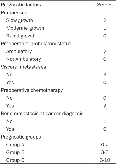
Previously, we retrospectively developed a scor-ing system by analyzscor-ing a series of 206 cases in a single institution to estimate the survival and function outcome of patients with MESCC. This scoring system included the following five prognostic factors. Primary site, preoperative ambulatory status, visceral metastases, preop-erative chemotherapy, and bone metastasis at cancer diagnosis. For each of the above men-tioned five prognostic characteristics, the scor-ing points were obtained from the hazard ratios based on the multiple Cox proportional hazards regression model (round values). The total prognostic score for each patient was the sum of the scoring points of the five significant prog-nostic characteristics. The total progprog-nostic scores were ranged from 0 to 10 points, and three risk groups were designed according to 6-month survival rate and median survival time of each prognostic score. Patients with 0 to 2 points were regarded as group A, patients with 3 to 5 points were considered as group B, and 6 to 10 points were group C (Table 1). Patients with scores more than 3 points were recom-mended to surgery (Case report was shown in
Figure 1). Kaplan-Meier method and log-rank test were used to compare the survival progno-sis in the above mentioned three groups, and Chi-square test was used to estimate differ-ence in ambulatory rate according to the scor-ing system.
The scoring system would be applied in the same way in the present study to validate whether the scoring system was reproducible. Three prognostic groups were designed to be consistent with the previous scoring system. Besides, in order to evaluate and compare the capabilities of the scoring system and other scoring systems (Perhaps, the Tomita scoring system was the most widely used scoring mod-Table 1. The new scoring system for patients
with MESCC
Prognostic factors Scores
Primary site
Slow growth 2
Moderate growth 1
Rapid growth 0
Preoperative ambulatory status
Ambulatory 2
Not Ambulatory 0
Visceral metastases No 3 Yes 0 Preoperative chemotherapy No 0 Yes 2
Bone metastasis at cancer diagnosis
No 1
Yes 0
Prognostic groups
Group A 0-2
Group B 3-5
Group C 6-10
Abbreviations: MESCC, Metastatic epidural spinal cord compression. Slow growth: hormone-dependent breast cancer, hormone-dependent prostate cancer, thyroid can-cer, multiple myeloma, and malignant lymphoma. Moder-ate growth: lung cancer treModer-ated with molecularly targeted drugs, independent breast cancer, hormone-independent prostate cancer, renal cell carcinoma, endometrial cancer, ovarian cancer, and sarcoma. Rapid growth: lung cancer without molecularly targeted drugs, colorectal cancer, gastric cancer, pancreatic cancer, esophageal cancer, other urological cancers, hepato-cellular carcinoma, head and neck cancer, melanoma, malignant thymoma and cancers of unknown origin.
the capability of the score was calculated in the study.
Patients and methods
Patients
[image:2.612.91.289.96.374.2]Figure 1. A 67-year-old man who unable to walk due to metastatic epidural spinal cord compression (MESCC) re-sulted from prostate cancer. A and B. Preoperative X-ray presented vertebral collapse at T5. C. Preoperative MRI showed spinal cord compression at T5. D. Preoperative CT showed bone destruction at T5. E. Preoperative MRI showed spinal cord compression at T5. F and G. Following laminectomy at T4 and T5, and pedicle screw fixation was conducted at T3, T4, T6, and T7 to spine stabilization. He died at postoperative 9.2 months and spine stability was maintained throughout the survival period.
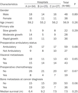
Table 2. Patient’s characteristics of the three hospitals
Characteristics Hospitals (N=86)Total P A (n=34) B (n=25) C (n=27)
Gender
Male 18 14 16 48 0.89
Female 16 11 11 38
Age (mean) 59.2 55.2 56.2 56.9 0.26
Primary site
Slow growth 5 9 8 22 0.29
Moderate growth 14 5 9 28
Rapid growth 15 11 10 36
Preoperative ambulatory status
Ambulatory 25 17 17 59 0.68
Not Ambulatory 9 8 10 27
Visceral metastases
No 19 11 13 43 0.65
Yes 15 14 14 43
Preoperative chemotherapy
No 26 21 20 67 0.67
Yes 8 4 7 19
Bone metastasis at cancer diagnosis
No 15 15 20 50 0.06
Yes 19 10 7 36
Median survival (m) 6.4 9.2 7.5 7.5 0.25
Hospital A: the First People’s Hospital of Shanghai Jiao Tong University, B: Xiangya Hospital Central South University, and C: the Affiliated Hospital of Academy of Military Medical Sciences.
els in clinical routine), the receiver operating characteristic (ROC) curve c-statistics were
cal-culated in the study. When we ini-tially designed this study, visceral metastasis was recorded as being present or not present. In the analy-sis of the c-statistics for the Tomita score, if the patients presented with visceral metastasis, they were re- garded as “non-removable”.
Statistical analysis
[image:3.612.92.347.356.658.2]was determined as P<0.05, and statistical analysis was carried out using SAS 9.2 software.
Results
Patient’s characteristics
As a result, 89 patients were enrolled in the study, but three of them were lost follow-up within a month. Thus, 86 patients were ana-lyzed in this present study (The First People’s Hospital of Shanghai Jiao Tong University: 34 cases, the Xiangya Hospital Central South University: 25 cases, and the Affiliated Hospital of Academy of Military Medical Sciences: 27 cases). In the entire cohort of patients, 22 patients with slow growth cancer, 28 patients with moderate growth cancer, and 36 patients with rapid growth cancer. Lung were the most common primary site, and lung cancer occurred in 33 patients. Sixty-three patients were treat-ed with surgery, and twenty-three patients were treated with non-operative treatments. In the surgically treated patients, 54 patients were operated with posterior decompressive surgery and spine stabilization, 6 patients were oper-ated with anterior procedures, and 3 patients were operated with anterior and posterior pro-cedures. The median overall survival was 7.5 months (95% CI, 6.5-8.6 months), and 6-month and 12-month survival rates were 69.2% and 24%, respectively. At the latest follow up, 25 patients were alive with a mean follow-up of 9.7 months (range, 4.1-15.3 months), and 6 patients who were lost follow-up had a mean follow-up of 5.9 months (range, 2.3-7.8 months). Table 2 showed the patient’s characteristics, which demonstrated that the distribution of characteristics was similar in three hospitals (P>0.05).
Validation of the scores
In the present study, the median survival time was 3.9 months (95% CI, 2.7-6.1 months) for patients with 0-2 points, 6.7 months (95% CI, 6.4-8.5 months) for those with 3-5 points, and 12 months (The lower limit of 95% CI: 8.2 months) for those with 6-9 points, respectively. The corresponding 6-months survival rates was 27.8%, 66.0%, and 88.0%, respectively (P< 0.01, Figure 2). In the previous study, the cor-responding median survival time was 3.3 months, 6.6 months, and 16.4 months, respec-tively, and the corresponding 6-months survival rates were 8.2%, 56.5%, and 91.5%, respec- tively.
In the current study, the ROC curve c-statistic for the scores as a predictor of 3 months sur-vival rate was 0.75 (OR, 0.66, 95% CI: 0.49-0.88, P<0.01), c-statistic as a predictor of 6 months survival rate was 0.74 (OR, 0.65, 95% CI: 0.52-0.82), and c-statistic as a predictor of 12 months survival rate was 0.70 (OR, 0.73, 95% CI: 0.53-1.01) (Supplementary Figures 1, 2, 3). The accuracy rates for predicting 3, 6, and 12 months survival were 69.1%, 69.2%, and 64.9%, respectively. The ROC curve c-sta-tistics for the Tomita scores as a predictor of 3 months, 6 months, and 12 months survival rate were 0.70 (OR, 1.35, 95% CI: 1.05-1.75), 0.68 (OR, 1.31, 95% CI: 1.09-1.59), and 0.66 (OR, 1.26, 95% CI: 0.95-1.67), respectively (Supplementary Figures 4, 5, 6). The accuracy rates for predicting 3, 6, and 12 months sur-vival were 58.4%, 58.4%, and 56.4 %, respec- tively.
The postoperative ambulatory rates were 55.6% in patients with 0-2 points, 73.5% in patients with 3-5 points, and 94.1% in patients with 6-10 points, respectively (P<0.01, Table 3). In the previous study, the corresponding ambulatory rates were 35.7%, 73.3%, and 95.9%, respectively. In the entire cohort of patients, 77.9% patients (67/86) had the ability to walk after surgery, and 51.9% (14/27) of nonambulatory patients before operation be- came ambulatory after surgery.
Discussion
Personalized treatment has been widely stud-ied in the palliative context of MESCC. Rades et al. [8] clearly showed that the outcome of radio-therapy alone appeared similar to those of sur-Figure 2. Survival curves for the three prognostic
[image:4.612.89.288.70.194.2]gery plus radiotherapy. However, many other investigations revealed that direct decompres-sive and stabilized surgery followed by radio-therapy was superior to radioradio-therapy alone in terms of postoperative ambulatory status, pain outcome, and even survival prognosis for selected patients [9-11]. The personalized treatment for patients with MESCC should con-sider the patient’s survival and function out-come [4-7]. In general, patients with shortest survival time and poorest function outcome appear to be best treated with radiotherapy or best supportive care alone. Patients with pref-erable survival prognosis and function outcome can be treated with surgery to realize better local control of disease and improvement of patient’s quality of remaining life.
Previously, we retrospectively developed a new scoring system to estimate the survival progno-sis and functional outcome by analyzing 206 patients [7]. This scoring system included five prognostic factors: primary site, preoperative ambulatory status, visceral metastases, preop-erative chemotherapy, and bone metastasis at cancer diagnosis. The prognostic scores ranged from 0 to 10 points, and three risk groups were designed. There were those with 0 to 2 points (Group A), those with 3 to 5 points (Group B), and those with 6 to 10 points (Group C). The corresponding median overall survival time was 3.3 months, 6.6 months, and 16.4 months, respectively, the 6-month survival rates were 8.2%, 56.5%, and 91.5%, respectively, and the corresponding ambulatory rates were 35.7%, 73.3%, and 95.9%, respectively.
However, this scoring system was not validated. In the present study, we validated the scoring system in a prospective and multicenter data set. Eighty-six patients who were from three dif-ferent hospitals were included in the study. The median survival time was 3.9 months for patients with 0-2 points, 6.7 months for those
[image:5.612.89.354.111.176.2]with 3-5 points, and 12 months for those with 6-9 points. The corre-sponding 6-months survival rates was 27.8%, 66.0%, and 88.0%, respectively. The results were simi-lar to those in our previous study. The ROC curve c-statistic for the scores as a predictor of 3 months survival rate was 0.75, c-statistic as a predictor of 6 months survival Table 3. Ambulatory status of the patients in the three
prog-nostic groups 4 weeks after surgery or conservative treat-ments. P-value was obtained from Chi-square test
Groups Scores Patients (n) Neurological status P-value Not ambulatory ambulatory
A 0-2 18 8 10 <0.01
B 3-5 34 9 25
C 6-10 34 2 32
rate was 0.74, and c-statistic as a predictor of 12 months survival rate was 0.70. The accura-cy rates for predicting 3, 6, and 12 months sur-vival were 69.1%, 69.2%, and 64.9%, respec-tively. The ROC curve c-statistics for the Tomita scores as a predictor of 3 months, 6 months, and 12 months survival rate were 0.70, 0.68, and 0.66, respectively. The corresponding accuracy rates for predicting 3, 6, and 12 months survival were 58.4%, 58.4%, and 56.4 %, respectively. Those founding showed that the capability of the scores to predict survival prognosis was relatively better than the Tomita scoring system.
In conclusion, this scoring system should be considered valid and reproducible to estimate the survival prognosis and functional outcome. This scoring model can help select the optimal therapy for patients with MESCC, and its capa-bility to predict survival prognosis was relatively better than the Tomita scoring system.
Acknowledgements
The work is supported by Application Study of Capital Clinical Characteristics of China (NO. Z131107002213052 and NO. Z161100000- 516101).
Disclosure of conflict of interest
None.
jon-son8920@sina.com; Yaosheng Liu, Department of Orthopedic Surgery, Affiliated Hospital of Academy of Military Medical Sciences, 8 Fengtaidongda Road, Beijing 100071, People’s Republic of China. Tel: +86-10-66947317; Fax: +86-10-66947317; E-mail: 632763246@qq.com
References
[1] Shiue K, Sahgal A, Chow E, Lutz ST, Chang EL, Mayr NA, Wang JZ, Cavaliere R, Mendel E, Lo SS. Management of metastatic spinal cord compression. Expert Rev Anticancer Ther 2010; 10: 697-708.
[2] Robson P. Metastatic spinal cord compression: a rare but important complication of cancer. Clin Med 2014; 14: 542-5.
[3] Vajkoczy P, Wehofsky R, Czabanka M, Woitzik J, Onken J. Surgery of spinal metastases. Der Onkologe 2016; 22: 299-315.
[4] Tokuhashi Y, Matsuzaki H, Oda H, Oshima M, Ryu J. A revised scoring system for preopera-tive evaluation of metastatic spine tumor prog-nosis. Spine (Phila Pa 1976) 2005; 30: 2186-91.
[5] Lei M, Liu Y, Tang C, Yang S, Liu S, Zhou S. Pre-diction of survival prognosis after surgery in patients with symptomatic metastatic spinal cord compression from non-small cell lung cancer. BMC Cancer 2015; 15: 853.
[6] Lei M, Liu Y, Liu S, Wang L, Zhou S, Zhou J. In-dividual strategy for lung cancer patients with metastatic spinal cord compression. Eur J Surg Oncol 2016; 42: 728-34.
[7] Lei M, Li J, Liu Y, Jiang W, Liu S, Zhou S. Who are the best candidates for decompressive surgery and spine stabilization in patients with metastatic spinal cord compression?: A new scoring system. Spine (Phila Pa 1976) 2016; 41: 1469-76.
[8] Rades D, Huttenlocher S, Dunst J, Bajrovic A, Karstens JH, Rudat V, Schild SE. Matched pair analysis comparing surgery followed by radio-therapy and radioradio-therapy alone for metastatic spinal cord compression. J Clin Oncol 2010; 28: 3597-604.
[9] Patchell RA, Tibbs PA, Regine WF, Payne R, Sa-ris S, Kryscio RJ, Mohiuddin M, Young B. Direct decompressive surgical resection in the treat-ment of spinal cord compression caused by metastatic cancer: a randomised trial. Lancet 2005; 366: 643-8.
[10] Lee CH, Kwon JW, Lee J, Hyun SJ, Kim KJ, Jahng TA, Kim HJ. Direct decompressive sur-gery followed by radiotherapy versus radiother-apy alone for metastatic epidural spinal cord compression: a meta-analysis. Spine 2014; 39: E587-92.
Supplementary Figure 1. ROC curve for the scores as a predictor of 3 months survival rate.
Supplementary Figure 3. ROC curve for the scores as a predictor of 12 months survival rate.
Supplementary Figure 5. ROC curve for the Tomita scores as a predictor of 6 months survival rate.