Original Article
Construction of a three-dimensional interactive digital
atlas of the dural sinus and deep veins based on
human head magnetic resonance images by
a comprehensive modeling protocol
Zhirong Yang, Zhilin Guo
Department of Neurosurgical, The Ninth People Hospital, Medical School, Shanghai Jiaotong University, Shanghai 200011, China
Received August 7, 2017; Accepted January 25, 2018; Epub April 15, 2018; Published April 30, 2018
Abstract: Objectives: To design a three-dimensional (3D) interactive digital atlas of the human dural sinus and deep veins for assisting neurosurgeons in preoperative planning and neurosurgical training. Methods: Sagittal head mag-netic resonance (MR) images were obtained of a 54-year-old female who suffered from left posterior fossa tumor. A comprehensive modeling protocol consisting of five steps including thresholding, crop mask, region growing, 3D calculating and 3D editing was used to develop a 3D digital atlas of the dural sinuses and deep veins based on the MR images. The accuracy of the atlas was also evaluated. Results: The 3D digital atlas of the human dural sinus and deep veins was successfully constructed using 176 sagittal head MR images. The contours of the acquired model matched very well with the corresponding structures of the original images in axial and oblique view of MR cross-sections. The atlas can be arbitrarily rotated and viewed from any direction. It can also be zoomed in and out directly using the zoom function. Conclusion: A 3D digital atlas of human dural sinus and deep veins was successfully cre-ated, it can be used for repeated observations and research purposes without limitations of time and shortage of corpses. In addition, the atlas can potentially be used for preoperative planning and surgical training.
Keywords: Three-dimensional (3D), human dural sinus, deep veins
Introduction
The dural sinuses form a channel system that functions primarily as collection and drainage of the intracranial venous blood [1]. The deep venous system connects with the straight sinus by the vein of Galen behind the splenium of the corpus callosum. The dural sinuses and deep veins also serve as important landmarks in neurosurgery [2-9], and understanding of their complex anatomical features facilitates preop-erative planning and navigation through the neurosurgical landscape. They also gain clinical significance in cases of trauma, meningioma and, though rare, dural sinus thrombosis [10]. However, the spatial structure of the dural sinuses and deep veins cannot be visualized directly by gross observation or head magnetic
Table 1. The operating parameters of sinuses and cerebral veins
Thresholding Crop mask Operating
Mix Max x × y × z, mm3 Region growing (t) 3D calculate 3D edit
Head MRI 0 2287 - - -
-Block
A 571 1403 10 × 19.5 × 36 1
B 696 1342 49 × 62 × 113 1
C 815 1441 27 × 37 × 106 2
D 517 1004 51 × 40 × 106 3 Manual
E 855 1750 29 × 41 × 46 1 Optimal -Bilateral superior petrosal sinus 477 1024 16 × 47 × 104 2 Manual Left inferior petrosal sinus 556 2098 25 × 35 × 37 1 Manual Deep cerebral veins 576 894 31 × 60 × 49 3 Manual
sinuses; block IV: bilateral sphenoparietal sinus; block V: cavernous sinus; block VI: bilat-eral superiorpetrosal sinus and the left inferior petrosal sinus. The right inferior petrosal sinus was not investigated because it failed to be visualized in head MRI.
Model construction
The thresholding procedure was used to gener-ate masks from original head MRI data. The Mimics software (Materialise’s, Belgium) was used, it presented segmentation functions based on image density thresholds. The thresh-old jail of the image mask fell within the range from 0 to 2287 (Table 1). The threshold jail value of 0 was taken as background and the values above 2287 were considered as noises, excluding normal anatomic structures. The block including target structures was segment-ed from the thresholding mask to obtain block mask. Generated masks were further pro-cessed using an embedded region growing algorithm in Mimics and then reconstructed into a 3D model. The surface model was refined automatically and further checked by cross-sectioning the surface models with original head MRI and manually revised using 3ds Max (version 11.0, Autodesk, USA) until it accorded with the correct anatomy. The dural sinus model was outputted in the SLT format and imported into 3-matic viewer for format conversion. The 3D dural sinus model was exported from the 3-matic viewer into Adobe Acrobat 11 Pro (http://www.adobe.com) to create interactive 3D portable document format (PDF) files, which can be displayed and rotated in any directions using any structure as the focal point of rotation.
Model validation
For evaluation of the accuracy of the superior sagittal sinus and inferior sagittal sinus, we imported the 3D dural sinus atlas into the 3D data system of the raw head MRI images, and the 3D dural sinus model was validated by matching the cross-section of the surface mod-els with the original head MRI. Furthermore, a random marker was placed in the 3D dural sinus atlas and the corresponding coordinates of the marker in head MRI images were verified by naked eye. The accuracy of the dural sinus and deep veins was evaluated by separately examining the midline and the skull base
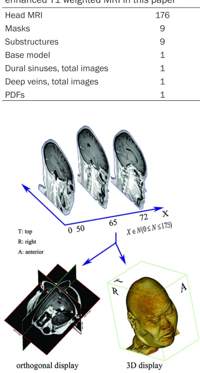
struc-Table 2. List of structures based on contrast-enhanced T1 weighted MRI in this paper
Head MRI 176
Masks 9
Substructures 9
Base model 1
Dural sinuses, total images 1 Deep veins, total images 1
PDFs 1
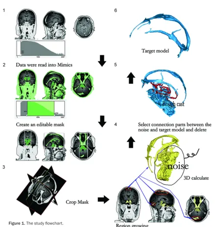
Figure 2. Image organizing sequence, a 3-D data or-ganized by the 176 sagittal images.
Methods
Data acquisition
[image:3.612.88.287.90.460.2]tures. In addition, for evalua-tion of the transverse sinus and the superior petrosal vein in the skull base, a reference model was created of the anterior, middle and posterior skull base with head MRI data and a dural mater reference model was also constructed; these models were compared with the dural sinus and deep vein atlas for consistency of contours in the reference mo- del and the atlas. Furthermore, a reference model was estab-lished in the 3D Max and the dural sinus model in.stl format was imported into the 3D Max and registered with the refer-ence model for observation. We checked up the accuracy of our 3D dural sinus and deep venous model by cross-sec-tioning the surface models and matching original head MRI images and by consisten-cy between the placed marker and its corresponding coordi-nates in the three orthogonal slices in Mimics. The coordi-nates of the base model was set the same as the original scan coordinate.
Results
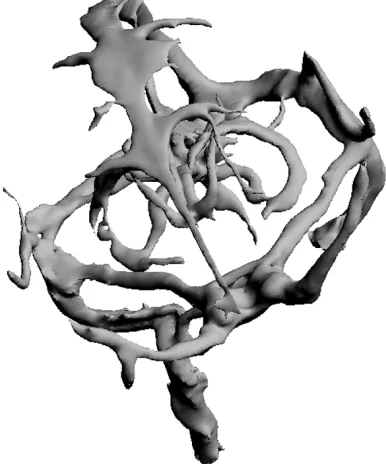
[image:4.612.89.373.74.319.2]The study flowchart is shown in Figure 1. We acquired total-ly 176 head MR sagittal imag-es with a matrix size of 256 × 256 using a 3.0 T MR scanner. All data sets of head MRI, masks, substructures, the ba- se reference model, dural si- nus and deep vein panoramic images and their PDF’s are listed in Table 2. The main dural sinus structure was clearly visualized in most head MRI images. The slice dis-tance was 1.0 mm. These sag-ittal images were successfully organized into a single 3-D Figure 3. This figure shows the dural sinus and deep veins, surrounded by
the decomposition model from various angles.
[image:4.612.91.374.375.601.2]Figure 5. A base model with head MRI information. The five green arrows in left picture indicate the contours of the dural sinus and deep veins model match very well on the corresponding structure edge in axial and oblique view) MRI cross-sections.
entity (Figure 2). The dural sinus and deep vein atlas was created and can be separated from the main cerebral struc-tures and was observable from different angles (Figure 3).
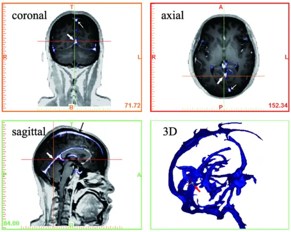
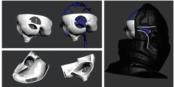
The superior sagittal sinus was viewed in the midline area (Figure 4). The superior and inferior sagittal sinus contour lines agreed very well with the MRI image edges of the corre-sponding structure. The coor-dinates of the random marker placed on the inferior sagittal sinus was automatically creat-ed on the front surface and shown as a cross sign (Figure 4). The cross-sectional con-tour of the sphenoparietal sinus, cavernous sinus and superior petrosal sinus ma- tched very well with MRI image edges of the reference model (Figure 5). The dural sinus and deep vein were adapted into the corresponding parts in the reference model (Figure 6). The contour lines of the venous sinuses, deep veins and the reference model matched very well with their counterparts in the original head MRI images. In the 3D environment, the venous sin- uses and deep veins are con-sistent with the original head data (Figure 7).
Figure 7. The consistency of dural sinus and deep vein atlas with the cor-responding parts in reference model
Figure 8. An example demonstration of the assistance of our 3D atlas for a preoperative precise surgical approach planning.
sinuses (Figure 9). The model of the venous sinuses and deep veins in PDF format can be rotated 360 degrees in any plane using any structure as the focus of rotation (Figure 10). An additional human dural sinus atlas obtained by using similar materials and the same meth-od is shown in Supplementary Figure 1. Discussion
Understanding anatomical structures is of fun-damental importance for neurosurgeons and an interactive and intuitive atlas provides a readily understandable and maneuverable tool for neuroanatomists and neurosurgeons. In this study, by using a novel approach, we con-structed a 3D digital atlas of the dural sinuses and deep veins using contrast-enhanced head
also provide valuable information, particularly in diagnosing diseases and guiding therapies for clinicians. Early atlases consist of autopsy photographs and manual drawings of histologi-cal sections, and lack 3D information and are not interactive. Digital atlas is characterized by digitalization and visualization of anatomical information [12] and offers an interactive tool for users. Though numerous digital atlases are available of the brain, cerebrovascular, skull based and cranial nerves [13-17], few atlases of the dural sinus have been published. Our digital atlas is well suited to the task of select-ing an appropriate surgical approach that can avoid injuries to specified regions. It also facili-tates preoperative planning, as in the case of resecting a posterior fossa tumor, the atlas could be employed to calculate the position of the flap and bone window.
[image:6.612.87.376.309.454.2]method has proven to work well even for imag-es with rather poor quality. The current method is semi-automatic: the noise of the rough cast from 3D calculating can be deleted manually by 3D edit in the 3ds-Max and noises can be fil-tered out after careful screening.
[image:7.612.91.358.68.369.2]Accuracy of digital atlas is a key issue. We eval-uated the atlas accuracy in several ways. On the one hand, we evaluated the accuracy of our 3D atlas by cross-sectioning the surface mod-els with the original head MR image. On the other hand, we evaluated consistency between the marker point and its corresponding coordi-nates in the three orthogonal slices. The model contours match very well on the venous sinus and deep venous walls of raw head MRI images in three orthogonal (axial, coronal and sagittal view) MRI cross-sections. By clicking a point in the 3D model, the three orthogonal slices com-prising this point will display, and the corre-sponding coordinates will be automatically adjusted. The atlas can be made into a small Figure 9. The comparison of dural sinus atlas constructed by different
meth-ods.
In 3D model reconstruction, segmentation of target imag-es is based on the gray con-trast or boundaries of differ-ent tissues. However, seg- mentation of some structure is not successful, particularly when the gray contrast be- tween target tissue and the surrounding in the original image is too low. The dural sinus wall is very thin and its density is very close to the sur-rounding tissues, and its boundary is not clear. Conse- quently, it is very challenging to segment the dural sinus by just using a single method. In the current study, we devel-oped a comprehensive model-ing protocol consistmodel-ing of five steps including thresholding, crop mask, region growing, 3D calculating and 3D editing. The protocol has successfully yielded an accurate and smooth surface model of the venous sinus and deep veins. An atlas is required to be of very high accuracy, and our
[image:7.612.92.285.440.672.2]movie to play or can be installed on the PC for viewing. The atlas can be arbitrarily zoomed, rotated and cut according to the needs of the observers. Moreover, the atlas can be displayed in a computer in the operating room for intraop-erative observation, while printed atlases are not convenient for such purposes. However, no matter how elegant the 3D atlas obtained from head MRI is, it may be still not sufficient for pre-senting the full anatomic detail of the venous sinuses and deep veins and we believe that our 3D digital atlas could be further improved in the future with advancement of imaging and com-puting technologies.
Conclusion
An orderly synthesis method for building a 3D digital atlas of the dural sinus and deep veins was developed. The accuracy of the atlas was proved to be sufficient for the purpose of medi-cal education, and guidance for surgimedi-cal op- eration.
Disclosure of conflict of interest
None.
Address correspondence to: Drs. Zhirong Yang and Zhilin Guo, Department of Neurosurgical, The Ninth People Hospital, Medical School, Shanghai Jiaotong University, 639 Zhizaoju Road, Shanghai 200011, China. Tel: +86 021-23271699-5155; E-mail: [email protected] (ZRY); [email protected] (ZLG)
References
[1] Browder J, Browder A, Kaplan HA. Anatomical relationships of the cerebral and dural venous systems in the parasagittal area. Anat Rec 1973; 176: 329-32.
[2] Bonnal J, Brotchi J. Surgery of the superior sag-ittal sinus in parasagsag-ittal meningiomas. J Neu-rosurg 1978; 48: 935-45.
[3] Giombini S, Solero CL, Lasio G, Morello G. Im-mediate and late outcome of operations for Parasagittal and falx meningiomas. Report of 342 cases. Surg Neurol 1984; 21: 427-35. [4] Ransohoff J. Removal of convexity,
parasagit-tal, and falcine meningiomas. Neurosurg Clin N Am 1994; 5: 293-7.
[5] Kondziolka D, Flickinger JC, Perez B. Judicious resection and/or radiosurgery for parasagittal meningiomas: outcomes from a multice- nter review. Gamma Knife Meningioma Study Group. Neurosurgery 1998; 43: 405-13; dis-cussion 13-4.
[6] DiMeco F, Li KW, Casali C, Ciceri E, Giombini S, Filippini G, Broggi G, Solero CL. Meningiomas invading the superior sagittal sinus: surgical experience in 108 cases. Neurosurgery 2004; 55: 1263-72; discussion 72-4.
[7] Bassiouni H, Hunold A, Asgari S, Stolke D. Ten-torial meningiomas: clinical results in 81 pa-tients treated microsurgically. Neurosurgery 2004; 55: 108-16; discussion 16-8.
[8] Gabibov G KA, Kozlov A, Korshanov A,Timirgaz V, Kalinina E. Surgical results and prognostic factors in parasagittal meningiomas. Experi-ence in 1546 operations. Abstract, 10th Euro-pean Congress of Neurosurgery 1995, Berlin; May 7-12.
[9] H O. The surgical treatment of intracranial tu-mors. Handbuch der Neurochirurgie 1967; 4: 1-191.
[10] Kemp WJ 3rd, Tubbs RS, Cohen-Gadol AA. The innervation of the cranial dura mater: neuro-surgical case correlates and a review of the literature. World Neurosurg 2012; 78: 505-10. [11] Yang Z, Guo Z. A three-dimensional digital atlas
of the dura mater based on human head MRI. Brain Res 2015; 1602: 160-7.
[12] Toga AW, Thompson PM, Sowell ER. Mapping brain maturation. Trends Neurosci 2006; 29: 148-59.
[13] Haber SN, Calzavara R. The cortico-basal gan-glia integrative network: the role of the thala-mus. Brain Res Bull 2009; 78: 69-74. [14] Saikali S, Meurice P, Sauleau P, Eliat PA,
Bel-laud P, Randuineau G, Vérin M, Malbert CH. A three-dimensional digital segmented and de-formable brain atlas of the domestic pig. J Neurosci Methods 2010; 192: 102-9.
[15] Ganser KA, Dickhaus H, Metzner R, Wirtz CR. A deformable digital brain atlas system accord-ing to Talairach and Tournoux. Med Image Anal 2004; 8: 3-22.
[16] Calzavara R, Mailly P, Haber SN. Relationship between the corticostriatal terminals from ar-eas 9 and 46, and those from area 8A, dorsal and rostral premotor cortex and area 24c: an anatomical substrate for cognition to action. Eur J Neurosci 2007; 26: 2005-24.