Abstract-There is evidence that both the herb Sanicle and the cytokine TGF- β3 can be beneficial in enhancing wound repair. In this study 3T3 fibroblast cells were cultured and the confluent monolayers were wounded (scarred) using a disposable plastic pipette. Various amounts of TGF-β3 (a
growth factor) and Sanicle extract were applied to the cell
monolayers. TGF-β3 was applied at concentrations of 50ng/ml, 5ng/ml, 500pg/ml, 50pg/ml and 5pg/ml to five different culture flasks with one additional flask acting as control. Sanicle was applied at concentrations of 100μg/ml, 10μg/ml, 1μg/ml, 100ng/ml and 10ng/ml with one additional flask as a control. The cells were imaged over a period of 20 hours with or without presence of TGF-β3 and Sanicle. The results indicated that although there were no significant increases in the rate of wound closure in relation to application of TGF-β3, there is an indication that TGF-β3 may enhance model wound closure at optimum working concentration between 5ng/ml and 50ng/ml. However, the sanicle extract did not stimulate enhanced repair
of the model in vitro wound, but instead seemed to promote cell
death along the wound margin. These results indicate that sanicle may be used in the care of wounds, but not as a growth promoter, but because it acts as an antibiotic agent, and possibly
because it aids wound debridement.
Index Terms: 3T3 Fibroblast Cell Culture; Growth Factor TGF-β3; Sanicle; Astringent; Anti-Scarring Effect (Wound healing).
Manuscript received March 15, 2010. This work was supported by school of Engineering and School of Pharmacy, University of Bradford, UK.
F. Sefat, (Corresponding Author) is with the School of Engineering, Design and Technology-Medical Engineering and Institute of Pharmaceutical Innovation (ipi), University of Bradford, Bradford, BD7 1DP, United Kingdom. (Phone: 01274 234533; Fax: 01274 234525; e-mail: [email protected] or [email protected]).
C.B. Beggs is with the School of Engineering, Design and Technology-Medical Engineering, University of Bradford, Bradford, BD7 1DP, United Kingdom. (Phone: 01274 233679; Fax: 01274 234525; e-mail: [email protected]).
A. Lemmerz, is with the School of Engineering, Design and Technology-Medical Engineering, University of Bradford, Bradford, BD7 1DP, United Kingdom. (Phone: 01274 234533; Fax: 01274 234525; e-mail: [email protected]).
M.C.T Denyer is with the School of Life Science, Institute of Pharmaceutical Innovation (ipi), University of Bradford, Bradford, BD7 1DP, United Kingdom. (Phone: 01274 234747; Fax: 01274 236060; e-mail: [email protected]).
C.Wright is with the School of Life Science, University of Bradford, Bradford, BD7 1DP, United Kingdom. (Phone: 01274 234739; e-mail: [email protected]).
M. Youseffi is with the School of Engineering, Design and Technology-Medical Engineering, University of Bradford, Bradford, BD7 1DP, United Kingdom. (Phone: 01274 234533; Fax: 01274 234525; e-mail: [email protected]).
I.INTRODUCTION
Sanicle, Sanicula europaea (Umbelliferaea) is a perennial herb found in Northern and Central Europe including the British Isles [1, 2]. Although there appears to be little call for Sanicle in herbal medicine today, the plant was formerly an important medicinal plant sought after especially for its wound healing properties. The name of the plant is derived from the Latin Sano meaning “I heal or cure” and in medieval times this was reflected in the popular saying: “Celui qui sanicle a De mire affaire il n‟a”, which translated means “He that hath sanicle needeth no surgeon”. Modern medicine has moved away from herbal remedies in wound repair, and focused more on methods of engineering enhanced wound repair via combinations of implantable constructs and therapeutic use of cell growth factors and cytokine. One such cytokine currently gaining a reputation in promoting scarless healing is TGF- 3 [3-7].
Growth factors, and cytokines, are associated with a number of cell and tissue repair mechanisms functioning as signaling molecules in the inflammatory response and in the modulation of cell proliferation and differentiation. Transforming growth factor beta (TGFß) enhances fibroplasia and the production of ECM components such as collagen, elastin and laminin whilst suppressing the ECM breakdown. It is also apparent that the relative proportions of cytokines can modulate the process of wound repair. For example, it has been reported [3] that foetal wounds heal with little or no scarring and that this is due to variations in the concentration of different TGF- isoforms. In adults, it is evident that TGF-β1 and TGF-β2 predominate in the wound site and function through activation of the immune system which in turn promotes scarring. However, in the embryo TGF-β3 is up-regulated and TGF-β1 and TGF-β2 concentrations are low, indicating that TGF-β3 plays a role in scarless healing. This view has been confirmed in experiments looking at wound healing in adults, where virtually scarless healing can be induced by the introduction of between 5 and 50ng/ml of TGF-β3 into the wound site [3, 4, 5, 6].
Although cell and tissue engineering based systems predominate in the generation of new therapeutic treatments in wound repair there is a growing realisation that these may be augmented by aspects of historical treatments. In the case of sanicle [1, 2, 8], its use in wound healing has ceased and little is known about how sanicle extracts improved wound prognosis. This study aims at comparing the effects of a sanicle extract and TGF-β3 on the repair of an in vitro model wound.
The Effect of Transforming Growth Factor Beta
(TGF-β3) and Sanicle on Wound Healing
II.AIMS AND OBJECTIVES
Main objective was to investigate the effect of TGF-β3 and Sanicle on wound closure in cultured monolayers of 3T3 fibroblast cells. The lab-based experimental work investigated and compared the wound closure properties of TGF-β3 and sanicle in cultured dish environment using cultured monolayers of 3T3 fibroblast cells. Other cellular responses such as proliferation and detachment have also been investigated along with different stages of cell behaviour and morphology during wound healing.
III.MATERIALS ANDMETHODS
A.Preparation of extracts:
Dried S. europaea was obtained from the Herbal Apothecary, Leicester, UK, and its identity was confirmed microscopically by comparison with an authentic specimen. An aqueous extract was prepared by shaking the powdered herb (5 g), with water (150 ml), filtering and then freeze drying using a Dura-Stop MP Lyophiliser (FTS Systems, Stoney Ridge, New York). For the preparation of methanolic extracts, plant material was shaken with methanol and the filtered extract was concentrated using a rotary evaporator and dried in vacuo at room temperature.
A crude glyoside extract was prepared using the following method: Plant material (28 g) was defatted with hexane and then extracted 3 times with separate volumes of methanol 80% for 1 hour in a Soxhlet extractor. The combined extracts were concentrated to a small volume on a rotary evaporator, diluted to 60 ml with water and extracted with chloroform 3 times. The aqueous phase was then extracted with butanol and the butanol extract was dried on a rotary evaporator. The residue was taken up into a small volume of methanol and dropped into ether. The precipitate (crude glycoside fraction) was collected, washed with ether and dried.
The experiments involved harvesting and maintenance of a 3T3 fibroblast cell line. 3T3 cells were cultured in culture medium consisting of high glucose DMEM (SIGMA) supplemented with 10% newborn calf serum, 2.5 mM U/ml L-Glutamine, 100 U/ml Penicillin, 0.1 mg/ml streptomycin and 1µg amphoterysin B Fungizone (Sigma Aldrich-UK). The 3T3 cells were cultured in standard Plug seal cap 25cm2 culture flasks and bathed in the culture media. The cells spread and formed a monolayer at the bottom of the culture flasks. Prior to plating, 6 parallel lines were drawn on the underside of each culture flask. Following plating and the cells reaching confluence a model wound was made by scratching through the middle of the monolayer using a sterile long-nosed plastic pipette. The model wound was orientated so that it bisected the parallel lines drawn on the underside of the flask at 90 degrees. This facilitated orientation while imaging and allowed rapid identification and re-identifiaction of specific sites of the wound at different time points. Once scratched the cells were maintained at 37oC in culture media containing either sanicle extract or TGF-β3. Six culture flasks were used for the TGF-β3 treatment (1 control and 5 with different TGF-β3 concentrations) and another 6 flasks for the
Sanicle treatment (1 control and 5 with different Sanicle concentrations). All experiments were repeated 6 times.
B.TGF-β3 and Sanicle Dilution
Purified recombinant human TGF-β3 (SIGMA) was reconstitutioned in 0.2μm-filtered 4mM HCl solution containing 1mg/ml of bovine serum albumin (BSA) to produce a TGF-β3 stock solution of 1μg/ml. TGF-β3 was used at 5 dilutions, 50ng/ml, 5ng/ml, 500pg/ml, 50pg/ml and 5pg/ml. A sanicle stock solution of 1mg/ml was made up in culture media and was diluted in culture media to 100μg/ml, 10μg/ml, 1μg/ml, 100ng/ml and 10ng/ml working dilutions.
C.Data acquisition
After wounding the culture flasks were incubated for 20 hours at 37oC and photomicrographs of 6 wound sites per culture flask were taken every 5 hours. The wound sites imaged were repeatedly identified at the different time points by only photographing the wound site positioned to the right of each parallel drawn on the underside of the flask. Images acquired were stored as tiff files and analysed using Image J (NIH).
[image:2.595.307.554.573.745.2]Quantitative analyses of the acquired images involved determining the smallest mean distance between wound margins (Figure 1) derived from 6 points per wound per time period per replicate experiment (Time 0, 5 hours, 10 hours, 15 hours and 20 hours). These replicate treatment means were then averaged to determine the overall treatment means. Initial measurements of the wound indicated a reasonably high degree of variability in wound width, from about 267µm to 454µm, thus to compare the wound closure in response to different treatments. The acquired data was normalized by converting mean distances between margins to percentage wound closure values, where a value of 100% represents no wound closure and a percentage wound closure value of 0% represents full wound closure. Percentage wound closure was then plotted against time per treatment. All mean values were recorded SE and statistically analysed using Independent Student T-Tests.
IV.RESULTS
Qualitative analyses of wound closure revealed treatment related differences in model wound repair. Control wounds initially had mean widths of 380±21 µm and 400±12 µm, respectively, and these widths were not significantly different from one another (Figure 2). After five hours the cells at the wound margins had acquired an elongated, spread morphology, and had started to replicate and migrated into the wound site perpendicularly to the wound axis. This process continued so that by the 15 hour period the cells had migrated into the wound site and elongated in order to meet cells from the opposite side of the wound. These cells then formed bridges connecting the wound margins together. As soon as the bridges formed in the wound site the wound began to close quite rapidly such that after 20 hours the wound was filled with loosely arranged cells.
In comparison treatment with sanicle, even at concentration of 5µg/ml seemed to modify the model wound repair process (Figure 3). After five hours the wound width seemed to have increased and the number of cells along the margins of the wound decreased. This process seemed to be associated with migration of cells away from the wound margin and an increased cell death, resulting in a decreased cell density extending from the wound margins into the un-wounded cell monolayers. The cells at the wound margin like those in the controls took on an elongated morphology but seemed not to migrate into the wound site, but instead only formed bridges with the neighboring cells along the wound edge. After 20 hours (Figure 3) the cells that had migrated across the wound site, were less densely arranged when compared with control treatment cells.
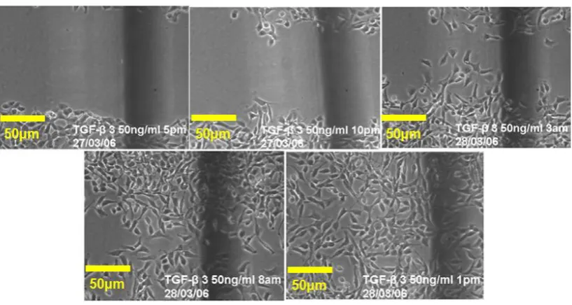
Application of TGF- 3at different concentrations induced qualitative enhancements in wound repair. When applied at 50 ng/ml (see Figure 4), TGF- 3 promoted rapid morphological changes in the cells along the wound margin. Within 5 hours cells took on a migratory phenotype and had started to migrate into the wound perpendicularly to the wound axis. After 10 hours (3am) the cells had already covered a great part of the wound site. Cells from both wound edges had migrated and elongated sufficiently to form bridges between the wound margins and cells had started to migrate across these bridges to fill the wound site. After 20 hours (1pm) the wound was covered by cells, although these were slightly less densely arranged than those in the unwounded monolayers.
Differences in the cell density between control treatments and TGF- 3 treatments were also apparent. The TGF- 3 treated cells at the 20 hour point had a more elongated morphology and were less densely arranged than those cells in the wound site of control treatments, possibly suggesting that the TGF- 3 cells had migrated faster into the wound site at the cost of cell replication.
Quantitative analyses supported the qualitative analyses. After 20 hours in culture all the culture flasks treated with TGF-β3 (even the lowest concentration of 500pg/ml) showed a high percentage of wound closure (Figure 5). In comparison wound closure in the control cultures were reduced with the wound having a mean wound opening of 35% after 20 hours in culture. Of the different TGF-β3 treatments, the wound at the 20 hour time-point for the highest TGF-β3 concentration (50ng/ml) was only 20% open, i.e. an improvement of 15% in comparison to the control. Similarly the lowest TGF-β3 concentration (500pg/ml) also induced a reduction of the wound opening, with the wound opening dropping to 27% after 20 hours in culture. These results suggest that the TGF-β3 addition increased the rate of wound repair. However, statistical analyses indicated that there were no significant differences (p > 0.05) between the wound widths at the 20 hour point between the control wound and wounds subjected to any of the TGF-β3 treatments.
In comparison, sanicle extract seemed to inhibit model wound closure (Figure 6). In these experiments model wounds in the control cultures reached a mean wound opening of 20% at the 20 hour time point (see Figure 3). This degree of wound healing was matched by treatment with sanicle extract at concentrations of 10ng/ml, 100ng/ml and 1µg/ml (Figure 6). This indicates that sanicle extract had no real effect on wound closure at these concentrations. However, sanicle extract treatment at higher concentrations of 10µg/ml and 100µg/ml had a profound inhibitory effect on wound closure. After 20 hours in culture with sanicle extract at a concentration of 100µg/ml the wound remained 90% open. Similarly the sanicle extract at a concentration of 10µg/ml also inhibited wound repair resulting in model wound width after 20 hours in culture remaining 64% open. This evidence was supported by statistical analyses which revealed that there were no significant differences in wound closure between the control and sanicle extract treatments at concentrations of less than 10µg/ml, but that sanicle extracts treatment at 10µg/ml and 100µg/ml significantly inhibited wound closure (P 0.05 and P 0.01 respectively)
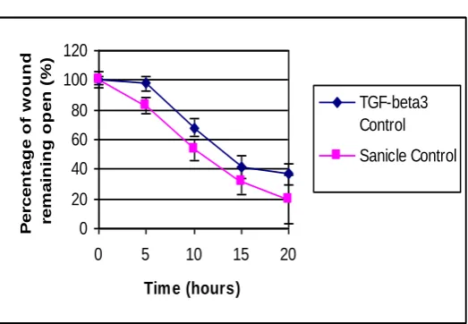
To ensure that the TGF-β3 and sanicle extract related experiments were comparable; the controls for each treatment were compared at each time point (Figure 7). No significant differences between the controls at each time point were found (P> 0.05).
[image:4.595.83.501.285.494.2]
Figure 2. Photomicrographs showing wound closure in the sanicle control at time points 0 houurs, 5 hours, 10 hours 15 hours and 20. At the twenty hour point the wound is almost completely filled by cells.
Figure 3. Effect of Sanicle addition (5µg/ml) on wound closure over a period of 20 hours.
[image:4.595.87.496.516.734.2]0 20 40 60 80 100 120
0 5 10 15 20
Time (hours) P e rc e n ta g e o f w o u n d re m a in in g o p e n ( %
) 50 ng/ml
5 ng/ml 500 pg/ml 50 pg/ml 5 pg/ml control -20 0 20 40 60 80 100 120
0 5 10 15 20
Tim e (hours)
P e rc e n ta g e o f w o u n d re m a in in g o p e n ( % ) 100 microgram/ml 10 microgram/ml 1 microgram/ml 100 ng/ml 10 ng/ml control 0 20 40 60 80 100 120
0 5 10 15 20
Time (hours) P e rc e n ta g e o f w o u n d re m a in in g o p e n ( % ) TGF-beta3 Control Sanicle Control
-10
0
10
20
30
40
0
5
10
15
20
25
Time (hours)
S pe e d of W ou nd C los ur e (m ic r o-m e te r /ho ur)
50 ng/ml
5 ng/ml
500 pg/ml
50 pg/ml
5 pg/ml
control
-5 0 5 10 15 20 25 300 5 10 15 20 25
Time (hours) S pe e d of W ou nd C los ur e (m ic r o-m e te r /ho ur ) 100 microgram/ml 10 microgram/ml 1 microgram/ml 100 ng/ml 10 ng/ml control
Figure 5. Time related percentage of wound remaining open following control treatment and treatment with 5pg/ml, 50pg/ml,
500ml/pg, 5ng/ml and 50ng/ml concentrations of TGF- 3. (Error Bar = SE)
Figure 6. Time related percentage of wound remaining open following control treatment and treatment with 10ng/ml, 100ng/ml, 1µg/ml, 10µg/ml and 100µg/ml concentrations of Sanicle. (Error Bar = SE)
Figure 7. Comparison of time related percentage of wound remaining open for both sanicle and TGF- 3 controls. (Error Bar = SE)
[image:5.595.44.574.42.224.2]Figure 8. Speed of wound closure with TGF- 3 addition.
[image:5.595.40.573.283.463.2]
Figure 9. Speed of wound closure with Sanicle addition.
V.DISCUSSION
Qualitative analysis indicates that TGF- 3 even when applied at the lowest concentration of 500pg/ml enhanced the model wound healing process in relation to the controls. The results also indicated that this improvement in wound repair was at its best when applied at concentrations of 50ng/ml. This is in good agreement with evidence from the literature in relation to that effect of TGF-β3 on dermal wounds [5]. However, statistical analyses of the TGF- 3 related data showed that although there was trend indicating that TGF- 3 enhanced wound closure, there were no significant differences between TGF- 3 treatments and the controls. This could be associated with TGF- 3 inducing a small yet consistent effect that was indiscernible because of the large standard error induced by a relatively small sample size. To verify this further experiment with larger samples would be required.
[image:5.595.35.297.515.696.2]applied at high doses of 100µg/ml seems to be positively cytotoxic, inducing pronounced cell death and even some early wound enlargement. This resulted in wounds remaining open for significantly longer periods of time.
It is known [1, 2, 8] that Sanicle is an astringent, i.e. a chemical compound that constricts body tissues. Internal use of astringents allows for restraint of discharge of blood serum or mucous secretions that occur for example with sore throats, haemorrhages or diarrhoea. External application of astringents causes coagulation of skin proteins as well as drying, and hardening of skin [1, 2, 8]. This indicates that sanicle is potentially cytotoxic, and could explain why during the first five hours of sanicle application cell death rates increased and cells failed to replicate and migrate into the wound site.
However, even at high doses, sanicle did not kill all of the cells in the culture, and after a certain period of time some cells did begin the process of repair by migrating into the model wound. This indicates that cells in the high density regions away from the wound margins are able to metabolise sufficient of the toxic contents of the sanicle extract to enable cell survival. In terms of wound repair and the beneficial effects of sanicle this could be very valuable. Numerous cytotoxins at low concentrations can be tolerated by eukaryotic cells but may not be tolerated by prokaryotic cells. This is certainly the case for traditional antibiotics. So in terms of sanicle‟s value in wound repair, sanicle may in fact function like an antibiotic, promoting the destruction of invading pathogens whilst at the same time only proving moderately toxic to the healthy cells in the wound. This moderate eukaryotic cytotoxic property may also be beneficial in that it could potentially aid wound debridement. The experiments using sanicle in this study indicated that sanicle promoted a greater cell death at the wound margin when compared with unwounded regions of the cell monolayers. So in a wound sanicle may function as an antibiotic and thus decrease infection rates, whilst at the same time promoting the death of those cells undergoing physiological stress at the wound margin without having a profound effect on underlying tissue. As a result of this the dead and dying cells may slough off more readily and sanicle metabolised by the cells would enable the toxic dose to be limited to a relatively short time period at the wound margin, after which healthy cells could migrate into and repair a cleaner more sterile wound.
VI. CONCLUSIONS
The herbal remedy Sancile has had a “long standing reputation” for healing wounds which acts as a detoxicant and helps to stop internal bleeding. It is a potentially valuable herb, but not often used in modern herbalism. However, no clinical research has yet been undertaken on Sanicle thus not much is known about the way it acts in relation to the wound closure and the healing process. The in vitro experiments performed during this study indicate that Sanicle does not aid wound closure in a pure cell culture environment instead acts as a cytoxic agent that may function as antibiotic. To verify this, further experiments need to be performed in terms of
examining the effects of sanicle on bacterial cultures and on eukaryotic cell cultures infected with bacterial cells.
REFERENCES
[1] M. McGuffin, C. Hobbs, R. Upton, A. Goldberg, eds. (1997), “American Herbal Products Association's Botanical Safety Handbook”, Boca Raton, FL: CRC Press, LLC.
[2] The Herb Directory, Orbis Direct (1997), Sanicle.
[3] M. Francesca Cordeiro, (2002), “Beyond mitomycin: TGF beta and wound healing”, Progress in Retinal and Eye Research 21, pp 75-89. [4] Laurence Rosenberg and Jorge de la Torre, (2003), “Wound Healing and Growth Factors”, available online at „emedicine – Instant access to the minds of medicine‟.
[5] D.P.P. Vooijs, X.F. Walboomers, J.A.T.C. Parker, J.W. Von den Hoff, J.A. Jansen, (2004), “Transforming growth factor beta 3-loaded microtextured membranes for skin regeneration in dermal wounds”, Journal of Biomedical Materials Research, Part A, Vol. 70A, Issue 3, pp 402-411.
[6] M. Shah, D.M. Foreman, M.W. Ferguson, (1995), “Neutralization of TGF-β1 and 2 or exogenous addition of TGF-β3 to cutaneous rat wounds reduces scarring”, Journal of Cell Sciences, 108, pp 985-1002.
[7] L. Wu, A. Siddiqui, D.E. Morris, D.A. Cox, S.I. Roth, and T.A. Mustoe, (1997), “Transforming growth factor beta 3 (TGF beta 3) accelerates wound healing without alteration of scar prominence; Histological and competitive reverse-transcription-polymerase chain reaction studies”, Archives of Surgery, Vol. 132, No. 7, pp 753-760. [8] The Complete Herbal, Nicholas Culpeper, Wordsworth Editions, pp